Although chemotherapy remains a cornerstone of oncological treatment, its clinical application is severely hindered by the systemic toxicity and poor targeting efficiency inherent in conventional drug delivery systems. Given the relatively low levels of reactive oxygen species (ROS) in pathological tissues (<100 μM), there is an urgent need to develop stimuli-responsive carriers with superior sensitivity. In this study, we developed ditelluride-bridged carboxymethyl chitosan nanoparticles (DTeC NPs) as an innovative, ROS-triggered doxorubicin (DOX) release platform, using disulfide-crosslinked nanoparticles (DSC NPs) as a comparative control. Both nanocarriers exhibited a uniform particle size of approximately 200 nm and achieved high drug loading (∼25%) via electrostatic interactions with the polysaccharide backbone. Owing to the exceptional sensitivity of ditelluride bonds to oxidative environments, DTeC NPs demonstrated significantly accelerated degradation and drug release; notably, the cumulative DOX release reached 98.1% within 48 h in the presence of 100 μM H2O2. Furthermore,in vitroassays confirmed that DTeC/DOX NPs effectively enhanced cellular uptake. At a DOX concentration of 8 μg Ml-1, DTeC/DOX NPs reduced the viability of HepG2 and H22 cells to 27.4% and 33.9%, respectively, demonstrating significantly higher cytotoxicity compared to DSC/DOX NPs (35.9% for HepG2 and 45.8% for H22).In vivostudies further revealed that DTeC/DOX NPs achieved a tumor growth inhibition (TGI) rate of 72.29% in H22 tumor-bearing mice, markedly outperforming both free DOX (42.19%) and the disulfide-crosslinked control (60.81%). In summary, these findings underscore the immense potential of the ditelluride-crosslinking strategy for enhancing the precision and therapeutic efficacy of cancer chemotherapy.
Figure S2: OCRC and CRC differed substantially in ECM constitution, biomechanics, and collagen characteristics. (A) Masson staining of OCRC, CRC and normal tissues (Scale bars = 1mm, 50μm). (B) Quantification of collagen fiber content in OCRC, CRC and normal tissues by Masson staining (n=15). (C) Mean E-moduli measurements in OCRC, CRC and normal tissues (n=10). (D) Raman spectroscopy analysis of collagen fibers in OCRC and CRC tissues (n=10). (E) Immunohistochemistry staining and semi-quantitative analysis of CD44 in the ECM of OCRC and CRC tissues (n = 9, Scale bars = 1mm, 50μm). (F) Safranin O-fast green staining to assess chondroitin sulfate levels in OCRC and CRC tissues (Scale bars = 1mm, 50μm). (G) Analysis of collagen fiber alignment in OCRC and CRC ECM (n=15). (H) Raman spectroscopy analysis of collagen fiber orientation in OCRC and CRC ECM (n=10). (I) Immunohistochemical staining analysis of T cells (CD4, CD8) and macrophages (CD68) in the tumor microenvironments of OCRC and CRC (Scale bars = 1mm, 50μm). Statistical significance was determined by unpaired Student t test. *P <0.05, **P <0.01, ***P <0.001, ****P <0.0001, ns, no significance.
Marine biofouling initiates with microbial attachment and early biofilm formation, posing challenges for titanium alloys. Herein, CeO2 nano-octahedra with exposed {1 1 1} facets and haloperoxidase (HPO)-like activity were synthesized and incorporated into a TiO2-based coating on TA1 titanium by one-step plasma electrolytic oxidation (PEO). Structural analyses showed increased Ce incorporation and outward Ce enrichment, together with locally observed CeO2(1 1 1)-related lattice features in Ce-6.Increasing the CeO2 content from 0 to 6 g·L−1 increased the coating thickness from 18.7 ± 1.6 to 24.9 ± 2.1 μm and the surface roughness from 0.81 ± 0.01 to 1.35 ± 0.08 μm. The surface Ce3+ fraction increased from 26.9% in the CeO2 precursor to 50.1% in Ce-6, accompanied by increased oxygen-vacancy-related species; these changes were associated with enhanced H2O2 activation and bromide oxidation. Ce-6 exhibited the strongest HPO-like activity, as supported by phenol red bromination and celestine blue bleaching. In the fifth consecutive 24-h catalytic cycle, Ce-6 retained 96.1% of its first-cycle activity. Under conditions containing both H2O2 and Br-, Ce-6 reduced the survival rates of E. coli and S. aureus to 9.4% and 11.9%, respectively. This work provides a one-step surface-engineering strategy for constructing antibacterial titanium surfaces and supports their further evaluation for controlling early-stage microbial fouling
Figure S14: PA promotes mCAF activation and regulates cytokine secretion via the NF-κB pathway. (A) Immunofluorescence staining detection of mCAFs in subcutaneous and orthotopic CRC models of C57BL/6 and Apcmin/+ mice, green: FN1, red: α-SMA (Scale bars = 1mm and 50μm). (B) FN1-positive α-SMA cell ratio in tumors of C57BL/6, BALB/c, and Apcmin/+ mice. (C) CCK-8 assay determine the optimal inhibitory concentration of the NF-κB pathway inhibitor QNZ in CRC cells. (D) QNZ inhibition of PA-induced CSF-1, TGF-β1, and CXCL8 secretion in CRC cells. Statistical significance was determined by unpaired Student t test. *P < 0.05, **P < 0.01, ***P < 0.001, ****P < 0.0001, ns, no significance.
Aiming to enhance the hydride-transfer ability of the active Mn-H species, molecular-level tuning of Mn-PN complexes was investigated by substituting an amino hydrogen with an electron-donating alkyl group. This strategy affords a new class of secondary amine-based Mn-PN catalysts, which significantly improves the catalytic efficiency in the hydrogenation of ketones and aldehydes compared to their primary amine-based counterparts. Herein, the complex with a methyl substituent, (o-Ph2PC6H4NHMe)Mn(CO)3Br, shows the best catalytic performance with a wide scope of substrates and mild reaction conditions.
Figure S16: Vanillylacetone inhibited PA-induced CAF activation, ECM stiffness, and OCRC progression. (A) Tumor growth and mass measurements in C57BL/6 and BALB/c mice subcutaneous tumors following vanillylacetone treatment. (B-C) Analysis of lipid deposition, collagen fiber content, CAFs abundance, and ECM biomechanics properties in BALB/c tumors after vanillylacetone treatment (Scale bars = 1mm and 50μm; AFM 10μm). (D) Quantification of PA content in subcutaneous tumor from C57BL/6 and BALB/c mice after vanillylacetone treatment. Statistical significance was determined by unpaired Student t test and two-way ANOVA. *P < 0.05, **P < 0.01, ***P < 0.001, ****P < 0.0001, ns, no significance.
Bacterial infections, particularly those caused by multidrug-resistant pathogens and biofilm formation, have significantly undermined the efficacy of conventional antibiotics, highlighting the urgent need for novel antibacterial strategies. NIR-driven photothermal (PTT) and photodynamic therapies (PDT) have emerged as promising non-antibiotic approaches due to their noninvasive nature and precise spatiotemporal controllability. However, single-modality NIR treatments remain constrained by potential thermal damage to healthy tissues and reduced efficacy within the infection microenvironment. To address these limitations, researchers have developed NIR-mediated multimodal strategies that integrate complementary mechanisms for synergistic antimicrobial effects. Despite increasing studies, a systematic overview of NIR-based synergistic antibacterial strategies remains lacking. This review provides the first comprehensive focus on the combined application of NIR-mediated PTT/PDT with gas therapy (GT), chemodynamic therapy (CDT), immunotherapy, antibiotic adjuvants, and sonodynamic therapy (SDT). We summarize recent advances, discuss how these approaches overcome resistance, disrupt biofilm barriers, enhance antibacterial efficiency, and promote wound healing, and highlight the current challenges and prospects for clinical translation in this rapidly evolving field.
With the fast development of precision medicine, porphyrin-based photosensitizers (PSs) have been extensively employed in photodynamic therapy (PDT). However, their poor water solubility and aggregation-induced self-quenching during reactive oxygen species (ROS) generation result in less satisfactory therapeutic outcomes. Herein, three novel porphyrin derivatives have been successfully synthesized through adjusting the ratios of hydrophilic and hydrophobic side chains, and ultimately three different sizes of carrier-free porphyrin nanoparticles (NPs) have been prepared by nanoprecipitation. The three porphyrin NPs exhibited good water dispersion, stable photophysical properties, and showed a higher efficiency in singlet oxygen (1O2) generation than traditional porphyrin-based PSs under 660 nm laser irradiation. Among them, T2 NPs exhibit the highest phototoxicity during in vitro cell experiments, attributed to their effective cellular uptake, high intracellular ROS yields, and specific localization in the mitochondria and lysosomes of T2 NPs in tumor cells. Moreover, in vivo animal experiments further confirmed the outstanding antitumor activity of T2 NPs under PDT treatment, along with excellent biocompatibility and biosafety. This study provides a promising strategy for utilizing modified water-soluble porphyrin NPs as highly effective PSs, demonstrating great potential in enhancing PDT efficacy.
Figure S3: Analysis of stromal cell content in the OCRC and CRC tumor microenvironment. (A) Semi-quantitative analysis of epithelial cells, CAFs, microvessels, and lymphatic vessels in OCRC and CRC stromal regions. (B) Tissue multiplex immunofluorescence staining analysis of CAFs and immune cells in OCRC and CRC stromal (Scale bars = 1mm, 20μm). (C) Semi-quantitative analysis of CAFs and immune cells in OCRC and CRC stromal regions. Statistical significance was determined by unpaired Student t test. *P <0.05, **P <0.01, ****P <0.0001.
Nucleic acid-based therapeutics hold great promise for cancer immunotherapy but remain limited by the lack of safe, stable, and effective delivery systems. Herein, we report a multifunctional siRNA vesicular delivery platform induced by sulfonium-containing random heteropolypeptoids. These polymers integrate bioinspired polypeptoids with permanent cationic sulfonium groups, enabling efficient nucleic acid complexation. Optimization of side chains yielded SG37OG36-O-Bn, which uniquely drives siRNA assembly into well-defined SG37OG36-O-Bn/siRNA vesicular nanostructures, termed PS siRNA, rather than conventional polymer–siRNA nanospheres. The resulting nanovesicles demonstrated remarkable storage stability, with high transfection efficiency preserved after long-term ambient storage. In tumor-bearing mice, PS siCD47 effectively silenced CD47, enhancing macrophage phagocytosis, dendritic cell maturation, and T cell-mediated anti-tumor immunity. Moreover, the vesicular architectures enabled co-encapsulation of catalase to decompose tumor-associated H2O2, remodel the immunosuppressive tumor microenvironment, achieved synergistic tumor inhibition with potent systemic anti-tumor immunity. This platform enables synergistic gene and enzyme therapy for next-generation cancer immunotherapy.
Messenger RNA (mRNA) vaccine is undoubtedly a medical breakthrough in drug development, however, its clinical application remains limited by inefficient delivery to target tissues and cells. In this study, we proposed a two-step screening strategy to optimize in vivo mRNA delivery system. First, we used the clinically approved cationic lipid, ionizable lipid and amphiphilic polymer to construct an initial library of lipid-polymer particles (LPP) with 60 various formulations for in vivo evaluation of their transfection efficiencies. Based on the results, we further constructed another library of 15 formulations to screen more effective LPPs. Then, the optimized LPP was selected and proved to be capable of effectively delivering mRNA to antigen-presenting cells (APCs), activating immune effector cells to trigger Th1/Th2 immune response, and promoting the formation of antigen-specific immune memory T cells. More importantly, LPP loaded with mRNA vaccine exhibited potent antitumor effects in both B16F10-OVA tumor model and human papillomavirus (HPV)-related TC-1 tumor model, exhibited comparable therapeutic activity to that of lipid nanoparticles (LNP) following intravenous injection. This study provides an innovative paradigm for the development of efficient mRNA delivery systems with high efficacy, safety, and clinical translation potential.
Luminescence platinum(II) complexes have demonstrated significant potential as highly efficient theranostic probes that integrate bioimaging and chemotherapeutic capabilities. The construction of platinum(II) complexes-based supramolecular nanoprobes that are capable of both deep-penetrating NIR bioimaging (>800 nm) and tumor chemotherapy is a highly attractive but significantly challenging task. In this study, a series of polymer micelles exhibiting enhanced NIR phosphorescence emission were prepared via electrostatic self-assembly between cationic platinum(II) complexes with anionic poly(acrylate sodium) blocks in water. The platinum(II) complexes in the micellar cores exhibited high aggregation tendency in aqueous solution, which is driven by Pt(II)···Pt(II) and π-π stacking interactions and further promoted by the electrostatic interactions. Consequently, the platinum(II)-containing polymer micelles E136A28-1e and E136A28-3 showed intense NIR emission bands at 836 and 898 nm with high quantum yields and large Stokes shifts, respectively. Benefiting from their superior NIR phosphorescence and anticancer activity in vitro, E136A28-1e and E136A28-3 displayed excellent NIR imaging of tumor tissues in vivo, permitting the visualization of drug accumulation in tumor areas and the in situ killing of cancer cells without harming healthy organs. This work provides an extraordinary opportunity to create Pt-based nanomedicines with NIR luminescence applied for cancer theranostics.
Figure S4: Functional analyses on α-SMA-positive CAFs and NFs from OCRC, CRC, and normal tissues. (A) KEGG and GO enrichment analysis of α-SMA positive cells from OCRC and CRC tissues. (B) KEGG and GO enrichment analysis of α-SMA positive cells from OCRC and Normal tissues. (C) KEGG and GO enrichment analysis of α-SMA positive cells from CRC and Normal tissues.
Proteolysis-targeting chimeras (PROTACs) offer a powerful strategy for targeted protein degradation but suffer from poor solubility, bioavailability, and in vivo distribution due to their "beyond rule-of-five" physicochemical properties, severely limiting clinical translation. Here, we transform these intrinsic drug-likeness liabilities into a driving force for molecular self-assembly by developing a carrier-free nanoplatform in which PROTACs spontaneously co-assemble with cyanine dyes, including IR783 and the clinically approved indocyanine green (ICG). This strategy generates stable supramolecular assemblies with ultra-high PROTAC loading (up to 70 wt%) without the need for exogenous carriers, while imparting intrinsic NIR fluorescence for real-time, non-invasive in vivo tracking. The assemblies undergo stimuli-responsive disassembly upon ultrasound or X-ray irradiation, enabling spatiotemporally controlled PROTAC release within tumors. Incorporation of diselenide-bridged cyanine derivatives further confers radiosensitization capability, allowing synergistic combination with radiotherapy. In vivo studies demonstrate efficient tumor accumulation, robust target protein degradation, and potent antitumor efficacy. Collectively, this work establishes a versatile supramolecular strategy that directly addresses the long-standing delivery challenges of PROTACs and advances precise, controllable oncological therapy.
Thermally activated delayed fluorescence (TADF) emitters show great potential in photodynamic therapy (PDT) and bioimaging, leveraging their structural adaptability, efficient reverse intersystem crossing (RISC), robust photosensitizing capability, and high photoluminescence quantum yields (PLQYs). Herein, we developed a new class of donor-acceptor-donor (D-A-D)-type TADF materials by connecting the highly twisted indolizine-benzophenone electron acceptors with a series of electron donors including phenoxazine, phenothiazine and 9,9-dimethyl-9,10-dihydroacridine. These materials exhibit enhanced TADF properties, aggregation-induced emission (AIE), alongside high reactive oxygen species (ROS) generation efficiency, effectively mitigating aggregation-caused quenching observed in traditional fluorophores. Among them, IDP-p-PXZ, incorporating the phenoxazine donor, stands out with the smallest singlet-triplet splitting energy ( REST ) and the highest spin-orbit coupling matrix elements (SOCMEs). Upon encapsulation into 1,2-distearoyl-sn-glycero-3-phosphoethanolamine-N-[methoxy(polyethylene glycol)-20 0 0] (DSPE-PEG2000) nanoparticles (NPs), IDP-p-PXZ demonstrates extended delayed fluorescence lifetimes in air, an exceptionally fast intersystem crossing (ISC) rate constant (kISC) of 3.4 x 10⁷s-1, and a radiative rate constant (kr) of 5.05 x 10⁶ s-1 . These NPs exhibit superior biocompatibility, efficient cellular internalization, and potent ROS production, enabling effective simultaneous PDT and confocal fluorescence imaging in HeLa cells. (c) 2025 Published by Elsevier B.V. on behalf of Chinese Chemical Society and Institute of Materia Medica, Chinese Academy of Medical Sciences.
Abstract In vivo CAR-T engineering using mRNA-LNP, lentiviral (LV), or AAV vectors requires analytical methods that are rapid, scalable, and mechanistically informative across CMC and bioassay workflows. We assembled an integrated luminescent toolkit to quantify vector input, functional potency, and innate-immune risk. For mRNA-LNP, a Lumit® dsRNA detection assay uses a split luciferase system to quantify double-stranded RNA impurities that can trigger innate sensing and diminish transfection. For broader assessment of immunogenicity, cell-based TLR reporter assays profile vector- or formulation-induced pathway activation. For quantification of LV vectors, the homogeneous, no-wash Lumit® p24 Immunoassay measures the LV p24 capsid protein as a surrogate for viral particle count with a wide linear range in ∼60 minutes. Functional potency can be measured using a cell-based reporter bioassay in which Jurkat T cells stably expressing an NFAT-Luc2 reporter are transduced with CAR LV and co-cultured with antigen-positive targets to generate an antigen-dependent luminescent signal that reflects LV identity and potency. Together, these assays streamline development, release, and comparability for in vivo CAR-T delivery systems. Citation Format: Julia K. Gilden, Pete Stecha, Rich Moravec, Jun Wang, Rod Flemming, Jim Harnett, Steven Edenson, Kristin Riching, Jamison Grailer, Mei Cong. Bioluminescent tools for quantification of in vivo CAR-T delivery systems [abstract]. In: Proceedings of the American Association for Cancer Research Annual Meeting 2026; Part 1 (Regular Abstracts); 2026 Apr 17-22; San Diego, CA. Philadelphia (PA): AACR; Cancer Res 2026;86(7 Suppl):Abstract nr 275.
[This corrects the article DOI: 10.3389/fmed.2026.1910496.].
 Xianzhu Yang (杨显珠)合作论文数School of Biomedical Sciences and Engineering, South China University of Technology;Laboratory for Nanomedicine and Biomaterials, South China University of Technology79
 Jinzhi Du (杜金志)合作论文数Laboratory of Nanomedicine and Biomaterials, School of Biomedical Science and Engineering, South China University of Technology62
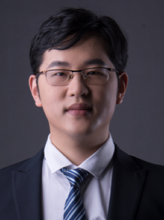 Song Shen (沈松)合作论文数School of Biomedical Sciences and Engineering, South China University of Technology;Laboratory for Nanomedicine and Biomaterials, South China University of Technology60
 Congfei Xu (许从飞)合作论文数华南理工大学生物医学科学与工程学院55
 Yucai Wang (王育才)合作论文数School of Life Sciences, University of Science and Technology of China;The CAS Key Laboratory of Innate Immunity and chronic Disease53
 Rupei Tang (唐汝培)合作论文数School of Life Sciences, Anhui University45
 Xin Wang (王鑫)合作论文数School of Life Sciences, Anhui University43Du Xiaojiao合作论文数40Shenghe Deng合作论文数35
 Kailin Cai (蔡开琳)合作论文数Union Hospital, Tongji Medical College, Huazhong University of Science and Technology35Junnan Gu合作论文数34Yinghao Cao合作论文数32
 Youyong Yuan (袁友永)合作论文数华南理工大学生物医学科学与工程学院31Fuwei Mao (毛富巍)合作论文数29
 Menghua Xiong (熊梦华)合作论文数华南理工大学生物医学科学与工程学院29Denglong Cheng (程登龙)合作论文数28Chunyang Sun (孙春阳)合作论文数28
 Hongjun Li (李洪军)合作论文数School of Pharmacy, Zhejiang University;The Second Affiliated Hospital, School of Medicine, Zhejiang University26
 Yang Liu合作论文数School of Life Sciences and Medical Center, University of Science and Technology of China26Le Qin合作论文数25
 Guoqing Yan (闫国卿)合作论文数School of Life Sciences, Anhui University24Ying-Li Luo合作论文数Univ Sci & Technol China, Sch Life Sci, Hefei 230027, Anhui, Peoples R China23
 Zhiting Cao (曹志婷)合作论文数School of Biopharmacy, China Pharmaceutical University23Zhu Yan-Hua合作论文数23
 Li Liu (刘莉)合作论文数Union Hospital, Tongji Medical College, Huazhong University of Science and Technology23
 Tianmeng Sun (孙天盟)合作论文数International Center of Future Science, Jilin University;Bethune First Hospital of Jilin University23
 Xiu Nie (聂秀)合作论文数Union Hospital, Tongji Medical College, Huazhong University of Science and Technology23Jia Yang合作论文数21
 Kam W. Leong (梁锦荣)合作论文数Department of Biomedical Engineering, School of Engineering and Applied Science, Columbia University;Department of Systems Biology, Columbia University19Liu Dahai合作论文数18
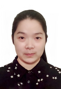 Chengqiong Mao (毛成琼)合作论文数School of Biomedical Engineering, Guangzhou Medical University17Zi-Dong Lu合作论文数Institutes for Life Sciences and School of Medicine, South China University of Technology, Guangzhou, Guangdong 510006, People's Republic of China16
 Yandan Yao (姚燕丹)合作论文数Sun Yat-sen Memorial Hospital of Sun Yat-sen University15
 Feng Wang (王峰)合作论文数School of Food and Biological Engineering, Hefei University of Technology15Shuang Dou合作论文数15Xu Cheng合作论文数13Ji-Long Wang合作论文数13
 Hai-Quan Mao (毛海泉)合作论文数Department of Materials Science and Engineering, Whiting School of Engineering, Johns Hopkins University;Institute of NanoBioTechnology, Johns Hopkins University;LifeSprout, Inc.13Gui Zhao合作论文数12Yue Zhang (张越)合作论文数College of Chemical and Pharmaceutical Engineering,Hebei University of Science and Technology12Hou-Bing Zhang合作论文数Sch Biomed Sci & Engn, South China Univ Technol11
 Erwei Song (宋尔卫)合作论文数Sun Yat-sen Memorial Hospital, Sun Yat-sen University;Health Science Center, Sun Yat-sen University11Shengxiang Fu合作论文数10
 Hongxia Wang (王洪霞)合作论文数College of Food Science, Southwest University10
 Yangzhong Liu (刘扬中)合作论文数School of Chemistry and Materials Science, University of Science and Technology of China10Dao-Qiang Lu合作论文数9
 Rong Sun合作论文数9
 Xiqiu Liu (刘熙秋)合作论文数School of Pharmacy, Tongji Medical College, Huazhong University of Science and Technology9Guanqing Yang合作论文数Key Lab Polymer Ecomat, Chinese Acad Sci8




