RNA-seq gene set enrichment analysis for siRNA KDM4 A, B, C, A-C, and non-targeting control in WiT49 cells
Gene set enrichment analysis demonstrated significant alteration of the GO_CHROMATIN_SILENCING_AT_RDNA pathways in WiT49 and HEK293 cells treated with QC6352.
QC6352 treatment does not result in decreased cell viability or growth as determined by crystal violet assay in the anaplastic Wilms tumor cell line 17.94, or the favorable histology Wilms tumor cell lines PDM182 and COG-W-408.
QC6352 treatment (25nM x 72 hours) reduces nucleoli in WiT49 and HEK293. (A) KDM4A (red) and the nucleolar marker fibrillarin (green) co-localize in the nucleoli [also represented by areas of reduced DAPI staining (blue) in the nucleus due to incompact chromatin in nucleoli] in WiT49 cells. (B) QC6352 treatment caused a reduction in the fibrillarin/nuclear area (determined by the area of fibrillarin/area DAPI) in WiT49 cells (p<0.0001), representing a reduction of nucleoli. (C) Similar findings were found in QC6352-treated HEK293 cells, which also exhibited a (D) reduction of the fibrillarin/nuclear area (p<0.0001).
Gene set enrichment analysis demonstrated significant downregulation of the REACTOME_EUKARYOTIC_TRANSLATION_INITIATION and REACTOME_PRC2_METHYLATES_HISTONES_AND_DNA pathways in QC6352-treated WiT49 cells and the REACTOME_EUKARYOTIC_TRANSLATION_INITIATION pathway in HEK293 cells.
Children with favorable-histology Wilms tumor (FHWT) who relapse or whose tumors show blastemal predominance post-chemotherapy often face poor outcomes. The purpose of this study is to identify mechanisms of chemotherapy resistance in FHWT. We induce a patient-derived xenograft model (KT-47) to develop blastemal predominance after chemotherapy and to become resistant to vincristine, actinomycin-D, and doxorubicin (VAD). Multi-omics analyses reveal chromatin and transcriptional changes, including increased H3K4me3 and decreased H3K27me3 at stem cell and nephrogenesis gene loci. LIN28B is the most upregulated resistance-associated gene, linked to MYCN copy gain/upregulation and chromatin remodeling. ABCB1 expression correlates with interchromosomal enhancer interactions and functions as the mediator of chemotherapy resistance in vitro. These findings are validated in additional Wilms tumor models. Overall, resistance is associated with de-differentiation to a stem-like state and is driven by ABCB1 upregulation, suggesting that therapeutic strategies targeting chromatin regulation and drug efflux may be relevant in therapy-resistant Wilms tumor.
(A) Log2CPM RNA-seq values demonstrate that KDM4A transcription and protein levels are not directly correlated and that high KDM4A expression by RNA-seq does not positively correlate with sensitivity to QC6352. (B) QC6352 treatment did not alter protein levels of KDM4A, B, or C in COG-W-408, PDM182, or 17.94 cells, all of which are insensitive to QC6352.
QC6352 does not cause a biologically significant increase in cell death or apoptosis in WiT49 or HEK293 cells. The percent difference in cell death was statistically significant in HEK293 cells, but the absolute difference was less than 1%. ***paired two-tailed t-test p value <0.001; ns=non-significant.
Dose-dependent decrease in ribosomal protein expression in response to QC6352 treatment in WiT49 and HEK293. Treatment of WiT49 and HEK293 cells with the above concentrations of QC6352 for 72 hours resulted in a dose-dependent decrease in KDM4A-C and the ribosomal proteins RPS6, RPS3, RPL7a, RPL26, and RPS27a by western blot. In addition, a dose dependent decrease in P70 S6Kinase, which phosphorylates the ribosomal protein RPS6 to initiate protein translation, was observed.
Transmission electron microscopy demonstrates that 25 nM QC6352 treatment for 72 hours caused a structural change resulting in less compact nucleoli WiT49 and HEK293 cells. More compact nucleoli with dense/compact fibrillar centers are known to be more active. Nuclear membranes indicated with black arrows and nucleoli are labeled.
RNA-seq gene set enrichment analysis for QC6352-treated WiT49 and HEK293 cells and controls
RNA-seq of QC6352-treated WiT49 and HEK293 cells (25nM x 72 hours) did not demonstrate a change in KDM4 A, B, or C transcript levels.
siRNA mediated knockdown of KDM4A, B, C, and KDM4A-C did not reduce ribosomal protein expression in WiT49 and HEK293 cells.
Knockdown of KDM4A alters QC6352 sensitivity. (A) The PrestoBlue assay demonstrated that siRNA-mediated knockdown of KDM4A and C was associated with resistance to QC6352 when compared to parental cells and empty vector control in WiT49. (B) The PrestoBlue assay demonstrated that siRNA-mediated knockdown of KDM4A was associated with resistance to QC6352 when compared to parental cells and empty vector control in HEK293. (C)IC50 values for WiT49 and HEK293 with siKDM4A-C and NTC siRNA.
PRISM gene set analysis. (A) PRISM multiplexed cell line profiling RNA expression and compound sensitivity correlation analysis from 931 cancer cell lines was utilized to determine genes with basal RNA expression that correlated with QC6352 sensitivity. Analysis of the top 100 genes with basal expression levels associated with QC6352 sensitivity using the Enrichr database demonstrated that 9 of the top 10 pathways identified on gene set analysis were associated with the ribosome. (B) Tabular view of the top 10 gene sets. (C) Individual ribosomal protein genes comprising the gene sets from A and B that were among the top 100 genes in the PRISM analysis.
High-risk neuroblastoma (NB) is driven by the amplification of MYCN in conjunction with additional oncogenic mutations in genes encoding kinases such as ALK. NB cells require antioxidant responses to maintain redox balance and are highly sensitive to ferroptosis. Here, we show that metabolites derived from infiltrating immune cells expressing IL4i1, a secreted oxidoreductase, are potent suppressors of NB ferroptosis. IL4i1 metabolites (indole-3-pyruvate and 4-hydroxyphenylpyruvate) blocked ferroptosis in all human NB cell lines via a mechanism that depended on free radical scavenging and NRF2 activation but did not require the aryl hydrocarbon receptor. Supernatant transfer experiments confirmed that IL4i1 creates a milieu that protects NB cells from oxidative cell death. Importantly, mice lacking IL4i1 were protected from NB in a high-penetrance MYCN and mutant ALK-driven autochthonous cancer model. Therefore, we propose that immune IL4i1 is permissive for NB growth and survival. IL4i1 produces context-dependent oncometabolites and, as a secreted enzyme, represents a target for cell death manipulation in cancers sensitive to oxidative stress-driven cell death.
 Davidoff Andrew M合作论文数38Jie Fang合作论文数St Jude Childrens Res Hosp, Dept Dev Neurobiol, Memphis, TN 38105 USA35Christopher L. Morton合作论文数26Prahalathan Pichavaram合作论文数Dept Clin & Adm Pharm, Univ Georgia25
 Hyea Jin Gil合作论文数St Jude Childrens Res Hosp, Dept Genet, Memphis, TN 38105 USA24
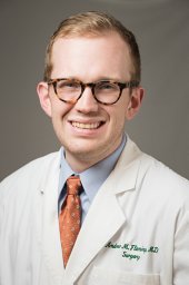 Andrew M Fleming合作论文数Health Science Center, The University of Tennessee24Melissa M. Ronan合作论文数23
 Andrew Boghossian合作论文数23Jennifer A Roth合作论文数Broad Inst MIT & Harvard, Cambridge, MA 02142 USA23
 Matt Rees合作论文数23
 Carolyn Jablonowski合作论文数17
 Gerard P. Zambetti合作论文数Comprehensive Cancer Center, St. Jude Children's Research Hospital16
 Hong-Jian Jin合作论文数College of Life Sciences, Zhejiang University12Murphy Andrew W合作论文数10
 Xiang Chen合作论文数Department of Computational Biology, Graduate School of Biomedical Sciences, St. Jude Children's Research Hospital10
 ruoning wang (王若宁)合作论文数9Glazer Evan S合作论文数Ctr Hlth Sci, Univ Tennessee9
 Sivaraman Natarajan合作论文数9
 Xiaotu Ma合作论文数Department of Computational Biology, St. Jude Children's Research Hospital;Comprehensive Cancer Center, St. Jude Children's Research Hospital7
 H. Sheppard合作论文数Research Department, CIBA Pharmaceutical Products Inc.7
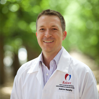 Murphy Andrew James合作论文数Baker IDI Heart and Diabetes Institute6
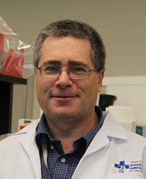 David J. Picketts合作论文数Ottawa Health Research Institute;University of Ottawa Centre for Neuromuscular Disease and Departments of Medicine, Biochemistry, Microbiology and Immunology, Ottawa, ON, K1H 8M5, Canada;Ottawa Health Research Institute, University of Ottawa6Tingting Wang合作论文数Nationwide Childrens Hosp, Ohio State Univ5
 Jeffrey D Steinberg合作论文数5Shivendra V. Singh合作论文数5
 Richa Jain合作论文数4
 Shondra M Pruett-Miller合作论文数St Jude Childrens Res Hosp, Dept Cell & Mol Biol, 332 N Lauderdale St, Memphis, TN 38105 USA3
 Quarni Waise合作论文数H. Lee Moffitt Cancer Center, University of South Florida3
 Paul G. Thomas合作论文数Vaccine and Infectious Disease Division, Fred Hutchinson Cancer Center3
 Taosheng Chen合作论文数Department of Chemical Biology and Therapeutics, St. Jude Children's Research Hospital3Meifen Lu合作论文数2Qiong Wu合作论文数2Thomas Confer合作论文数2
 Geoffrey Neale合作论文数2Nour Yadak合作论文数2Ti-Cheng Chang合作论文数2Dongli Hu合作论文数Department of Genetics and Tumor Cell Biology, St. Jude Children's Research Hospital, Memphis, Tennessee 38105, USA.2Lingling Liu合作论文数Ohio State Univ, Nationwide Childrens Hosp, Res Inst, Ctr Childhood Canc & Blood Dis, Columbus, OH 43205 USA2
 Peter Murray合作论文数Max-Planck-Institut für Biochemie;Technische University Munich2
 Beisi Xu合作论文数2Lee Ha Won合作论文数2
 Ahmed Abu-Zaid合作论文数2
 Gang Wu合作论文数Department of Pathology, St. Jude Children’s Research Hospital;Center for Applied Bioinformatics, St. Jude Children’s Research Hospital2Teresa Santiago合作论文数2
 Jerold Rehg合作论文数St. Jude Children's Res. Hospital, St Jude Children's Research Hospital2Melissa M. Johnson合作论文数2
 Changde Cheng合作论文数Dept Integrat Biol, Univ Texas Austin2
 Kevin Freeman合作论文数Department of Immunology, Baylor College of Medicine2David Shibata合作论文数2




