GSDMA, the primary member of the gasdermin family found in the skin, is critical for pathogen-induced pyroptosis during infection. Recent studies revealed that another gasdermin, GSDMD, undergoes palmitoylation during pyroptosis. However, whether and how the other gasdermin members undergo lipid modification remain poorly understood. Here, we demonstrate that GSDMA is S-acylated at the conserved cysteine residues in its N-terminal domain. We show that the S-acylation of GSDMA promotes pyroptosis by facilitating its membrane anchoring and protein oligomerization, a mechanism distinct from the palmitoylation of GSDMD at the N-terminal C191 residue. In addition, we present evidence that recombinant proteins of GSDMA and GSDMD can undergo S-acylation in vitro independent of palmitoyl transferases via direct interaction with palmitoyl-CoA. Furthermore, we identify ABHD17A as one of the deacylating enzymes that regulate the dynamic fatty acylation cycle of GSDMA. Overall, our studies reveal new molecular mechanisms underlying GSDMA function through S-acylation and underscore its important role in regulating pyroptosis mediated by GSDMA.
Transcriptional-enhanced associate (TEA)/transcriptional enhanced factor (TEF) domain transcription factors (TEAD1-4) regulate the transcriptional output of Hippo signaling by interacting with the coactivators Yes-associated protein (YAP) and transcriptional coactivator with PDZ-binding motif (TAZ) and play a crucial role in animal development and tumorigenesis. Much of the regulation of the TEAD-YAP/TAZ complex is through the regulation of nuclear translocation and degradation of YAP/TAZ by the upstream Hippo pathway regulators. However, TEADs undergo several posttranslational modifications, which are mostly independent of upstream Hippo pathway components. Alternative splicing of TEAD1 has also been reported to modulate TEAD activities posttranscriptionally. In addition, TEADs can bind to Vestigial-like (VGLL) proteins, which regulate distinct transcription programs that influence tumor growth, immunity, and development. VGLL1-3 may act as transcriptional activators or repressors, while VGLL4 functions primarily as a repressor by competing with YAP/TAZ for TEAD binding. Here, we discuss the Hippo-independent regulations and functions of TEADs, as well as the role of the TEAD-VGLL complex in development and disease, shedding light on therapeutic strategies of targeting the TEAD-VGLL complex.
Supplementary Figure 5 shows the IHC results looking at CD4 and CD8 expression in KC and KCRG mice. The results show no difference in the immune landscape between these mouse models.
Supplementary Figure 6 describes RNA-seq shows differential gene expression induced by oncogenic KRAS.
Supplementary Figure 2 describes the characterization of the impact Gli2 overexpression in pancreas development. showing that Gli2 loss has no impact on pancreas development or survival in vivo.
Supplementary Figure 10 shows not differences in H3K4me1 enrichment at Ccnd1 promoter in mutant KRAS cells.
Supplementary Figure 4 shows the validation of Kras signaling activation and chronic pancreatitis phenotypic examples in KC and KCRG mice.
Supplementary Figure 8 shows the expression of GLI target genes in all experimental groups.
The failure to clear dysfunctional mitochondria, cell death and inflammation have been linked in neurodegenerative disease, but their relationship and role in these conditions is not fully understood. Loss of Vps13d prevents clearance of mitochondria, and mutations in human VPS13D have been associated with neurological movement disorders. To investigate the relationship between mitochondrial health, inflammation and neurodegeneration, we created a conditional Vps13d-knockout mouse. Loss of Vps13d in excitatory neurons resulted in behavioral changes and neurodegeneration. Vacuolar protein sorting 13D (VPS13D) deficiency also caused mitochondrial ultrastructural defects and dysfunction in neurons followed by gasdermin E processing, cyclic GMP-AMP synthase (cGAS)-stimulator of interferon response cGAMP interactor (STING) signaling, microglial activation and cell death. Gasdermin E localization with mitochondria in Vps13d-mutant neurons was required for elevated extracellular mitochondrial DNA that promoted activation of microglia. Depletion of microglia suppressed cell death and behavioral phenotypes but not mitochondrial changes in the neuron-specific Vps13d-knockout model, indicating that microglia promote cell death in this model of neurodegenerative disease.
Supplementary Figure 7 shows how oncogenic KRAS modulates GLI target gene expression.
Supplementary Table 1 describes the characteristics of the patients used in our study.
Intestinal homeostasis is tightly regulated by the reciprocal interaction between the gut epithelium and adjacent mesenchyme. The Hippo pathway is intimately associated with intestinal epithelial homeostasis and regeneration; however, its role in postnatal gut mesenchyme remains poorly defined. Here, we find that removal of the core Hippo kinases Lats1/2 or activation of YAP in adult intestinal smooth muscle layers has largely no effect; however, Hippo-YAP signaling in the niche-forming Gli1+ mesenchymal cells plays intrinsic roles in regulating intestinal homeostasis. We find that Lats1/2 deletion drives robust mesenchymal over-proliferation, and YAP activation in Gli1+ pericryptal cells disrupts the intestinal epithelial-mesenchymal crosstalk via promoting Wnt ligand production. We show that YAP is upregulated in the stroma during dextran sodium sulfate (DSS)-induced injury, and mesenchymal YAP activation facilitates intestinal epithelial regeneration. Altogether, our data suggest an important role for mesenchymal Hippo-YAP signaling in the stem cell niche during intestinal homeostasis and pathogenesis.
The Hippo signaling pathway plays crucial roles in various processes related to development and tissue homeostasis. This study demonstrates that sustained YAP1 activity can influence cell fate within the reproductive system, specifically in lutein and Müllerian mesenchymal cells, causing them to transdifferentiate into myofibroblasts. Recent reports have suggested that the Hippo intracellular signaling pathway is required for homeostasis in a variety of tissues, including the ovary and female reproductive tract. To further define the role of the Hippo effector YAP1 in the female reproductive system, transgenic mouse models were designed to direct the expression of a dominant stable mutant form of YAP1, termed YAP5SA, to the granulosa cells of antral follicles (R26YAP5SA ;CYP19-cre) and Müllerian mesenchymal cells (R26YAP5SA ;Amhr2 cre/+). Unexpectedly, YAP5SA expression in the ovaries of R26YAP5SA ;CYP19-cre mice was not detected in granulosa cells, but rather in a subset of lutein cells. This caused the lutein cells to transdifferentiate into cells having the morphologic and functional properties of myofibroblasts, including collagen deposition. These cells coalesced into roughly spherical lesions that persisted in the ovaries, but did not interfere with ovarian function or fertility. Seminiferous tubule-like structures also formed in the ovaries of adult R26YAP5SA ;CYP19-cre mice, containing SOX9-positive Sertoli-like cells but no germ cells. Although multi-lineage transdifferentiation had been reported in mice lacking the Hippo kinases Lats1 and -2 in their granulosa cells, comparative transcriptomic analyses of granulosa cells expressing YAP5SA vs granulosa cells lacking Lats1/2 showed few similarities in transcriptome alterations. R26YAP5SA ;Amhr2 cre/+ mice had severe developmental defects of their reproductive tracts, which were attributed to the transdifferentiation of Müllerian mesenchymal cells into myofibroblasts during embryogenesis. Together, these results indicate that sustained YAP1 signaling induces transdifferentiation in lutein and Müllerian mesenchymal cells, and further underscores the role of Hippo signaling in the maintenance of their fates.
Supplementary Figure 3 shows Gli2 expression in CRG and KCRG mice and Gli luciferase activity in ΔNGli2-transfected cells.
Studies on Hippo pathway regulation of tumorigenesis largely center on YAP and TAZ, the transcriptional co-regulators of TEADs. Here, we present an oncogenic mechanism involving VGLL and TEAD fusions that is Hippo pathway-related but YAP/TAZ-independent. We characterize two recurrent fusions, VGLL2-NCOA2 and TEAD1-NCOA2 , recently identified in human spindle cell rhabdomyosarcoma. We demonstrate that in contrast to VGLL2 and TEAD1 the fusion proteins are potent activators of TEAD-dependent transcription, and the function of these fusion proteins does not require YAP/TAZ. Furthermore, we identify that VGLL2 and TEAD1 fusions engage specific epigenetic regulation by recruiting histone acetyltransferase EP300 to control TEAD-mediated transcriptional and epigenetic landscapes. We show that small-molecule EP300 inhibition can suppress fusion protein-induced oncogenic transformation both in vitro and in vivo in mouse models. Overall, our study reveals a molecular basis for VGLL involvement in cancer and provides a framework for targeting tumors carrying VGLL , TEAD , or NCOA translocations.
Supplementary Table 2 includes the SNP analysis of components of the Hedgehog pathway.
Supplementary Figure 9 shows that Gli2 does not change H3K27Ac enrichment at Ccnd1 promoter downstream of oncogenic KRAS.
Supplementary Figure 1 shows correlation of SNP rs1992901 and GLI2 Transcript Expression.
Aberrant activation of GLI transcription factors has been implicated in the pathogenesis of different tumor types including pancreatic ductal adenocarcinoma. However, the mechanistic link with established drivers of this disease remains in part elusive. In this study, using a new genetically engineered mouse model overexpressing constitutively active mouse form of GLI2 and a combination of genome-wide assays, we provide evidence of a novel mechanism underlying the interplay between KRAS, a major driver of pancreatic ductal adenocarcinoma development, and GLI2 to control oncogenic gene expression. These mice, also expressing KrasG12D, show significantly reduced median survival rate and accelerated tumorigenesis compared with the KrasG12D only expressing mice. Analysis of the mechanism using RNA sequencing demonstrate higher levels of GLI2 targets, particularly tumor growth-promoting genes, including Ccnd1, N-Myc, and Bcl2, in KrasG12D mutant cells. Furthermore, chromatin immunoprecipitation sequencing studies showed that in these cells KrasG12D increases the levels of trimethylation of lysine 4 of the histone 3 (H3K4me3) at the promoter of GLI2 targets without affecting significantly the levels of other major active chromatin marks. Importantly, Gli2 knockdown reduces H3K4me3 enrichment and gene expression induced by mutant Kras. In summary, we demonstrate that Gli2 plays a significant role in pancreatic carcinogenesis by acting as a downstream effector of KrasG12D to control gene expression.
 Mihir Rajurkar合作论文数Dept Canc Biol, Univ Massachusetts23
 Martin E. Fernandez-Zapico合作论文数Department of Oncology, Mayo Clinic;Division of Gastroenterology and Hepatology, Department of Internal Medicine, Mayo Clinic22Jennifer L. Cotton合作论文数21
 Maite García Fernández-Barrena合作论文数Cima Universidad de Navarra21
 Andrew McMahon合作论文数Division of Biology and Biological Engineering, Caltech20Kayla C. LaRue-Nolan合作论文数18
 Luis Flores合作论文数18Anne M Vrabel合作论文数18Fatemeh Mousavi合作论文数18Sandhya Sen合作论文数18
 Tara Hogenson合作论文数18Renzo E Vera合作论文数Mayo Clinic Schulze Center for Novel Therapeutics18
 Glancis Luzeena Raja合作论文数Graduate School of Biomedical Sciences, Mayo Clinic18
 David R. Pease合作论文数18
 Ryan M. Carr合作论文数Mayo Clin, Dept Internal Med, Rochester, MN USA18
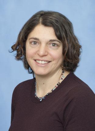 Marina Pasca Di Magliano合作论文数Department of Surgery, University of Michigan18
 William R Bamlet合作论文数18
 Rondell P. Graham合作论文数Mayo Clinic18Hagen Catherine E合作论文数18
 Tolosa Ezequiel J合作论文数Schulze Center for Novel Therapeutics, Mayo Clinic18
 Thomas Smyrk合作论文数Mayo Clinic;Mayo Medical School18Sigafoos Ashley N合作论文数18
 Ann L. Oberg合作论文数Mayo Clinic Comprehensive Cancer Center;Center for Clinical and Translational Science, Mayo Clinic;Division of Computational Biology, Department of Quantitative Health Sciences, Mayo Clinic18
 Robert R. McWilliams合作论文数Department of Oncology, Mayo Clinic;Mayo Clinic Comprehensive Cancer Center18
 Peter G. Zaphiropoulos合作论文数Department of Biosciences and Nutrition, Karolinska Institutet18Lizhi Zhang合作论文数18Kari G. Rabe合作论文数18Luciana L. Almada合作论文数18
 Hugues Sicotte合作论文数Joseph Henry Laboratories;Princeton University;Joseph Henry Laboratories, Princeton University18
 Eric Chevet合作论文数Anatomy and Cell Biology, McGill University18
 Sherine F Elsawa合作论文数Mayo Clinic, Mayo Clinic College of Medicine18Christopher L. Pin合作论文数Department of Paediatrics, The University of Western Ontario18
 Erin L. Schenk合作论文数Eppley Institute for Research in Cancer and Allied Diseases, University of Nebraska Medical Center18David L. Marks合作论文数18Qi Li合作论文数14
 Yicktung T Ip合作论文数Medical School, University of Massachusetts13Brian C. Lewis合作论文数12Xu Wu合作论文数12Kyvan Dang合作论文数10
 Lihua Julie Zhu合作论文数Dept Mol Cell & Canc Biol, Univ Massachusetts8
 Chen Wu (吴晨)合作论文数Cancer Hospital, Chinese Academy of Medical Sciences;Peking Union Medical College, Chinese Academy of Medical Sciences;Changping Laboratory7Wilfredo E. De Jesus-Monge合作论文数7Zhipeng Tao合作论文数Harvard Medical School, Massachusetts General Hospital6
 Yang Sun合作论文数Harvard Medical School and Brigham and Women's Hospital, Harvard Medical School6
 Lan Xu (徐岚)合作论文数School of Biology & Basic Medical Sciences, Soochow University6Byeong-Moo Kim合作论文数5
 Arthur M. Mercurio合作论文数The Mercurio Laboratory, Department of Molecular, Cell and Cancer Biology, University of Massachusetts Chan Medical School5
 Randy Johnson合作论文数Department of Cancer Biology, Graduate School of Biomedical Sciences, The University of Texas MD Anderson Cancer Center5Singh Alka合作论文数5




