Introduction: Oral Submucous Fibrosis (OSF) is an oral precancerous condition with highest potential for malignant transformation. When OSF advances to oral cancer, epithelial cells undergo several dysplastic changes that alter the epithelial properties and architecture. Analysis of these features can provide useful diagnostic information to assess malignant potentiality of the disease process to avoid progression to oral cancer. Aim: The aim of the study was to assess the progression of non dysplastic and dysplastic OSF to malignancy using histomorphometry and E-cadherin expression in different layers of epithelium. Materials and Methods: The present cross-sectional study was conducted in the Department of Oral and Maxillofacial Pathology, Guru Nanak Institute of Dental Sciences and Research (GNIDSR), Panihati, Kolkata, and School of Medical Science and Technology (SMST), IIT, Kharagpur, West Bengal, India during the period of December 2019 to August 2021. It included 50 subjects divided into two groups, with 43 individuals suffering from OSF and seven individuals without disease process. Biopsy was conducted to establish diagnosis of OSF and stained sections were classified into nondysplastic and dysplastic category. Some sections were also prepared on lysine coated slides for immunohistochemical analysis. Finally, both sections were taken to SMST, IIT, Kharagpur, India, for staining with E-cadherin antibody and for procurement of photomicrographs using inverted microscope to undergo histomorphometrical analysis in basal-parabasal layers of epithelium and for assessing/comparing expression of adhesion molecule (E-cadherin) in basal-parabasal and spinous layers of non dysplastic and dysplastic OSF tissue using Image J software. Results: The mean cell area was found to be gradually increased from Normal Oral Mucosa (NOM) (35.60±3.26) to OSF with dysplastic (wd (39.12±4.99) followed by OSF without dysplastic (wtd) (64.11±9.90). The mean value of major axis was highest in OSFwtd (14.53±3.20) in comparison to OSFwd (11.23±1.98) and NOM (8.14±0.99), whereas the mean of minor axis was found to be decreased in OSFwd (4.24±0.89) and increased in case of OSFwtd (6.62±1.11) when compared with NOM (6.60±0.83). aspect ratio was highest in OSFwd (2.82±0.86), which showed gradual decrease in OSFwtd (2.37±0.71) and NOM (1.24±0.08). The gray scale value of membranous expression of E-cadherin in basal-parabasal layers was found to be highest in OSFwd (124.6±14.8) whereas it showed decrease in cytoplasmic expression in OSFwd (89.85±20.08) indicating loss of E-cadherin expression in cell membrane and simultaneous accumulation in cell cytoplasm (as gray scale value is inversely proportional to E-cadherin expression).The spinous cell layer also showed increased membranous gray scale value in OSFwd (143.17±15.5) and decreased cytoplasmic gray scale value in OSFwd (93.03±6.98). Conclusion: The study concluded that semiquantitative light microscopic histomorphometrical parameters like cellular size (area, major axis, minor axis) and shape (aspect ratio) depicting various statistically significant alterations, along with membranous loss of E-cadherin with concomitant cytoplasmic accumulation, both in basal-parabasal and spinous layers of the surface epithelium can be regarded as a significant indicator in predicting the disease progression of OSF to malignancy.
Diabetic wound healing presents a major challenge due to impaired vascular function, chronic inflammation, and a defective immune response caused by hyperglycemia. Current wound care technologies, including hydrogels, growth factor therapies, and bioengineered skin substitutes, have inherent limitations and fail to address the complex healing requirements of diabetic wounds. In this study, we report the development of a silk fibroin-based scaffold that exhibits controlled drug delivery modulated by the presence of bioinspired surface patterns. We demonstrate remarkable in vivo efficacy of the patterned scaffold in a diabetic rat model. Wound healing and tissue regeneration were tracked using Swept-Source Optical Coherence Tomography (SS-OCT), histopathology, and molecular analysis (immunohistochemistry and quantitative PCR). We show that the scaffold significantly accelerates wound healing, as evidenced by enhanced cellular differentiation, angiogenesis, and extracellular matrix remodeling. These findings highlight the scaffold's ability to overcome the limitations of current wound care technologies by integrating structural support with sustained drug release. The approach not only improves wound closure and tissue regeneration but also provides a promising, bioinspired solution for targeted diabetic wound management with high potential for clinical translation.
INTRODUCTION:In Traditional Chinese Medicine (TCM), tongue diagnosis plays an important role. Besides shape, colour and textural attributes of tongue regions have significance due to their association with different organs (viz. heart, lung, kidney-bladder, liver-gallbladder and stomach-spleen) in health and disease. Although numerous quantitative methods have been proposed to reduce inter and intra observer variability in tongue diagnosis, almost all of them considered only global colour/textural features i.e. from entire body of tongue. In this exploratory work, regional colour and texture information of tongue have been analysed for 16 healthy subjects (NOM) and 18 patients suffering from chronic acidity and indigestion (ACD). METHODS:Tongue boundaries are marked manually in each image and the tongue body is automatically partitioned into five regions linked with said internal organs. Rough assessments of region boundaries are obtained from literature. The boundaries are fine-tuned according to experts' advice. Several first and second order statistical features based on grey scale intensities and a∗ values of LAB colour space (indicative of redness) have been extracted from five different regions of tongue images. Intragroup analysis using Wilcoxon signed rank test compared different regions within individual study groups. Additionally, intergroup analysis, using Wilcoxon rank sum test, compared tongues of NOM with ACD for each region separately. p values were adjusted using the Benjamini-Hochberg false discovery rate procedure with q = 0.05. RESULTS:Intragroup analysis reveals a∗ values are statistically different (p ≤ 0.05) between stomach-spleen and liver-gallbladder regions for ACD while no significant change of a∗ are noted between these regions of normal tongue. It was found from intergroup analysis that correlation (one of the second order texture features extracted from grey scale images) of heart region has significantly decreased while correlation of kidney, liver-gallbladder and stomach-spleen regions significantly increased from NOM to ACD. CONCLUSION:This exploratory study sheds light on importance of inclusion of regional textural analysis of tongue in quantitative studies and can be applied to automated tongue diagnosis for other diseases.
The manuscript concentrates on spatial distribution and various nucleomorphometric parameters of epithelium to diagnose Oral Submucous Fibrosis (OSF). Histologically confirmed OSF and normal submucosa tissue samples were procured and stained with diamidino phenylindole (DAPI) to visualize nuclei. E-cadherins and p63 were also immunohistochemically stained. Microphotographs were analyzed to quantify the spatial distance among the nuclei of the stained tissue samples. Intensity of the DAPI stained nuclei and p63 was quantified. In addition, morphometric analysis of nuclei was done with the help of ImageJ software to quantify the geometric alterations in OSF tissue. Spatial distances among the nuclei of OSF tissue samples were found to be significantly higher than that of normal tissue. We also observed a significant decrease in the mean intensity of DAPI and p63 in OSF tissue samples. In addition, we have found statistically significant alterations of various morphometric quantifications in OSF tissue nuclei. There was a considerable change in the spatial distribution of nuclei, as well as some distinct changes in the nucleogeometry of OSF tissue, which corresponds to histological abnormalities. Decreased intensity of DAPI and p63 advocate disease progression. The biomarkers in this study are the accountable role-players for early detection of oral carcinoma.
Traditional drug testing via polystyrene or glass-based cell culture platforms exposes cells to static drug doses and mechanically rigid environments [stiffness in gigapascals (GPa)], which do not accurately replicate physiological conditions. To address these limitations, we developed a polydimethylsiloxane (PDMS)-based microfluidic concentration gradient generator (μCGG) with six integrated cell culture chambers, using a cost-effective and frugal micro-hydrogel molding-assisted technique that eliminates the need for cleanroom infrastructure, specialized equipment, or advanced expertise. This platform facilitates dynamic drug exposure to cells cultured in chambers with flexible PDMS bases [stiffness in kilopascal (kPa) range], providing a scalable and accessible approach for drug dose-response analysis under physiologically relevant conditions, thereby improving accuracy. μCGG utilized a pressure-driven flow design that repeatedly split, mixed, and recombined fluid streams owing to the presence of the mesh-like geometry of the microchannels. This generated a stable and predictable drug concentration gradient across six outlet chambers, as validated through COMSOL simulations, fluorescence microscopy, and UV-Vis spectroscopy using 5-fluorouracil (5-Fu) as a model drug. MDA-MB-231 breast cancer cells were then cultured in the outlet chambers and exposed to six distinct dynamically generated concentrations of 5-Fu. Cellular viability assessed via live/dead assays yielded an IC50 value of 41 ± 4 μM, closely matching the results from conventional multiwell plates using manually pipetted gradients under static conditions (IC50: 36 ± 3 μM). Additional validation was carried out using immunocytochemistry and flow cytometry to assess apoptotic markers and treatment responses. Overall, our study presents a simple, frugal, and scalable microfluidic platform that addresses the major limitations of traditional drug testing platforms by incorporating dynamic chemical gradients, physiologically relevant mechanical environments, and low-barrier fabrication methods, paving its way for broader adoption in preclinical drug evaluation and dose-response assays.
Emotions are a vital semantic part of human correspondence. Emotions are significant for human correspondence as well as basic for human–computer cooperation. Viable correspondence between people is possibly achieved when both the importance and the emotion of the correspondence are perceived by all groups included. Understanding the significance of language has generally been concentrated on in natural language processing (NLP) as a semantic examination. In NLP, the text can be handled appropriately for classification. Emotion detection from facial emotion is the subfield of social signal processing applied in a wide assortment of regions, explicitly for human and PC collaboration. Many researchers have proposed various approaches, generally utilizing machine learning concepts. Automatic emotion recognition (AER) is significant for working with consistent intuitiveness between a person and a smart device toward fully acknowledging an intelligent society. Many researchers examined cross-lingual and multilingual speech emotion as a stage toward language-free emotion acknowledgment in natural speech. In the present work, we are proposing a deep learning-based AER system using four openly accessible datasets, namely Basic Arabic Vocal Emotions Dataset (BAVED), Acted Emotional Speech Dynamic Database (AESDD), Urdu written in Latin/Roman Script (URDU), and Toronto Emotional Speech Set (TESS), by utilizing the Jupyter notebook and a Python library for music and audio synthesis named Librosa. The experimental results exhibited that the proposed approach achieves better than the existing approaches, i.e., the accuracy of the proposed system with the URDU dataset is 96.24
The Internet of Things (IoT) is being prominently used in smart cities and a wide range of applications in society. The benefits of IoT are evident, but cyber terrorism and security concerns inhibit many organizations and users from deploying it. Cyber-physical systems that are IoT-enabled might be difficult to secure since security solutions designed for general information/operational technology systems may not work as well in an environment. Thus, deep learning (DL) can assist as a powerful tool for building IoT-enabled cyber-physical systems with automatic anomaly detection. In this paper, two distinct DL models have been employed i.e., Deep Belief Network (DBN) and Convolutional Neural Network (CNN), considered hybrid classifiers, to create a framework for detecting attacks in IoT-enabled cyber-physical systems. However, DL models need to be trained in such a way that will increase their classification accuracy. Therefore, this paper also aims to present a new hybrid optimization algorithm called “Seagull Adapted Elephant Herding Optimization” (SAEHO) to tune the weights of the hybrid classifier. The “Hybrid Classifier + SAEHO” framework takes the feature extracted dataset as an input and classifies the network as either attack or benign. Using sensitivity, precision, accuracy, and specificity, two datasets were compared. In every performance metric, the proposed framework outperforms conventional methods.
Magnetic drug targeting (MDT) leverages external magnetic fields to guide magnetic drug carriers (MDCs) to diseased sites. However, its clinical use is hindered by challenges such as the weakening of magnetic fields with tissue depth, MDC size/instability issues, and hydrodynamic shear. Despite years of research, progress remains limited due to the absence of reliable disease models, as animal models pose ethical and interspecies concerns, while current synthetic platforms struggle to replicate the tumor microenvironment (TME) and assess cellular responses to magnetic stimuli accurately. To address these limitations, we present an on-chip model developed using a lithography-free fabrication method to recreate physiologically relevant tumor conditions for evaluating MDC-assisted therapy. Our model closely replicates the breast TME by using an MDA-MB-231 cell-embedded hydrogel matrix flanked by two HUVEC-lined deformable microchannels, facilitating endothelial-tumor cell interactions and pressure-driven perfusion on-chip. As a proof of concept for targeted therapy applications, the platform was used to investigate MDT using a 10-20 nm (diameter) chitosan-coated MDC by assessing its retention against critical parameters such as variable magnetic fields and shear stress conditions, enabling precise magnetic-field calibration for optimal targeting. Subsequently, high-resolution imaging captured dose-response effects of the magnetically targeted drug via live/dead assays and immunocytochemistry studies, while flow cytometry and gene expression analysis revealed apoptotic pathway activation and reduced invasion markers. Overall, we establish our bioengineered chip as a cost-effective, scalable, and first-of-its-kind biomimetic system for MDT research designed to facilitate preclinical screening of potential anticancer therapies.
Surface functionalization strategies to replicate extracellular matrix (ECM) properties often rely on chemical modification. However, issues such as cytotoxicity, poor degradation control, and sensitivity to physiological conditions limit their applicability in long-term and translational contexts. To address this, we report a material-independent, chemically inert approach that uses biomimetic surface topographies to direct cell behavior using physical cues alone. While natural surfaces offer a wealth of hierarchical micro/nano architectures, the functional distinctions among topographies derived from different phenotypes of the same biological origin remain underexplored. Here, we present a systematic comparison of polydimethylsiloxane (PDMS) replicas inspired by lotus leaves, red rose petals, and yellow rose petals to examine their influence on fibroblast behavior. Using soft lithography and UV-assisted replication, we fabricate high-fidelity surfaces and characterize them via atomic force microscopy (AFM) and scanning electron microscopy (SEM). Cellular responses were assessed through proliferation assays, morphological analysis, fluorescence imaging, and mechanosensing behavior. Our results reveal that each surface elicits distinct cell-substrate interaction profiles, with the yellow rose petal-inspired topography showing superior support for adhesion, spreading, and proliferation. This behavior is attributed to its unique topographical density and orientation. The study offers a novel framework for harnessing natural geometries in the design of reproducible and cytocompatible platforms for regenerative and in vitro biomedical applications.
[This corrects the article DOI: 10.1016/j.jtcme.2019.10.002.].
Cancer stands as a formidable adversary on the global stage, claiming a significant number of lives each year. Yet, amidst this sobering reality, the importance of early detection cannot be overstated. Vigilant screenings, educational initiatives, and advancements in diagnostic technologies have emerged as crucial allies in our fight against this disease, enabling the identification of cancer at its most treatable stages and bolstering the prospects of successful intervention. In this paper, we embark on a transformative journey in cancer data analysis, harnessing the power of competitive ensemble machine learning techniques. Through meticulous feature selection, hyperparameter tuning, and data preprocessing, our methodology seeks to transcend the limitations of individual models, striving for heightened accuracy and nuanced insights.Three dataset used in the work is taken from UCI Machine Learning Repository. Our experimental findings underscore the efficacy of this approach, with accuracy rates reaching impressive levels: 99
When recognizing underwater images, problems, including poor image quality and complicated backdrops, are significant. The main problem of underwater images is the blurriness and invisibility of objects present in an image. This study presents a unique object identification design built on a YOLOv8 (You Only Look Once) framework upgraded to address these problems and further improve the models' accuracy. The study also helps in identifying underwater trash. The model is a two-phase detector model. The first phase has an Underwater Image Enhancer (UIE) data augmentation technique that works with Laplacian pyramids and gamma correctness methods to enhance the underwater images. The second phase, the proposed refined, innovative YOLOv8 model for classification purposes, takes the output from the first stage as its input. The YOLOv8 model's existing feature extractor is replaced in this study with a new feature extractor technique, HEFA, that yields superior results and better detection accuracy. The introduction of the UIE and HEFA feature extractor method represents the significant novelty of this paper. The proposed model is pruned simultaneously to eliminate unnecessary parameters and further condense the model. Pruning causes the model's accuracy to decline. Thus, the transfer learning procedure is employed to raise it. The trials’ findings show that the technique can detect objects with an accuracy of 98.5% and a mAP@50 of 98.1% and that its real-time detection speed on the GPU is double that of the YOLOv8m model's baseline performance.
In general, a network that possesses numerous free or autonomous nodes is proffered as a Mobile Ad hoc Network (MANET). In this, to send along receive data, every single node acts as a router. The entire network’s performance is degraded with the existence of faulty Sensor Nodes (SNs); thus, to obtain better Quality of Service (QoS), the detection of faulty SNs is highly significant. Therefore, an Efficient Node Localization and Failure Node Detection in a MANET environment is proposed here. Here, first, the nodes are initialized. Next, distance estimation, position computation, and optimal localization are the ‘3’ steps utilizing which the SNs are localized. After that, via the network, the data packets are broadcasted. The best path is regarded for transmitting the data packet efficiently. It includes ‘2’ steps for this; first, Failure Probability (FP) calculation; second, estimation of error calculation. Lastly, by employing the Soft Taxicab Poisson Binomial-Reference Point Group Mobility Model (STPB-RPGM), the Faulty Nodes (FNs) are detected. In this, the Taxicab Distance (TD)-Fuzzy C-Means (TD-FCM) is utilized to detect the group members along with by utilizing the Poisson Binomial Distribution (PBD)-Emperor Penguin Optimization (PB-EPO), the group leader is selected. Consequently, the failure along with the non-failure node is detected effectually by the STPB-RPGM. Lastly, the proposed STPB-RPGM’s outcomes are analogized with the prevailing algorithms. The experiential outcomes displayed that the faulty SNs are detected with higher accurateness in the proposed methodology; thus, it surpassed the other state-of-the-art methodologies. The findings indicate that we were able to get 96.8% accuracy, 96.46% sensitivity, 96.88% precision, 97.12% specificity, 96.64% F-measure, and 96.46% recall using the STPB-RPGM. In 0.619 s, the TD-FCM finishes the clustering operation. The benefit of this investigation is that, compared to other clustering algorithms, the localization strategy decreases the number of dead nodes in data transmission, and the TD-FCM completes the clustering process quickly.
Adhesive dynamics of cells plays a critical role in determining different biophysical processes orchestrating health and disease in living systems. While the rolling of cells on functionalised substrates having similarity with biophysical pathways appears to be extensively discussed in the literature, the effect of an external stimulus in the form of an electric field on the same remains underemphasized. Here, we bring out the interplay of fluid shear and electric field on the rolling dynamics of adhesive cells in biofunctionalised micro-confinements. Our experimental results portray that an electric field, even restricted to low strengths within the physiologically relevant regimes, can significantly influence the cell adhesion dynamics. We quantify the electric field-mediated adhesive dynamics of the cells in terms of two key parameters, namely, the voltage-altered rolling velocity and the frequency of adhesion. The effect of the directionality of the electric field with respect to the flow direction is also analysed by studying cellular migration with electrical effects acting both along and against the flow. Our experiment, on one hand, demonstrates the importance of collagen functionalisation in the adhesive dynamics of cells through micro channels, while on the other hand, it reveals how the presence of an axial electric field can lead to significant alteration in the kinetic rate of bond breakage, thereby modifying the degree of cell-substrate adhesion and quantifying in terms of the adhesion frequency of the cells. Proceeding further forward, we offer a simple theoretical explanation towards deriving the kinetics of cellular bonding in the presence of an electric field, which corroborates favourably with our experimental outcome. These findings are likely to offer fundamental insights into the possibilities of local control of cellular adhesion via electric field mediated interactions, bearing critical implications in a wide variety of medical conditions ranging from wound healing to cancer metastasis.
To mitigate the shortcomings of manual vessel segmentation of bloods in retinal images, an automatic algorithm is proposed to enhance diagnosis accuracy and reduce ophthalmologists' workload. The developed procedure for segmenting blood vessels of retinal fundus images has two sections: Image Partitioning and Attribute Extraction. Comprehensive experiments using different (DRIVE, HRF) datasets assessed the algorithm's performance in detecting retinal vessels of blood. Five quantitative metric performances parameters—accuracy, sensitivity, specificity, positive predictive value and negative predictive value—were validated through calculations of its efficiency against current state-of-the-art methods on the dataset used (DRIVE).The introduced technique for extracting blood vessels of retinal fundus images has some mentionable improvement by scoring an Accuracy, Sensitivity, Specificity, PPV, and NPV.
Colorectal cancer (CC) is one of the most predominant malignancies in the world, with the current treatment regimen consisting of surgery, radiation therapy, and chemotherapy. Chemotherapeutic drugs, such as 5-fluorouracil (5-FU), have gained popularity as first-line antineoplastic agents against CC but have several drawbacks, including variable absorption through the gastrointestinal tract, inconsistent liver metabolism, short half-life, toxicological reactions in several organ systems, and others. Therefore, herein, we develop chitosan-coated zinc-substituted cobalt ferrite nanoparticles (CZCFNPs) for the pH-sensitive (triggered by chitosan degradation within acidic organelles of cells) and sustained delivery of 5-FU in CC cells in vitro. Additionally, the developed nanoplatform served as an excellent exogenous optical coherence tomography (OCT) contrast agent, enabling a significant improvement in the OCT image contrast in a CC tissue phantom model with a biomimetic microvasculature. Further, this study opens up new possibilities for using OCT for the non-invasive monitoring and/or optimization of magnetic targeting capabilities, as well as real-time tracking of magnetic nanoparticle-based therapeutic platforms for biomedical applications. Overall, the current study demonstrates the development of a CZCFNP-based theranostic platform capable of serving as a reliable drug delivery system as well as a superior OCT exogenous contrast agent for tissue imaging.
Extracellular matrix diseases like fibrosis are elusive to diagnose early on, to avoid complete loss of organ function or even cancer progression, making early diagnosis crucial. Imaging the matrix densities of proteins like collagen in fixed tissue sections with suitable stains and labels is a standard for diagnosis and staging. However, fine changes in matrix density are difficult to realize by conventional histological staining and microscopy as the matrix fibrils are finer than the resolving capacity of these microscopes. The dyes further blur the outline of the matrix and add a background that bottlenecks high-precision early diagnosis of matrix diseases. Here we demonstrate the multiple signal classification method-MUSICAL-otherwise a computational super-resolution microscopy technique to precisely estimate matrix density in fixed tissue sections using fibril autofluorescence with image stacks acquired on a conventional epifluorescence microscope. We validated the diagnostic and staging performance of the method in extracted collagen fibrils, mouse skin during repair, and pre-cancers in human oral mucosa. The method enables early high-precision label-free diagnosis of matrix-associated fibrotic diseases without needing additional infrastructure or rigorous clinical training.
Optical coherence tomography (OCT) is a medical imaging modality that allows us to probe deeper sub-structures of skin. The state-of-the-art wound care prediction and monitoring methods are based on visual evaluation and focus on surface information. However, research studies have shown that sub-surface information of the wound is critical for understanding the wound healing progression. This work demonstrated the use of OCT as an effective imaging tool for objective and non-invasive assessments of wound severity, the potential for healing, and healing progress by measuring the optical characteristics of skin components. We have demonstrated the efficacy of OCT in studying wound healing progress in vivo small animal models. Automated analysis of OCT datasets poses multiple challenges, such as limitations in the training dataset size, variation in data distribution induced by uncertainties in sample quality and experiment conditions. We have employed a U-Net-based model for segmentation of skin layers based on OCT images and to study epithelial and regenerated tissue thickness wound closure dynamics and thus quantify the progression of wound healing. In the experimental evaluation of the OCT skin image datasets, we achieved the objective of skin layer segmentation with an average intersection over union (IOU) of 0.9234. The results have been corroborated using gold-standard histology images and co-validated using inputs from pathologists.Clinical Relevance—To monitor wound healing progression without disrupting the healing procedure by superficial, non-invasive means via the identification of pixel characteristics of individual layers.
 Ranjan Rashmi Paul合作论文数56
 Mousumi Pal合作论文数Department of Oral and Maxillofacial Pathology, Guru Nanak Institute of Dental Science and Research50
 Ajoy Kumar Ray合作论文数National Metallurgical Laboratory44Debdoot Sheet合作论文数Indian Institute of Technology Kharagpur30
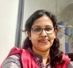 Sujatha Ramakrishnan合作论文数Vellore Institute of Technology21
 Das Raunak Kumar合作论文数School of Medical Science and Technology, Indian Institute of Technology Kharagpur21
 Amrita Chaudhary合作论文数School of Medical Science and Technology, Indian Institute of Technology Kharagpur21
 Sri Phani Krishna Karri合作论文数19
 Biswajoy Ghosh合作论文数UiT - The Arctic University of Norway18Barui Ananya合作论文数IIEST, Centre for Healthcare Science and Technology18
 Bag Swarnendu合作论文数School of Medical Science and Technology18
 Soumen Das合作论文数Sch Med Sci & Technol, Indian Inst Technol17
 Anji Anura合作论文数17
 Banerjee Provas合作论文数Banerjees׳ Biomedical Research Foundation17
 Raghvendra Kumar合作论文数16
 Sambuddha Ghosh合作论文数Regional Institute of Ophthalmology16
 Santanu Dhara合作论文数16
 A. Mukherjee合作论文数Fermi National Accelerator Laboratory Batavia IL 60510 USA14
 Manjunatha Mahadevappa合作论文数Sch Med Sci & Technol, Indian Inst Technol Kharagpur14
 Sanghamitra Sengupta合作论文数Biophysics Division;Indian Institute of Chemical Biology;Biophysics Division, Indian Institute of Chemical Biology14
 Hrushikesh Garud合作论文数13
 Debjani Chakraborty合作论文数Department of Mathematics, Indian Institute of Technology13Anurup Mukhopadhyay合作论文数12
 Mousumi Mandal合作论文数School of Medical Science and Technology, Indian Institute of Technology Kharagpur12Debaleena Nawn合作论文数Indian Inst Technol, Adv Technol Dev Ctr, Kharagpur 721302, W Bengal, India11
 Satarupa Banerjee合作论文数11
 Keya Chaudhuri合作论文数Human Genetics & Genomics Group;Indian Institute of Chemical Biology;Human Genetics & Genomics Group, Indian Institute of Chemical Biology11
 Abhishek Kumar合作论文数10
 Pramod Singh Rathore合作论文数Dept. of C.S, Gov. Eng. Coll., Ajmer, India;c;Dept. of C.S, Gov. Eng. Coll., Ajmer, India|c|10
 Monika Rajput合作论文数Dept. of Appl. Phys., Delhi Technol. Univ.;c;Dept. of Appl. Phys., Delhi Technol. Univ.10
 Preeti Gulia合作论文数MDU, Department of MCA;c;MDU, Department of MCA10
 Chandan Chakraborty合作论文数Electrical Engineering, Indian Institute of Technology Kharagpur10
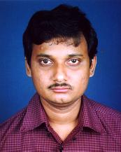 Pabitra Mitra合作论文数Department of Computer Science & Engineering, Indian Institute of Technology10
 Rabibrata Mukherjee合作论文数Department of Chemical Engineering, Indian Institute of Technology10
 Sailesh Conjeti合作论文数Technische Universität München10
 Nasib S. Gill合作论文数Maharshi Dayanand University10Das A K合作论文数9
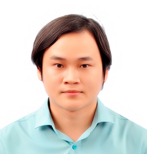 Dac-Nhuong Le合作论文数Faculty of Information Technology, Haiphong University9
 Anup Sadhu合作论文数Department of Radiology, Medical College9S. K. Basu合作论文数8Ayan Gope合作论文数School of Medical Science and Technology, Indian Institute of Technology8
 Atasi Sarkar合作论文数8
 Subhamoy Mandal合作论文数8
 Pranab Dutta合作论文数Department of Electrical Engineering, Indian Institute of Technology Kharagpur8
 Amin Katouzian合作论文数Vayo Inc.;GuideAI Health8
 Lopamudra Das合作论文数Sai University of Technology7
 Maitreya Maity合作论文数Sch. of Med. Sci. &Technol., Indian Inst. of Technol.-Kharagpur;c;Sch. of Med. Sci. & Technol., Indian Inst. of Technol.-Kharagpur7
 CHATTERJEE S N合作论文数7
 Nassir Navab合作论文数School of Computation, Information and Technology, Technische Universitat Munchen;Department of Computer Science, Technical University of Munich7




