When the Millennium Development Goals (MDGs) [1] were being developed, priority was given to the problems of the poorest billion people in the world. In terms of health, this was translated into a set of targets of indicators in health that give visibility to maternal and child health, (under) nutrition, acquired immunodeficiency syndrome (AIDS), malaria, and tuberculosis, and a vague catch-all category, 'and other diseases'. While progress has been made in developing specific plans with budgets to address the named MDG targets, no further work has been carried out on defining what constitute priority other diseases, or which interventions should be emphasized to address them.
Infection with hydatid cysts of domestic animals slaughtered by private individuals and at the Government Central Slaughterhouse is recorded. Both fertile and non-fertile cysts were seen in the infected animals. The home slaughtered sheep seems to play an important role in the dissemination of the parasite and the life cycle of Echinococcus granulosus in the State of Kuwait is assumed to be sheep--dog--sheep.
Infection of dogs with E. granulosus is recorded for the first time in the State of Kuwait and observations on 204 dogs from different suburbs showed an infection rate of 23.039%. Cattle, sheep, goats and camels killed for public consumption in the period 6th April to 6th May, 1975, in a slaughter house were inspected and the rate of infection with E. granulosus cysts was recorded. The possible methods of transmission from animals and dogs to man is discussed.
Mouse peritoneal macrophage cells suspended in a TC 199 calf serum medium and cultured in Leighton tubes, were infected with Trypanosoma (Schizotrypanum) cruzi culture forms in order to study the development of this parasite in vitro. Three cycles of development were thought to occur: 1. Epimastigotes or trypomastigotes were taken up by macrophages and transformed into amastigotes. These multiplied by binary fission and ruptured the infected cell after 4‐5 days. Released amastigotes were taken up by uninfected macrophages and the cycle was repeated. 2. A small proportion of intracellular amastigotes developed into ‘ovoid forms’ which transformed progressively into promastigotes, epimastigotes and trypomastigotes. 3. Some amastigotes became ‘round forms’ from which sphaeromastigotes developed. These transformed directly into trypomastigotes without the formation of epimastigotes.
 O. Hassounah合作论文数2
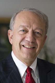 Pekka Puska合作论文数Finnish Institute for Health and Welfare (THL)2T. Khuhaprema合作论文数1J. Gordon McVie合作论文数1Luiz Antonio Santini合作论文数1Harry Burns合作论文数1david r mccance合作论文数1K-C Soo合作论文数1Zhao Ping合作论文数1J Bishop合作论文数1
 Twalib Ngoma合作论文数1T. Knezevic合作论文数1J-G Park合作论文数1T Türmen合作论文数1Vladimir Semiglazov合作论文数1Currow David C合作论文数1Belardelli Filippo合作论文数1
 Rajkumar Thangarajan合作论文数Dept Mol Oncol, Canc Inst WIA1Chan Margaret合作论文数1
 Andrzej Wojtyła合作论文数Department of Health Promotion, Food and Nutrition, Institute of Rural Health1Martin-Moreno J M合作论文数1
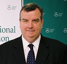 Peter Boyle合作论文数University of Strathclyde;International Prevention Research Institute;Strathclyde Institute for Global Public Health1
 David Kerr合作论文数University of Strathclyde;Radcliffe Department of Medicine, University of Oxford1
 Benjamin L Cox合作论文数Department of Medical Physics, University of Madison - Wisconsin;Medical Engineering Group, Morgridge Institute for Research;Laboratory for Optical and Computational Instrumentation, University of Wisconsin - Madison;University of Madison - Wisconsin, University of Wisconsin - Madison1
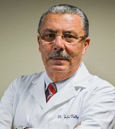 Carlos Vallejos合作论文数Instituto Nacional de Enfermedades Neoplásicas1
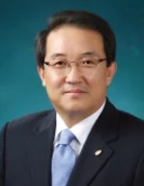 Keun-Young Yoo合作论文数College of Medicine, Seoul National University1
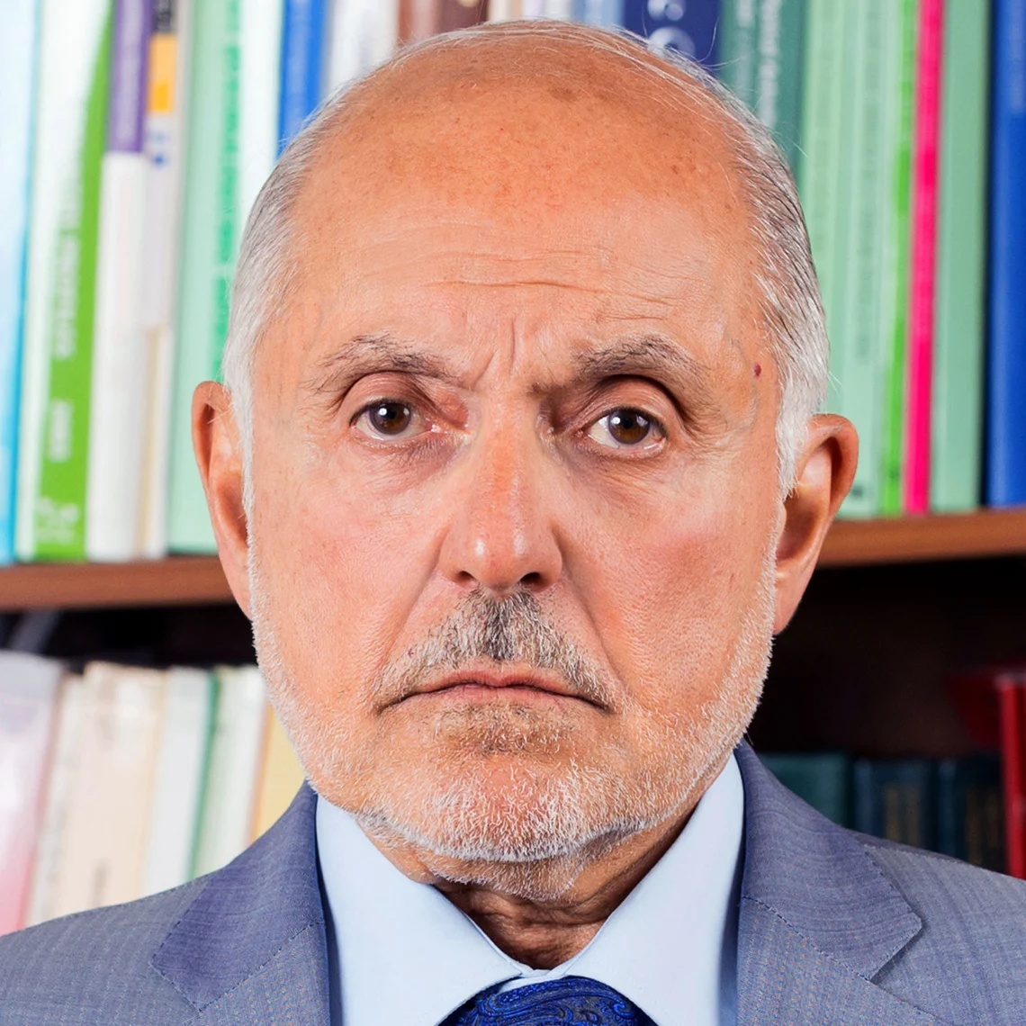 David Zaridze合作论文数Nuffield Department of Population Health, Medical Sciences Division, University of Oxford1Robertson Chris合作论文数1
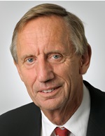 Harry Bartelink合作论文数Department of Radiation Oncology, The Netherlands Cancer Institute1Leif C. Andersson合作论文数Department of Pathology, University of Helsinki1
 Alexander Eggermont合作论文数Paris-Sud University;Erasmus University of Rotterdam;Louvain Catholic University1Ulrik Ringborg合作论文数1Derek Yach合作论文数1
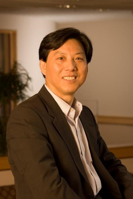 Tongzhang Zheng合作论文数Department of Epidemiology,School of Public Health, Brown University1
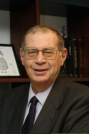 Thomas Tursz合作论文数Gustave Roussy;Medical Faculty, Paris-Sud University1John M. Fitzpatrick合作论文数University College Dublin School of Medicine & Medical Science1
 Marco Pierotti合作论文数Cogentech Srl;IFOM-IEO Campus;Fondazione IRCCS Istituto Nazionale Tumori of Milan1
 Benjamin O. Anderson合作论文数School of Medicine, University of Washington1
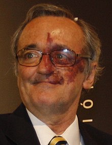 Mariano Barbacid合作论文数Spanish National Cancer Research Center1
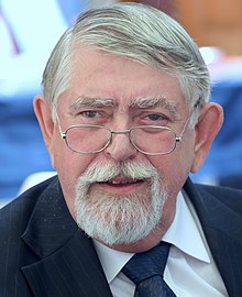 Miklós Kásler合作论文数National Institute of Oncology1
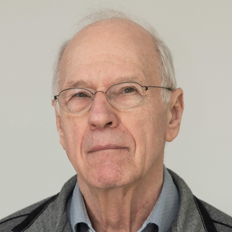 Anton Berns合作论文数The Netherlands Cancer Institute - Antoni van Leeuwenhoek Hospital1K Dinshaw合作论文数1
 Balakrishnan Rajan合作论文数Department of Academic Radiotherapy and Oncology, Royal Marsden NHS Trust and Institute of Cancer Research1Y. Ariyaratne合作论文数1
 Anna Gavin合作论文数Centre for Public Health, Queen’s University Belfast1
 Marek Nowacki合作论文数Institute of Applied Sciences, WSB Merito University in Poznan1G.-R. Auleley合作论文数1
 Pascoal Mocumbi合作论文数European & Developing Countries Clinical Trials Partnership (EDCTP)1
 Umberto Veronesi合作论文数European Institute of Oncology1




