Circadian rhythms have been shown to regulate sleep-wake timing across the lifespan, yet many questions remain about early childhood circadian physiology. Understanding dim-light melatonin onset (DLMO) and offset (DLMOff), established markers of circadian phase, is essential for characterizing circadian rhythms in early childhood. We examined the distribution of salivary DLMO and DLMOff and their relationship with actigraphic sleep timing across 20 healthy preschoolers (M = 4.31 ± 0.34 years, 45% female). After maintaining a consistent sleep schedule for 7 days, children completed an in-home circadian assessment. Children were awoken 1.5 h before habitual wake time, and saliva samples were collected in 20- to 30-min intervals throughout the morning to determine DLMOff, then in the evening until 50 min past habitual bedtime to assess DLMO. A 4-pg/ml threshold was used to calculate each phase marker. DLMO ranged from 17:22 to 20:40 (M = 18:55 ± 0:54) and was positively associated with bedtime, sleep onset, and midsleep. In contrast, morning melatonin levels were highly variable, allowing DLMOff calculation in only 8 participants. Within this small subsample, later DLMOff was associated with later chronotype (r = 0.81), sleep offset (r = 0.84), and midsleep (r = 0.80). Across the full sample, interpolated melatonin levels at habitual wake remained ≥ 4 pg/ml for 45% of children, a pattern broadly consistent with findings in adults, in which a majority of participants exhibit DLMOff after habitual wake time. These findings indicate that although evening melatonin profiles were consistently well-defined, permitting reliable calculations of DLMO across all participants, morning melatonin patterns were often irregular in young children. When able to be calculated, DLMOff showed strong associations with sleep timing, suggesting it could be a reliable marker of circadian phase. However, high variability and fluctuating morning melatonin patterns make DLMOff difficult to determine in many preschool-aged children.
Objective: Determine whether extending sleep improves insulin sensitivity in people with overweight/obesity, insulin resistance, and habitual short-sleep schedules (<7h/night). Research Design and Methods: Participants were randomized to habitual sleep (HS; n=15) or extended sleep (ES; n=14) for ~6 weeks. Multi-organ (whole-body [primarily muscle], hepatic and adipose tissue) insulin sensitivity (assessed by the hyperinsulinemic-euglycemic clamp procedure with tracer infusions) and glycemic control (assessed by 24-h serial plasma glucose and insulin concentrations during wakefulness). Results: Time-in-bed and sleep duration increased more in the ES group (1.3±0.6 and 1.1±0.5 h/night) than the HS group (0.3±0.8 and 0.0±0.4 h/night). Day-to-day variability in sleep and subjective measures of sleep health improved more in the ES than HS group, without differences in multi-organ insulin sensitivity or glycemic control between groups. Conclusions: Extending sleep by ~1 h/night for ~6 weeks in people with overweight/obesity and short-sleep schedules improves sleep health but not insulin sensitivity or glycemic control.
Abstract Introduction This guideline establishes recommendations for the treatment of shift work disorder (SWD). Methods The American Academy of Sleep Medicine (AASM) commissioned a task force of experts in sleep and circadian medicine to develop recommendations and assign strengths based on a systematic review of the literature and an assessment of the evidence using the GRADE process. The task force summarized the relevant literature and the quality of evidence, the balance of benefits and harms, patient values and preferences, and resource use considerations supporting the recommendations. The AASM Board of Directors approved the final recommendations. Results The following recommendations are intended as a guide for clinicians in choosing specific treatment(s) for symptoms of excessive sleepiness and/or insomnia, and for circadian adaptation in adults with SWD. Each recommendation statement is assigned a strength (e.g., “Strong” or “Conditional”). For SWD, there were only “conditional” recommendations for or against (i.e., “We suggest…”) and those requiring the clinician to use clinical knowledge and experience and consider the individual patient’s values and preferences to determine the best course of action. Interventions in recommendation statements were compared to no treatment. Conclusion Adults with SWD with symptoms of excessive sleepiness associated with work schedule: 1. In adults with SWD with excessive sleepiness, the AASM suggests the use of armodafinil or modafinil taken 30 to 60 minutes prior to the night shift over no armodafinil or modafinil (Conditional recommendation, moderate certainty evidence). 2. In adults with SWD with excessive sleepiness, the AASM suggests the use of bright light over no bright light during the night shift (Conditional recommendation, very low certainty evidence). Remark: Individuals with SWD who exhibit insomnia and daytime function impairments may also benefit from using bright light during their night shift. 3. In adults with SWD with excessive sleepiness, the AASM suggests the use of caffeine taken prior to and/or during the night shift over no caffeine (Conditional recommendation, very low certainty evidence). Remark: Caffeine intake close to bedtime can disrupt sleep onset and quality, which may exacerbate symptoms of shift work disorder. 4. In adults with SWD with excessive sleepiness, the AASM suggests the use of a clockwise rotating shift schedule over a counterclockwise rotating shift schedule (Conditional recommendation, very low certainty evidence). 5. In adults with SWD with excessive sleepiness, the AASM suggests taking a nap over no nap prior to the night shift (Conditional recommendation, very low certainty evidence). Remarks: Sleep inertia occurring soon after the nap may temporarily increase sleepiness, decrease cognitive performance, and increase accident risk. Allowing adequate time for resolution of sleep inertia prior to driving or performing other safety-sensitive behaviors may be needed to reduce these risks. 6. In adults with SWD with excessive sleepiness, the AASM suggests either not eating or eating a snack rather than eating a full meal during the night shift (Conditional recommendation, low certainty evidence). 7. In adults with SWD with excessive sleepiness, the AASM suggests the combination of caffeine and bright light over the combination of no caffeine and no bright light (Conditional recommendation, very low certainty evidence). Remark: Caffeine intake close to bedtime can disrupt sleep onset and quality, which may exacerbate symptoms of shift work disorder. 8. In adults with SWD with excessive sleepiness, the AASM suggests the combination of taking a nap and caffeine over the combination of no nap and no caffeine prior to the night shift (Conditional recommendation, very low certainty evidence). 9. In adults with SWD who are working either an 8-hour or 12-hour night shift, the AASM does not suggest working one shift duration over the other (Conditional recommendation, very low certainty evidence). Remark: This recommendation does not consider other shift durations, rotation, or the number of consecutive shifts. This guideline did not look at shifts over 12 hours. The total number of hours worked per week ranged from 36 to 52. Adults with SWD with sleep disturbance/insomnia: 10.9. In adults with SWD with daytime sleep disturbance/insomnia the AASM suggests the use of CBT-I over no CBT-I (Conditional recommendation, very low certainty evidence). 11.10. In adults with SWD with daytime sleep disturbance/insomnia following the night shift, the AASM suggests the use of melatonin over no melatonin (Conditional recommendation, very low certainty evidence). 12.11. In adults with SWD with sleep disturbance/insomnia, when transitioning from daytime to nighttime sleep, the AASM suggests the use of melatonin over no melatonin for night sleep following shift work (Conditional recommendation, low certainty evidence). 13.12. In adults with SWD who desire to take a nap prior to the first night shift, the AASM suggests the use of melatonin over no melatonin prior to the nap (Conditional recommendation, very low certainty evidence). Remarks: Melatonin may temporarily increase sleepiness, decrease cognitive performance, and increase accident risk. Allowing adequate time for resolution of sleepiness prior to driving, returning to work or performing other safety-sensitive behaviors may be needed to reduce these risks. 14.13. In adults with SWD with daytime sleep disturbance/insomnia, the AASM suggests the use of ramelteon or other melatonin receptor agonists over no melatonin receptor agonist (Conditional recommendation, low certainty evidence). 15.14. In adults with SWD with daytime sleep disturbance/insomnia, the AASM suggests the use of suvorexant or other dual orexin receptor antagonists over no dual orexin receptor antagonists (Conditional recommendation, low certainty evidence). 16.15. In adults with SWD with daytime sleep disturbance/insomnia, the AASM suggests against the use of triazolam and other benzodiazepines (conditional recommendation, very low certainty evidence). Adults with SWD seeking circadian adaptation 17.16. In adults with SWD seeking circadian adaptation to the night shift, the AASM suggests the use of bright light over no bright light during the night shift (Conditional recommendation, very low certainty). Remarks: For individuals who work rotating or frequently changing shifts, circadian adaptation is not a realistic goal of treatment and therefore they would not be expected to benefit from this intervention. The evidence supporting this recommendation came from studies in which individuals reached partial circadian adaptation to the night shift. 18.17. In adults with SWD seeking circadian adaptation to the night shift, the AASM suggests the combination of bright light at night and fixed daytime sleep timing over the combination of no bright light and no fixed sleep timing (Conditional recommendation, very low certainty evidence). Remarks: For individuals who work rotating or frequently changing shifts, circadian adaptation is not a realistic goal of treatment and therefore they would not be expected to benefit from this intervention. The evidence supporting this recommendation came from studies in which individuals reached partial circadian adaptation to the night shift. 19.18. In adults with SWD seeking circadian adaptation to the night shift, the AASM suggests the combination of bright light at night, fixed daytime sleep timing, and reduced-light-transmittance glasses in the morning over the combination of no bright light, no fixed sleep timing, and no reduced-light-transmittance glasses (Conditional recommendation, very low certainty evidence). Remark 1s : For individuals who work rotating or frequently changing shifts, circadian adaptation is not a realistic goal of treatment and therefore they would not be expected to benefit from this intervention. The evidence supporting this recommendation came from studies in which individuals reached partial circadian adaptation to the night shift. Remark 2: Attention to driving safety is important when wearing reduced-light-transmittance glasses Other considerations for Adults with SWD 19. In adults with SWD who are working either an 8-hour or 12-hour night shift, the AASM does not suggest working one shift duration over the other (Conditional recommendation, very low certainty evidence). Remark: This recommendation does not consider other shift durations, rotation, or the number of consecutive shifts. This guideline did not look at shift durations exceeding 12 hours. The total number of hours worked per week ranged from 36 to 52. Support (if any)
Abstract Accumulating evidence implicates sleep and circadian rhythm disruption in substance use disorders, including opioid use disorder (OUD). To understand whether weaker light exposure time cues are observed in patients with OUD, we compared the amplitudes of personal light exposure in patients with OUD ( n = 73) and healthy controls ( n = 49). Participants monitored personal light exposure and rest/activity via wrist-worn actigraphy for 1 week. We calculated light and physical activity amplitude for each day with non-orthogonal spectral analysis. Patients with OUD demonstrated significantly lower light exposure amplitudes and significantly higher physical activity amplitudes compared to healthy controls, suggesting lower light exposure amplitudes are not accounted for by sedentary behavior in patients with OUD. Blunted light exposure amplitudes, indicative of a weaker time cue to the circadian clock, characterize patients with OUD and may represent a novel target for improving sleep and circadian health in OUD.
Circadian misalignment, as experienced during shiftwork, impairs glucose metabolism and body weight regulation, yet the underlying biochemical mechanisms remain incompletely understood. Characterizing how circadian misalignment alters circulating metabolites provides a promising avenue to help identify these mechanisms. Although data from metabolomics studies have identified circulating metabolites with daily rhythms, it is not comprehensively known which rhythms shift during circadian misalignment and whether such shifts relate to metabolic impairment. We conducted 24-hour (h) metabolomic profiling every 4 h in 14 healthy adults (8 women) aged 26.4 ± 1.2 years (mean ± SD), undergoing a 6-day simulated night-shiftwork protocol. 24-h modeling analyses identified metabolite rhythms influenced by circadian versus behavioral cycles (sleep, food intake) and quantified internal circadian misalignment using acrophase shifts. Metabolic outcomes included glucose homeostasis (test meals) and energy expenditure (EE; whole-room calorimetry). Night-shiftwork produced widespread alterations in metabolite rhythms, with significant internal misalignment in multiple metabolites across pathways including pyrimidine metabolism, bile acid-microbiome signaling, and lipid metabolism. During misalignment, glucose and insulin area under the curve increased (p < 0.05) and EE decreased (p < 0.05). Internal misalignment of uridine and glycoursodeoxycholic acid was associated (p < 0.05) with impaired glucose tolerance, while their circulating concentrations were associated with decreased EE. Misalignment of uridine and glycoursodeoxycholic acid suggests dysregulated pyrimidine and bile acid-microbiome pathways as potential mechanisms linking circadian misalignment to cardiometabolic disease risk.
Abstract Introduction Prior literature implicates delayed circadian rhythms in obsessive-compulsive disorder (OCD), and later indicators of circadian rhythms are associated with more severe OCD symptoms. However, factors that contribute to delayed circadian rhythms in OCD remain unexplored. As light is the primary entrainment cue for the central circadian clock, and unhealthy light exposure patterns delay circadian rhythms, we examined associations between personal light exposure and circadian phase and sleep timing in individuals with OCD and delayed bedtimes. Methods Young adults with OCD and a bedtime of 01:00 or later (N = 35; 30 female, mean age = 22.18 ± 4.12) monitored personal light exposure and sleep for 2 weeks via wrist actigraphy (Actiwatch Spectrum, Phillips Respironics) and completed a 10-hour in-laboratory salivary dim light melatonin onset (DLMO) assessment. The following light exposure variables were calculated using the LightLogR package: time above threshold (TAT) 250, 500, and 1000 photopic lux; mean timing of light exposure (MLiT) above 250, 500, and 1000 lux; first and last timing of light exposure (FLiT; LLiT) above 50 lux and their within-subject standard deviation (FLiT, LLiT 50 lux SD). DLMO was defined as the linear interpolated point in time at which melatonin levels rose 2 SD above the mean of 3 baseline values. Midsleep was measured by actigraphy. We used linear regression models covarying for solar photoperiod to examine associations between personal light exposure, DLMO, and midsleep. All p-values were FDR-corrected. Results Lower TAT 250, 500, and 1000 lux, later MLiT 250, 500, and 1000 lux, later FLiT 50 lux, and higher FLiT 50 lux SD were associated with later midsleep (p< 0.05). Later MLiT 250, 500, and 1000 lux and FLiT 50 lux were associated with later DLMO (p< 0.05). Conclusion Lower, later, and irregular light exposure were associated with later sleep timing, whereas only later timed light exposure was associated with later circadian phase. Personal light exposure patterns, particularly light timing, may contribute to delayed circadian rhythms in OCD. Circadian medicine treatments to address delays in OCD may benefit from prioritizing advancing light exposure timing. Support (if any) NIH T32HL149646; NIH K23MH137376; American Academy of Sleep Medicine Foundation 270-FP-22; International OCD Foundation
Abstract Introduction Insufficient sleep increases obesity and diabetes risk, yet effective interventions to reduce this risk remain limited. Results from short-term experimental sleep restriction suggest physical activity may mitigate these adverse effects of sleep loss. However, it remains unclear if physical activity similarly mitigates risk among individuals with habitual insufficient sleep. Thus, we examined whether moderate-to-vigorous physical activity (MVPA) is associated with insulin sensitivity status in adults with insufficient sleep, and whether MVPA provides explanatory value beyond body mass index (BMI). This preliminary cross-sectional analysis leveraged data from two ongoing studies in adults with insufficient sleep and normal BMI (study A) or overweight/obese BMI (study B). Methods Participants in both studies reported habitual sleep < 6.5h/night; study A (n=24, age 23.4±4.4y, BMI=22.1±2.1kg/m2); study B (n=10, age 34.0±7.0y, BMI=29.3±3.1kg/m2). Across two weeks, MVPA was measured using ActivPAL™ devices and total sleep time (TST) was assessed by wrist-actigraphy. Insulin sensitivity was evaluated using Matsuda Index from oral glucose tolerance testing in study A, and glucose infusion rate/kg of body weight (GIR) from hyperinsulinemic–euglycemic clamps in study B. Using established thresholds, participants in both studies were classified as either insulin “sensitive” or “resistant”. Results Insulin sensitivity status differed with 76.9% in study A classified as “sensitive” versus 33.3% in study B (χ²(1)=5.64, p=0.018). MVPA was higher (p=0.03) in A (38.2±21.1min/day) versus B (23.1±14.6min/day). Using logistic regression, BMI, but not MVPA, significantly predicted insulin sensitivity status (BMI: OR=1.45, 95% CI:1.11–2.19 p=0.024). Furthermore, after adding MVPA, BMI remained significant (OR=1.32, 95% CI:1.05–1.85, p=0.049), and model fit was not significantly altered (p=0.77, Likelihood Ratio Test), indicating MVPA did not significantly attenuate the BMI-insulin sensitivity status association. Conclusion Among adults with habitual insufficient sleep, BMI was the strongest determinant of insulin sensitivity status, whereas MVPA did not independently predict insulin sensitivity status. Differences between study groups highlight variability in metabolic risk, suggesting individuals with insufficient sleep and higher BMI may face greater cardiometabolic vulnerability that MVPA alone may not counteract. Future research using randomized controlled trials is needed to confirm these observational analyses. Support (if any) NIH-R01HL178934, NIH-UL1TR002538; NIH-K01HL145099; NIH-R01HL166733; NIH-T32DK110966; Ben B. and Iris M. Margolis Foundation
Abstract Introduction Insufficient sleep, fragmented sleep, disrupted slow wave sleep are all associated with risk for diabetes. Conventional sleep staging methods of analyzing polysomnography do not fully encompass the depth of sleep. The odds ratio product (ORP) is a continuous measurement of sleep depth, but it is unknown if ORP is linked to insulin sensitivity. Thus, we assessed ORP and insulin sensitivity in healthy young adults with habitual insufficient sleep, hypothesizing deeper sleep will be linked to better insulin sensitivity. Methods We analyzed data from 16 participants (9 female; aged 21.1±3.5yr; BMI 21.4±2.1kg/m2 [mean±SD]) with habitual sleep < 6.5h per night. Participants completed 2-weeks of ambulatory monitoring with actigraphy. Following at home monitoring participants underwent overnight polysomnography to calculate ORP, a QEEG derived measure of sleep depth (ranging from 0 [deeply asleep] to 2.5 [fully awake]), and completed the Pittsburgh Sleep Quality Index (PSQI) survey to assess subjective sleep quality. Oral Glucose Tolerance Testing (OGTT) was completed to calculate Matsuda Index (MI) insulin sensitivity. Results Average PSQI was 5.1±2.4 (mean±SD), TST was 5.48±0.9h, ORP during the total recording time was 0.79±0.23, and MI was 7.6±4.4. Linear regression indicated both lower ORP and shorter TST were individually associated with worse MI (all p< 0.05). Yet, when included together in the model, neither were significant. Additionally, shorter TST was associated p< 0.05) with lower ORP. Conclusion Participants exhibited insufficient sleep indicated by short TST and average PSQI >5. As expected, shorter TST was associated with worse MI, indicating higher diabetes risk. Interestingly, shorter TST also associated with lower ORP, suggesting deeper sleep possibly driven by greater homeostatic sleep pressure. However, deeper sleep indicated, by ORP, was also associated with worse MI in this context of adults with habitual insufficient sleep, counter to our hypothesis. These preliminary analyses suggest among adults with insufficient sleep, deeper sleep depth may not confer metabolic benefit. Future analyses will compare ORP to MI among adults with >7h sleep/night to clarify whether the ORP–insulin sensitivity relationship differs under conditions of habitual adequate sleep. Support (if any) NIH-UM1TR004409; NIH-K01HL145099; NIH-T32DK110966; NIH-R01HL166733; University of Utah Seed Grant-10060570; University of Utah Diabetes and Metabolism Research Center Graduate Student Fellowship
Study Objectives:We examined the impact of a sleep extension intervention on multiple dimensions of sleep in adults with habitual short sleep duration. Methods:Thirty healthy participants (14 women; aged 23.1 ± 4.5 years; BMI 22.3 ± 2.2 kg/m2 [mean ± SD]) with <6.5 h sleep/night completed a 2-week baseline assessment followed by a 4-week sleep extension intervention (2 h/night increased time in bed). Wrist actigraphy, at-home electroencephalography (EEG; DREEM headband), Insomnia Severity Index (ISI), Pittsburgh Sleep Quality Index (PSQI), Epworth Sleepiness Scale (ESS), and daily quality/satisfaction and alertness Likert-scales quantified sleep. Results:Baseline total sleep time (TST) was 5.5 ± 0.7 h. During sleep extension, actigraphy time in bed and TST increased (p < .001) by 60.8 ± 46.7 and 46.6 ± 41.1 minutes, respectively, sleep onset shifted earlier (p < .001) by 51.9 ± 64.5 minutes, with regularity similar to baseline. EEG showed increases (all p < .05) in TST (61.8 ± 75.6 minutes), Stage N1 (5.09 ± 6.51 minutes), Stage N2 (39.54 ± 41.78 minutes), rapid eye movement sleep (13.58 ± 28.86 minutes), and wakefulness after sleep onset (4.86 ± 8.03 minutes), with a nonsignificant decrease (p = .07) in sleep efficiency (-1.38 ± 4.15%). Subjective alertness, ISI, PSQI, and ESS each improved (all p < .05) during sleep extension. Conclusion:Our findings help establish efficacy of sleep extension as an experimental intervention the sleep field can leverage across diverse contexts to study potential health benefits of increasing free-living TST. During sleep extension, the largest effects were observed for improved TST and ESS. Alternatively, some sleep dimensions including sleep regularity remained unchanged, highlighting a potential need for developing multi-component interventions that can improve more dimensions of sleep as both short and irregular sleep are linked with adverse health outcomes. Clinical Trials:Biomarkers of Increased Free Living Sleep Time. URL: https://clinicaltrials.gov/study/NCT04214184. ClinicalTRIALS.gov ID: NCT04214184.
Introduction:Patients with critical illness experience profound circadian disruption, driven in part by atypical light exposure in the intensive care unit (ICU). While prior studies characterize ICU light, the extent to which in patient, temporal, and structural factors determine light intensity remains unclear. Methods:We conducted an observational cohort study of light intensity in the rooms of patients admitted to one of thirteen medical ICU (MICU) rooms equipped with continuous light monitoring between February 2021 and 2022. Patients were included if they remained in the monitored MICU room for at least 48 h. We collected patient demographics and clinical features. We recorded light intensity (lux) every 60 s, and then calculated hourly mean lux, hourly proportion of measures ≥250 lux, and hourly proportion of measures ≤10 lux for the first 48 h of each patient's MICU admission. Intraclass correlation coefficients (ICC) quantified variance in each light outcome attributable to patient-level factors. Multivariable multilevel models assessed adjusted associations between patient, temporal and structural factors and light outcomes. Both ICCs and multilevel models were generated separately for day (05:00-22:59) and night (23:00-04:59). Results:Among the 337 patient-room pairs, the mean (± standard deviation) age of patients was 66.1 ± 14.8 years; mean SOFA score was 6.8 ± 3.6; and daytime and nighttime mean lux were 173.8 ± 94.6 and 63.7 ± 88.2, respectively. ICCs indicated that patient-level factors explained 26.3% of daytime and 46.9% of nighttime variance in mean lux; ICC results were similar for other light outcomes. In multivariate models we found that temporal (e.g., time of day, season of ICU admission) and structural factors (e.g., room direction) strongly influenced daytime light levels, whereas nighttime variation was more heavily driven by patient-level factors such as mechanical ventilation status. Conclusion:ICU light exposure shows substantial between-patient variability, particularly at night. In multilevel models, we found that this variability is driven by clinical, temporal and structural factors. Our findings suggest that structural design alone is insufficient to normalize ICU light exposure, and that interventions targeting nocturnal care behaviors and daytime light provision may be necessary to mitigate circadian disruption in patients with critical illness.
This systematic review synthesized findings from 41 human studies across the lifespan published between 2016 and 2025 examining associations of multidimensional sleep and circadian rhythm health with the gut microbiome (GM). The studies include experimental and observational designs of generally healthy participants, measuring sleep and circadian dimensions such as duration, quality, insomnia, and circadian disruptions (e.g., shift work). Results suggest sleep truncation, disturbances, and circadian misalignment may be linked to GM composition and function, though results were mixed on microbial diversity. The heterogeneous evidence suggests that the microbial family Oscillospiraceae/Ruminococcaceae is often associated with sleep and circadian metrics, warranting rigorous inquiry of taxa within this family. Functional outputs of the GM were infrequently assessed, but when measured, identified functions in amino acid and fatty acid metabolism, and vitamin, hormone/neurotransmitter, phosphate, short-chain fatty acid, and bile acid pathways. Inconsistencies in findings may reflect differing analytical methods, participants’ age, health status, and lifestyle (e.g., diet, physical activity). Future investigations should prioritize longitudinal and experimental, multi-omics studies to clarify causal pathways and assess interventions, especially in populations with sleep disruption. Findings underscore the potential for sleep and circadian interventions to support GM balance and improve health outcomes.
The circadian system maintains optimal biological functions at the appropriate time of day, and the disruption of this organization can contribute to the pathogenesis of cardiometabolic disorders. The timing of eating is a prominent external time cue that influences the circadian system. “Chrononutrition” is an emerging dimension of nutrition and active area of research that examines how timing‐related aspects of eating and nutrition impact circadian rhythms, biological processes, and disease pathogenesis. There is evidence to support chrononutrition as a form of chronotherapy, such that optimizing the timing of eating may serve as an actionable strategy to improve cardiometabolic health. This report summarizes key information from the National Heart, Lung, and Blood Institute's virtual workshop entitled “Chrononutrition: Elucidating the Role of Circadian Biology and Meal Timing in Cardiometabolic Health,” which convened on May 2 to 3, 2023, to review current literature and identify critical knowledge gaps and research opportunities. The speakers presented evidence highlighting the impact on cardiometabolic health of earlier and shorter eating windows and more consistent day‐to‐day eating patterns. The multidimensionality of chrononutrition was a common theme, as it encompasses multiple facets of eating along with the timing of other behaviors including sleep and physical activity. Advancing the emerging field of chrononutrition will require: (1) standardization of terminology and metrics; (2) scalable and precise tools for real‐world settings; (3) consideration of individual differences that may act as effect modifiers; and (4) deeper understanding of social, behavioral, and cultural influences. Ultimately, there is great potential for circadian‐based dietary interventions to improve cardiometabolic health.
We examined the association of recreational cannabis legalization (RCL) with frequency of using cannabis, alcohol, and sleep medication for sleep and with co-use of cannabis with other sleep aids. We used linear regression models to examine these associations in a population-based sample of adult twins (n = 3,141). Participants (Mage = 37 (SD = 5)) were primarily White (93%), with 5% Hispanic/Latinx, and female (61%). RCL was associated with using cannabis for sleep more frequently even after controlling for cohort, demographics, sleep quality, anxiety, depression, physical health, season, and pre-legalization cannabis use frequency (β = 0.123, p = .001). RCL was not associated with frequency of using alcohol or sleep medication for sleep, or with co-use of cannabis and other sleep aids. More research is needed to determine whether RCL leads to more frequent use of cannabis for sleep.
Despite significant public health efforts to counter the obesity epidemic, approximately 50% of US adults will have obesity by 2030. The cornerstone of obesity treatment is a behavioral intervention promoting negative energy balance via a reduced-calorie diet and increased physical activity. Behavioral treatment also requires additional support to promote adherence to these recommended lifestyle changes. More recently, sleep has been recognized as an important component of body weight regulation due to its effects on physiological and behavioral determinants of energy balance. Therefore, sleep may represent a modifiable behavior to target during the behavioral treatment of obesity. This review summarizes existing clinical evidence on the influence of sleep on energy balance and body weight regulation in adults. The results of controlled laboratory studies as well as pragmatic clinical trials are reviewed. In addition, we identify existing gaps and future areas of research that are necessary to understand the role of sleep in behavioral treatments for obesity.
Delayed sleep-wake phase disorder involves chronic difficulty going to bed and waking up at conventional times and often co-occurs with depression. This study compared sleep and circadian rhythms between patients with delayed sleep-wake phase disorder with depression (DSWPD-D) and without (DSWPD-ND) comorbid depression. Clinical records of 162 patients with delayed sleep-wake phase disorder (70 DSWPD-D, 92 DSWPD-ND) were analysed, including a subset of 76 patients with circadian phase determined by the dim light melatonin onset. Variables assessed included sleep behaviour on work and free days, weekly sleep duration, social jet lag, chronotype, and phase relationships between dim light melatonin onset and sleep/wake times. Mean (SD) or median [Q1-Q3] values were compared using t-tests or Mann-Whitney. Patients with DSWPD-D showed longer sleep on workdays (DSWPD-D = 7.63 hr [1.70] versus DSWPD-ND = 6.20 hr [1.59]; p < 0.001), but not on free days. DSWPD-D also showed later sleep onset (DSWPD-D = 03:30 14;hours [02:49 hours-04:23 hours], DSWPD-ND = 02:53 hours [02:00 hours-03:41 hours]; p = 0.02) and wake times (DSWPD-D = 11:30 hours [09:30 hours-13:00 hours], DSWPD-ND = 08:45 hours [07:20 hours-11:00 hours]; p < 0.01) on workdays. Furthermore, DSWPD-D showed less social jet lag (DSWPD-D = 0.38 [0.00-1.75] versus DSWPD-ND = 2.17 [1.25-3.03]; p < 0.01), and reported higher anxiety symptoms (DSWPD-D = 71.4% versus DSWPD-ND = 45.8%; p = 0.03) and medication use (DSWPD-D = 75.0% versus DSWPD-ND = 43.8%; p = 0.01). DSWPD-D also showed wider dim light melatonin onset phase relationships with dim light melatonin onset-mid-sleep (DSWPD-D = -5.77 [1.32] versus DSWPD-ND = -4.86 [1.53]; p = 0.01) and dim light melatonin onset-waketime (DSWPD-D = -9.46 [1.82]; DSWPD-ND = -8.13 [2.08]; p = 0.01). Multivariable Poisson regression, adjusted for age and sex, showed more medication use, less social jet lag, and longer weekly sleep duration as significantly associated with DSWPD-D. These findings suggest potential biopsychosocial protective factors linked to depression in delayed sleep-wake phase disorder. Further research is required to confirm these phenotypic differences and their relevance to delayed sleep-wake phase disorder aetiology and treatment.
Early childhood represents a period of profound developmental changes for sleep and circadian biology. Although the relationship between sleep and circadian timing has been well characterized in older populations, such data in young children remain limited. Here, we provide fundamental data on the relationship between endogenous circadian phase and sleep timing in a sample of preschool-aged children. Participants were 49 healthy children ages 3.1 to 6.0 years (M = 4.44 years, SD = 0.69 years, 27 female). After 7 days of maintaining a consistent, parent-selected sleep schedule, children completed an in-home, dim-light circadian assessment. Saliva samples were collected in 30-min intervals throughout the evening to determine the timing of children's dim-light melatonin onset (DLMO). Children's DLMOs occurred an average of 35.0 ± 35.3 min before their bedtimes, with parent-selected bedtime occurring before DLMO for 18.4% of children. Children with later DLMOs had significantly later bedtimes (r = 0.65), sleep onset times (r = 0.74), midsleep times (r = 0.74), and wake times (r = 0.66) (all p < 0.001). For every hour later that DLMO occurred, average bedtime and sleep onset time were 28.0 and 33.4 min later, respectively. In addition, children with later DLMOs had higher scores on a parent-reported measure of chronotype (r = 0.56, p < 0.001), indicating greater eveningness. No association between DLMO time and sleep duration or social jetlag was observed. These data extend previous findings in toddlers, demonstrating a consistent relationship between circadian phase and sleep timing, as well as chronotype, throughout early childhood.
Circadian rhythms, intrinsic 24-h cycles that drive rhythmic changes in behavior and physiology, are important for normal physiology and health. Previous work in adults has identified sex differences in circadian rhythms of melatonin, temperature, and the intrinsic period of the human circadian timing system. However, less is known about sex differences in circadian rhythms at other developmental stages. To address this gap, we considered a secondary analysis of sleep and circadian data from two studies involving adolescent participants during the academic year: (n=32, 15 females). We collected 1 week of in-home actigraphy data to calculate sleep-wake parameters and in-laboratory salivary melatonin data collection in dim-light conditions was used to compute dim-light melatonin onset (DLMO) and offset (DLMOff) using a threshold of 4 pg/mL. We found that DLMO was an average of 96 min earlier, the time between DLMO and bedtime was an average of 56 min greater, and the biological night (time between DLMO and DLMOff) was 60 min longer in females compared to males, even though bedtimes and waketimes were not statistically different between the groups. In addition, after accounting for differences in bedtime, sex was still a significant predictor of DLMO. Conversely, no evidence was found indicating a difference in DLMOff or the phase angle between DLMOff and waketime by sex. These findings suggest that sex differences in circadian rhythms are present in adolescents and may have implications for circadian health during this important developmental period.
Taxa level associations of the human gut microbiota have been reported in relation to sleep health; however, which taxa are associated with good sleep has yet to be determined. Many study methodologies rely on self-reported sleep metrics, 16S rRNA gene-amplicon sequencing or statistical methodologies not designed for gut microbiome assessments, which have limitations. We examined the association between sleep measures assessed by actigraphy followed by one night of in-laboratory polysomnography (PSG) and whole genome sequencing (WGS) gut microbiome taxa. Fifteen healthy participants aged 26±4.0(SD) were instructed to maintain a consistent 8h sleep schedule for fourteen days at home. Wrist actigraphy and time stamped call-ins were assessed for adherence. Following fourteen days of monitoring, participants underwent 8h overnight in-laboratory PSG. Fecal microbiome samples were collected at PSG visit. WGS at the operational genomic unit (OGU) were then referenced against Web of Life 2 for taxonomic assignment. Differential abundance testing (ANCOM-BC) was performed to detect the OGUs associated with individuals with high (≥85%) or low (< 85%) sleep efficiency (SE) assessed via actigraphy. Following, linear regression associations between SE, sleep onset latency (SOL) and wake after sleep onset (WASO) from PSG were tested against these differentially abundant OGUs to determine: 1) whether these associations were maintained with PSG SE and; 2) whether these taxa were associated with SOL or WASO. ANCOM-BC determined Bifidobacterium breve and Bifidobacterium saguini species were increased in the high SE wrist actigraphy group (log fold change 2.2, q=0.002 and log fold change 2.0, q=0.006, respectively). Linear regression indicated increased breve and saguini were associated with higher PSG SE (β=0.0003, p=0.03, R²=0.18 and β=0.0013, p=0.02, R²=0.46, respectively). Further investigation into PSG SOL and WASO indicated that the higher SE was due to these strains being associated with decreased WASO, specifically (β=–0.09, p=0.04, R²=0.38 and β=–0.58, p=0.04, R²=0.40, respectively). We find that higher SE, specifically by decreased WASO, is associated with higher levels of two Bifidobacterium species, breve and saguini. Bifidobacterium, commonly found in probiotics, may improve subjective sleep quality. Therefore, Bifidobacterium breve and saguini may be specific targets for improving human sleep. ONR N00014-15-1-2809, NIH-T32-HL149646, NIH/NCATS Colorado CTSA-UL1TR002535.
The duration between dim-light melatonin onset and offset represents the biological night, which tracks environmental darkness (i.e., scotoperiod). In humans, melatonin duration is longer after one week of exposure to natural winter (~9.33h light:14.67h dark) versus natural summer (~14.67h light:9.33h dark) light-dark cycles. Melatonin duration is also longer after one month of exposure to 14h versus habitual ~8h sleep opportunities; as well as in people who habitually sleep longer than 9h versus shorter than 6h. Our current aim was to determine whether a shorter difference in scotoperiod, 8h versus 9h sleep opportunities, impacts the duration of the biological night. Archival data from 76 healthy adult participants (aged 26.1±6.8[Mean±SD]; 44 females) from five research protocols were examined. Depending on the study protocol, participants maintained consistent 8h (n=40) or 9h (n=36) sleep schedules for one or two weeks at home, verified with wrist-actigraphy and bed/waketime call-ins. 24-hour plasma or salivary melatonin samples were then collected in the laboratory under dim-light conditions during wakefulness (< 10 lux max; ~1.9 lux, ~0.6 Watts/m2 in the angle of gaze) and darkness (0 lux) during sleep. Melatonin duration was calculated as the time between dim light melatonin onset (DLMO25%) and dim light melatonin offset (DLMOff25%). Melatonin duration was significantly longer in the 9h (11.1h±2.3) versus 8h (10.2h±1.2) (Mean±SD) sleep opportunity condition (p< 0.05). The current findings demonstrate that as little as one hour difference in scotoperiod is associated with an ~0.9h difference in the duration of the biological night, suggesting that melatonin duration tracks even relatively minor changes in environmental light-dark exposure. These findings have important implications for circadian and sleep research, including understanding how the environment contributes to differences in biological night. In future studies, it will be important to determine the impact of the duration of the biological night on human physiology and behavior. NIH HL085705, NIH HL109706, HL149646, DK111161, DK048520, TR000154, UL1TR002535 and TR001082; Sleep Research Society Foundation grant 011-JP-16; CurAegis Technologies Inc. (formerly Torvec, Inc); Office of Naval Research MURI N00014-15-1-2809; and Howard Hughes Medical Institute in collaboration with the Biological Sciences Initiative and Undergraduate Research Opportunities Grant University of Colorado Boulder
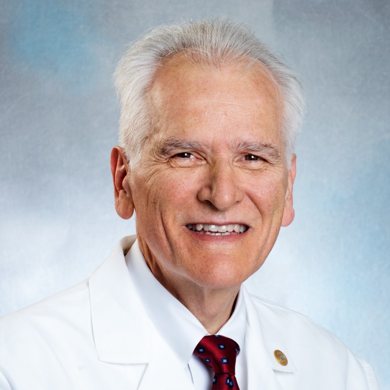 Charles A. Czeisler合作论文数Division of Sleep Medicine, Harvard Medical School;Division of Sleep and Circadian Disorders, Departments of Medicine and Neurology, Brigham and Women's Hospital64
 Chris M. Depner合作论文数Department of Health and Human Development, Montana State University41
 Edward Melanson合作论文数Energy Metabolism Lab, Division of Endocrinology, Metabolism and Diabetes, School of Medicine, University of Colorado Anschutz Medical Campus;University of Colorado Hospital36
 Andrew W. McHill合作论文数Sleep and Chronobiology Laboratory, University of Colorado Boulder27
 Tina Burke合作论文数Walter Reed Army Institute of Research25
 Monika R. Fleshner合作论文数Department of Integrative Physiology, College of Arts and Sciences, University of Colorado Boulder22
 Josiane L. Broussard合作论文数Diabetes and Obesity Research Institute and.21
 Christopher A. Lowry合作论文数Department of Integrative Physiology, College of Arts and Sciences, University of Colorado Boulder20
 Janine A Higgins合作论文数Center for Human Nutrition, University of Colorado Health Sciences Center19
 Monique K Lebourgeois合作论文数Dept Integrat Physiol, Univ Colorado17
 Fred Turek合作论文数Center for Circadian & Sleep Medicine, Feinberg School of Medicine, Northwestern University;Center for Sleep and Circadian Biology, Northwestern University;Department of Neurobiology, Northwestern University17
 Rob Knight合作论文数Rady Children's Hospital San Diego;Center for Microbiome Innovation, University of California, San Diego;Shu Chien-Gene Lay Department of Bioengineering, Jacobs School of Engineering, University of California, San Diego17
 Hannah Ritchie合作论文数16
 Martha Hotz Vitaterna合作论文数Department of Neurobiology, Weinberg College of Arts and Sciences, Northwestern University16
 Ellen Stothard合作论文数Sleep and Chronobiology Laboratory, University of Colorado Boulder;and Divisions of.16
 Steven W. Lockley合作论文数Department of Medicine, Brigham and Women's Hospital, Harvard Medical School14Dana Withrow合作论文数13
 Celine Vetter合作论文数13
 Kate Sprecher合作论文数13
 Thomas Roth合作论文数Sleep Disorders and Research Center, Henry Ford Hospital;Department of Psychiatry, School of Medicine, Wayne State University;Department of Psychiatry, School of Medicine, University of Michigan13Chinoy Evan D合作论文数Sleep and Chronobiology Laboratory, University of Colorado Boulder13
 Robert Eckel合作论文数Division of Endocrinology, Metabolism and Diabetes, School of Medicine, University of Colorado Anschutz;Lipid Clinic, University of Colorado Hospital;Department of Food Science and Human Nutrition, Colorado State University13
 Pieter C Dorrestein合作论文数Departments of Pharmacology and Pediatrics, Skaggs School of Pharmacy and Pharmaceutical Sciences, University of California, San Diego;Collaborative Mass Spectrometry Innovation Center, University of California, San Diego;Institute for Metabolomics Medicine, University of California, San Diego12
 Stacey L Simon合作论文数12
 Laura K. Barger合作论文数Division of Sleep Medicine, Brigham and Women’s Hospital, 221 Longwood Avenue, BLI 438, Boston, MA 02115, USA12
 Claude Gronfier合作论文数Division of Sleep Medicine;Brigham and Women ' s Hospital;Medical School, Harvard;Brigham and Women ' s Hospital, Harvard Medical School12
 Katie L. Stone合作论文数San Francisco Coordinating Center, University of California San Francisco;California Pacific Medical Center Research Institute11
 Cecilia G. Diniz Behn合作论文数11
 Kristen J Nadeau合作论文数11William C. Byrnes合作论文数Department of Exercise Science, University of Massachusetts11
 Leigh Perreault合作论文数Research Laboratory for Risk Management in Water Sciences, French Institute of Forestry Agricultural and Environmental Engineering (ENGREF),11D. J. Frey合作论文数10Fernando Vargas合作论文数Univ Calif San Diego, Skaggs Sch Pharm & Pharmaceut Sci, La Jolla, CA 92093 USA10
 Rachel Markwald合作论文数10Samuel J Bowers合作论文数9R. R. Markwald合作论文数9
 Gonzalez Antonio合作论文数Dept Pediat, Univ Calif San Diego9Rahman Shadab A合作论文数Mount Sinai Hospital, Samuel Lunenfeld Research Institute9
 Phyllis C Zee合作论文数Department of Neurology, Feinberg School of Medicine, Northwestern University;Division of Sleep Medicine, Northwestern Medicine;Center for Circadian and Sleep Medicine, Feinberg School of Medicine, Northwestern University9C. M. Jung合作论文数8
 Joseph M Ronda合作论文数8Sarah Morton合作论文数8
 Seth Creasy合作论文数Div Endocrinol Metab & Diabet, Univ Colorado8Jason P Sullivan合作论文数8
 O'Brien Conor S合作论文数Department of Biology, University of Maryland8
 Lameese D. Akacem合作论文数8
 David F. Dinges合作论文数Unit for Experimental Psychiatry, Division of Sleep and Chronobiology, University of Pennsylvania Perelman School of Medicine;Center for Sleep and Circadian Neurobiology, University of Pennsylvania Perelman School of Medicine8
 Christopher Landrigan合作论文数Division of General Pediatrics, Boston Children's Hospital;Harvard Medical School;Brigham and Women’s Hospital8Joseph M. Ronda合作论文数8




