Purpose: The intervertebral disc (IVD) is a spinal joint comprised of three main structures; the nucleus pulposus, annulus fibrosus and cartilage endplate. Intervertebral disc degeneration (IDD) is a progressive condition and a major cause of back pain. It primarily affects the nucleus pulposus associated with other structural changes, altering the biomechanics. These alterations are deeply connected to its extracellular matrix (ECM) composition. The human disc is matrix-rich embedded with few cells that are responsible for maintaining homeostasis, yet, cell numbers continually decline with aging/degeneration.
Conference Theme: Bones & Teeth: Translating Local Tissue Interactions and Systemic Interplays into New Therapies for Bones and Teeth
Introduction Asporin, a member of the small leucine rich proteins, is an extracellular matrix protein with elevated expression level in cartilage tissues with OA and degenerative disc disease. Aspartate(D) repeat polymorphism (from 7-21) can be found in human and D14 (14 aspartate repeat) has been identified as the risk factor for both OA and DDD, while D13 (13 aspartate repeat) is a common allele. It has been shown that asporin negatively regulates Tgf-β signaling in vitro by ATDC5 cell culture as well as contributes to collagen fibrillogensis. However, the in vivo role of asporin in development and pathogenesis in cartilage tissues is still unclear. Thus, in vivo study using mice as a model was performed in this study. Materials and Methods In order to study the correlation of asporin and Tgf-β signaling, immunostaining of asporin and P-Smad2/3 was performed on mice disc samples induced with injuries by looping or puncture. To further understand the cause and consequence relationship between asporin and Tgf-β/Smad signaling and the impact of overexpressing asporin in the disc, transgenic mice overexpressed with human asporin D13 and D14 were generated with a cartilage specific promoter Col11a2 using LacZ as reporter. Histological and molecular analysis were performed to study the effect of overexpressing asporin in the cartilage tissues. Results Immunohistochemical results showed that asporin and P-Smad2/3 are colocalized in the disc and articular cartilages. Moreover, asporin and P-Smad2/3 also present in the degenerative models including natural degeneration, disc puncture and tail looping. These results suggested the possible correlation between asporin and Tgf-β/Smad signaling as well as the contribution of asporin and Tgf-β/Smad in the pathologenesis of disc degeneration. Interestingly, in transgenic mice overexpressing asporin D13 and D14, overexpression of asporin induced glycosaminoglycans production in the transgenic nucleus pulposus, small cell clusters, and sparsely distributed cells with morphological differences comparing to clumps of vacuolated mouse nucleus pulposus cells in wildtype, indicated that overexpressing of asporin enhances differentiation or maturation process of notochordal-like cells in mouse nucleus pulposus toward the nucleus pulposus cells (NPCs) that is more indicative of human nucleus pulposus. In situ hybridization showed that the differentiated cell showed increased Col2a1 and Agc expression, which indicated characteristics of NPCs. Marker analysis further showed that characteristics of chondrocyte-like cells (CLCs) with the molecular signature of Sox9+T- and CK18- cells exhibited in the nucleus pulposus of D13 transgenic mice. However, fibrosis and hypertrophic differentiation, which are commonly observed in human disc degeneration, were absent in the D13 transgenic. Intriguingly, phosphorylation of Smad2/3 was found to be up-regulated in the transgene-expressing cells, suggesting that overexpression of Asporin promotes Tgf-β signaling. Conclusion It is proposed that Asporin, as a risk factor, enhances cellular differentiation of cell types that can lead to degenerative progression possibly though promoting Tgf-β signaling. Further study of Tgf-β related pathway are need to be investigated in order to gain more insight in the involvement of Tgf-β in causing disc degeneration. Disclosure of Interest None declared
Introduction Intervertebral disc (IVD) degeneration is a common world-wide occurrence and is classically associated with cellular, water and proteoglycan (PG) loss from the nucleus pulposus (NP) which can greatly affect IVD function. While several studies have demonstrated molecular changes during aging and degeneration, our understanding of the processes that occur during these events are limited. The identification and analysis of proteins that are present in the IVD during aging and degeneration may provide clues on the processes that occur during these two events, which was the aim of the present study. Materials and Methods Ethics was approved by the IRB board, and both degenerated and non-degenerated IVD specimens (seven samples of each) were acquired with patient consent. The NP and AF were dissected from the IVD, and proteins underwent extraction with 4M GuHCl buffer, fractionation using CsCl density gradient ultracentrifugation, and then subsequently separated into insoluble/soluble protein fractions. The proteins then underwent trypsin digestion and iTRAQ labeling for quantitative analysis using LCMS/MS. Data were analyzed using ProteinPilot 4.0 Results The proteins were first grouped into clusters to depict whether or not they were associated with increases, decreases or no change with increasing age. With increasing age, the AF of non-degenerated IVD had decreasing levels of collagen type I (soluble fraction) and types II and XI (soluble/insoluble fractions) which could impact on the tensile strength of the AF. A decreasing trend was also observed in the NP for collagen type II, however an increase in collagen type I was observed in the younger controls. In both AF and NP, increasing levels of decorin, lumican and PRELP were observed with increasing age, in addition to mimecan and biglycan in the NP. The identified proteins from the degenerated IVD was also clustered into increasing, decreasing, or no change in trend, and compared with the non-degenerated samples (controls) which indicated similar trends for both groups. Notable changes in the NP associated with degenerated samples included an accumulation of fibronectin and type I collagen (insoluble fraction) in younger degenerated samples, in addition to CILP and CILP2. In degenerated AF, there were increased levels of fibronectin, COMP and CILP, while aged and degenerated AP had increased aggrecan, decorin and PRELP (insoluble fraction). Conclusion The data in this study indicate that degeneration and aging share both similarities and differences in the static protein profile. Alterations in the matrix and shifts in protein content are likely impinge on cell-matrix interactions in addition to matrix turnover, and aging and degeneration both exhibit fibrotic-related proteins. Of particular interest is the accumulation of collagen type I and fibronectin (also associated with fibrosis) in young and degenerated IVD from the insoluble fraction, an indication of increased cross-linked molecules that have accumulated over time. Such differences may be an important indicator of degeneration-associated changes and warrants further investigation. Studying the dynamic protein profile using SILAC technology and ex vivo culture of IVD samples will identify actively synthesized proteins in the IVD. This will provide valuable information on cellular functions in the IVD during aging and degeneration, which will be the focus of future studies. Disclosure of Interest None declared
 Danny Chan (陳振勝)合作论文数School of Biomedical Sciences, LKS Faculty of Medicine, The University of Hong Kong75
 kkh leung合作论文数30
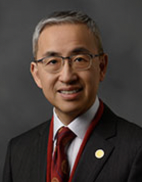 Man-Chee Kenneth Cheung (张文智)合作论文数Department of Orthopaedics & Traumatology, School of Clinical Medicine, The University of Hong Kong;Shenzhen Hospital, The University of Hong Kong26
 You-Qiang Song (宋又强)合作论文数School of Biomedical Sciences, Li Ka Shing Faculty of Medicine, The University of Hong Kong25SL Wynn合作论文数24
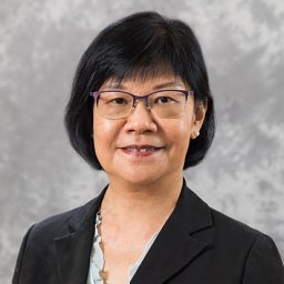 Mai Har Sham合作论文数Dept Biochem, Univ Hong Kong21y k au合作论文数20
 Jaro Karppinen合作论文数Department of Physical Medicine and Rehabilitation, University of Oulu;Research Unit of Health Sciences and Technology, University of Oulu20
 Pak Chung Sham (沈伯松)合作论文数Department of Psychiatry, School of Clinical Medicine, Li Ka Shing Faculty of Medicine, The University of Hong Kong;State Key Laboratory of Brain and Cognitive Sciences, The University of Hong Kong20J.C.Y. Leong合作论文数19i g melhado合作论文数17Ky Tsang合作论文数17
 Keith Dip-Kei Luk合作论文数The University of Hong Kong17k y tsang合作论文数16
 Bernd Fritzsch合作论文数Department of Neurological Sciences, College of Medicine, University of Nebraska Medical Center16
 Warren Chan合作论文数College of Engineering, Nanyang Technological University;School of Chemistry, Chemical Engineering and Biotechnology, Nanyang Technological University;Integrated Nanotechnology & Biomedical Sciences Laboratory, University of Toronto16mch cheung合作论文数15P.P.L. Tam合作论文数15R H Lovell-Badge合作论文数12
 Julian Alexander Tanner (唐柱霖)合作论文数School of Biomedical Sciences, Li Ka Shing Faculty of Medicine, The University of Hong Kong12
 Robin Lovell-Badge合作论文数Laboratory of Stem Cell Biology and Developmental Genetics, The Francis Crick Institute12pyp kao合作论文数11
 Yip Shea-Ping (叶社平)合作论文数Department of Health Technology and Informatics, Faculty of Health and Social Sciences, The Hong Kong Polytechnic University11
 Wood-Yee Woody Chan (陳活彜)合作论文数School of Biomedical Sciences, The Chinese University of Hong Kong;Institute for Tissue Engineering & Regenerative Medicine, The Chinese University of Hong Kong11s wynn合作论文数9Wong S Y合作论文数9AL Pelling合作论文数9kmc cheung合作论文数8asp tang合作论文数8jjt jim合作论文数8
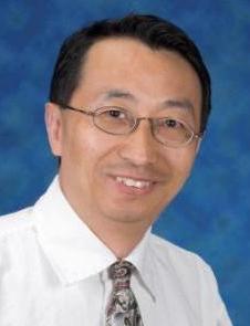 Henry H.Q. Heng合作论文数Center for Molecular Medicine and Genetics, School of Medicine, Wayne State University;Department of Pathology, School of Medicine, Wayne State University8
 Karen Steel合作论文数Wolfson Centre for Age-Related Diseases, King's College London8
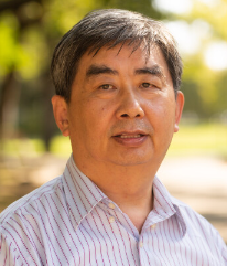 Michael Zhang合作论文数Department of Biological Sciences, School of Natural Sciences and Mathematics, The University of Texas at Dallas8vcw ng合作论文数7Lc Tsui合作论文数7ccw cheng合作论文数7anthony leung wai lun合作论文数7d chan合作论文数7
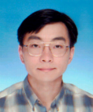 Kwan Kin Ming合作论文数School of Life Sciences,The Chinese University of Hong Kong7
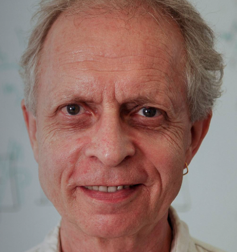 Jürg Ott合作论文数Laboratory of Statistical Genetics, Rockefeller University7Leena Ala-Kokko合作论文数7Denise A. Chan合作论文数6w f dung合作论文数6ccy chan合作论文数6w y chan合作论文数6s dong合作论文数6




