Bacteriophages are a promising tool for microbiome editing, yet their development has been constrained by limited insights into bacteriophage-host interactions within their shared mammalian body habitat. We isolated a lytic phage ΦKL11 that efficiently targets a disease-associated member of the human gut microbiota, Eggerthella lenta, during in vitro growth. However, ΦKL11 selects for a pre-existing and reversible bacteriophage-resistant sub-population in mice. Long-read sequencing revealed a massive genomic inversion event, representing >50% of the E. lenta genome, enriched in response to bacteriophage infection. Transcriptomics linked this inversion to the altered expression of three capsular polysaccharide synthesis (CPS) gene clusters and transmission electron microscopy confirmed differential capsule production. Finally, we show that ΦKL11 has a broad host range attributable to CPS and other strain-variable genes. These findings suggest a previously unrecognized strategy for phage evasion in the gut, involving megabase-scale genomic inversions and reversible capsule variation driving phage resistance.
A summary of the data from saturation binding assays used to calculate Bmax and receptor number per cell for bladder cancer cell lines. The data are representative of two independent assays. Data are reported as mean ± standard deviation.
Supplemental Movies 2a-2c: 22Rv1-NuclightGreen and SHP-77-NuclightRed mixed population co-cultures. Time-lapse movies of 22Rv1 cells (labeled with NuclightGreen) and SHP-77 cells (labeled with NuclightRed) co-cultured with T cells. Both the 22Rv1 and SHP-77 cells were plated at equal densities and treated with either NT control BiTE®, AMG 757, or the PSMA-targeting HLE BiTE, AMG 160. T cells were added to each well at an E:T ratio of 10:1. Images were captured every 4 hours at 4x magnification for 5 days. a. 22Rv1, SHP-77, and T cells co-cultured with NT control BiTE®. b. 22Rv1, SHP-77, and T cells co-cultured with AMG 757. c. 22Rv1, SHP-77, and T cells co-cultured with AMG 160.
CDCP1 mRNA expression by pathologic stage in the TCGA dataset. p=0.72 by Kruskal-Wallis test.
CDCP1 protein expression by clinical stage using the tissue microarray. p=0.32 by Kruskal-Wallis test.
A plot showing the mean tumor volume for the vehicle and treated arms from the antitumor assessment study in mice bearing UMUC3 xenografts. The differences in mean between treatment arms was found to be significant (P < 0.001) using an unpaired, two tailed t test by day 20. Data are reported as mean ± standard deviation.
CDCP1 mRNA expression in locally-advanced vs metastatic patients, from the IMvigor210 dataset. p=0.55 by Wilcoxon test, n=316 patients.
CDCP1 mRNA is overexpressed in basal/squamous subtypes of bladder cancer, related to Figure 1A-D. Pairwise comparisons of CDCP1 expression between bladder cancer consensus subtypes using the Wilcoxon rank-sum test across four different datasets are shown.
CDCP1 mRNA expression is not associated with overall survival. Kaplan–Meier survival analyses were performed on quartiles of CDCP1 mRNA expression with overall survival as the endpoint. p > 0.05 in both public datasets for which overall survival data were available.
A plot showing the mean tumor volume for the vehicle and treated arms from the antitumor assessment study in mice bearing UMUC3 xenografts. The differences in mean between treatment arms was found to be significant (P < 0.001) using an unpaired, two tailed t test by day 20. Data are reported as mean ± standard deviation.
 Jonathan Chou合作论文数bHelen Diller Family Comprehensive Cancer Center, University of California23
 Felix Y. Feng合作论文数Department of Radiation Oncology, University of California, San Francisco23
 Emily A Egusa合作论文数22Jun Zhu合作论文数21
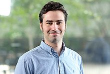 Evans Michael J合作论文数The Skaggs Institute for Chemical Biology and Department of Cell Biology, The Scripps Research Institute13Zhao Ning合作论文数Department of Chemistry, Louisiana State University13
 James A. Wells合作论文数Helen Diller Family Comprehensive Cancer Center, University of California, San Francisco;Department of Pharmaceutical Sciences, University of California, San Francisco13Jie Zhou合作论文数Univ Calif San Francisco, Dept Pharmaceut Chem, San Francisco, CA 94107 USA12
 Eric Small合作论文数Department of Urology, University of California, San Francisco;Division of Hematology and Oncology, Department of Medicine, University of California, San Francisco12
 Aggarwal Rahul合作论文数Helen Diller Family Comprehensive Cancer Center, University of California, San Francisco;Division of Hematology/Oncology, School of Medicine, University of California, San Francisco12Kevin K Leung合作论文数11Nima Hooshdaran合作论文数Department of Radiology and Biomedical Imaging, University of California11Emily Chan合作论文数11Chopra Shalini合作论文数11Sasank Sakhamuri合作论文数10Alberto Carretero-González合作论文数Department of Radiation Oncology, University of California10Michelle L Badura合作论文数9
 Jiaoti Huang合作论文数Department of Pathology, School of Medicine, Duke University9Bradley A. Stohr合作论文数9
 Peter J. Turnbaugh合作论文数Center for Genome Sciences, Washington University9Brian Yu合作论文数8Tanushree Shenoy合作论文数8Fei Lee合作论文数8Keegan Cooke合作论文数8Brant Delafontaine合作论文数8Nooshin Hashemi Sadraei合作论文数8Troy M Robinson合作论文数8Mark Salvati合作论文数8
 Sinan Wang合作论文数Univ Calif San Francisco8
 Anil P Bidkar合作论文数University of California8
 Sylvie Rottey合作论文数Department of Medical Oncology, Universitair Ziekenhuis Gent;Drug Research Unit Ghent, Universitair Ziekenhuis Gent8
 Veronica Steri合作论文数8
 Robert R. Flavell合作论文数Laboratory of Synthetic Protein Chemistry;Laboratory of Molecular Biophysics;UniVersity;Laboratory of Molecular Biophysics, Rockefeller UniVersity8
 Alan Ashworth合作论文数Division of Hematology and Oncology, Department of Medicine, UCSF;Helen Diller Family Comprehensive Cancer Center, UCSF8
 Yuzhuo Wang (王玉琢)合作论文数BC Cancer;Vancouver Prostate Centre;Department of Urologic Sciences, University of British Columbia;Living Tumor Laboratory8Julie M. Bailis合作论文数8
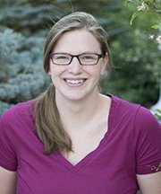 Margaret Alexander合作论文数Department of Pathology, University of Utah5
 Adam Foye合作论文数4
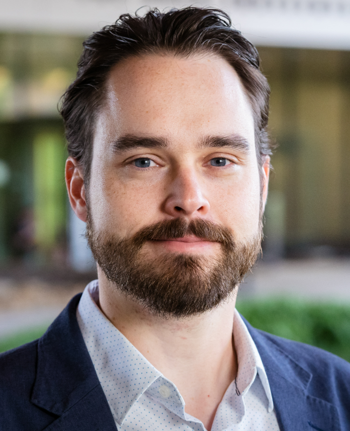 Jordan Bisanz合作论文数Department of Biochemistry and Molecular Biology, Eberly College of Science, The Pennsylvania State University;One Health Microbiome Center, Huck Institutes of the Life Sciences, The Pennsylvania State University4
 Li Zhang合作论文数4Noecker Cecilia A合作论文数Centro de Investigación Sobre Enfermedades Infecciosas, Instituto Nacional de Salud Pública;U.S.-Mexico Fulbright-García Robles Commission for Educational and Cultural Exchange;Centro de Investigación Sobre Enfermedades Infecciosas, Instituto Nacional de Salud Pública4
 Ye Tao (陶冶)合作论文数School of Art and Archaeology, Hangzhou City University;International Design Institute, Zhejiang University3Yung-Hua Wang合作论文数3
 Almut Heinken合作论文数3Shion A Lim合作论文数3
 Veronica Escalante合作论文数University of California San Francisco3Hyunjung Kim合作论文数3
 DeFelice Brian合作论文数Davis Genome Center, University of California3
 Dylan Dodd合作论文数Microbiology & Immunology Department, Stanford University/Department of Pathology, Stanford University3




