IntroductionOsteoarthritis (OA) is a degenerative joint disease marked by chronic inflammation, extracellular matrix degradation, and dysregulated cell death. The roles of apoptosis-, autophagy-, and ferroptosis-related genes in OA pathogenesis remain unclear.MethodsIntegrated bioinformatics analyses were conducted on public GEO datasets to identify apoptosis-autophagy-ferroptosis-related genes (AAFRGs). TLR3 and GLUL were identified using LASSO, random forest, and SVM-RFE algorithms. Immune infiltration analysis, immunohistochemistry, and functional assays in human chondrocytes were performed, and ACLT-induced rat OA models were used for in vivo validation.ResultsTLR3 was upregulated and associated with pro-inflammatory immune cells, while GLUL was downregulated and correlated with anti-inflammatory signatures. TLR3 knockdown reduced inflammation, apoptosis, and aberrant mineralization, partially restoring extracellular matrix integrity. GLUL overexpression promoted cellular homeostasis. In rats, TLR3 inhibition and PRP treatment decreased pro-inflammatory cytokines (IL-1β, TNF-α), reduced matrix-degrading enzymes (MMP3, MMP13), and restored GLUL and IL-10 levels.Discussion/ConclusionTLR3 and GLUL orchestrate inflammatory responses and homeostatic imbalance in OA, representing potential biomarkers and therapeutic targets for diagnosis and intervention.
Background/Aim: Diabetic retinopathy (DR) is a common comorbidity of diabetes involving the formation of abnormal vascular structures in the retina. Tissue inhibitor of metalloproteinases 2 (TIMP2), initially identified as a key mediator of extracellular matrix turnover, is pivotal for inflammatory processes and tissue homeostasis. The current study examined the influence of TIMP2 gene variations on the risk for DR. Materials and Methods: We investigated the association of TIMP2 gene variations with DR by analyzing four single-nucleotide polymorphisms (SNPs) of the TIMP2 gene (rs16971783, rs2889529, rs7220980, and rs8068674) in a cohort of 672 patients with DR and 919 diabetic controls with normal ophthalmoscopic findings. Results: Our results showed that rs16971783 of TIMP2 gene was associated with a higher risk for DR (TA vs. TT, AOR=1.445, p=0.028; TA+AA vs. TT, AOR=1.179, p=0.046). We further demonstrated that the association of rs16971783 with DR was exclusively observed in diabetic individuals with proliferative DR (TA vs. TT, AOR=1.827, p=0.035; TA+AA vs. TT, AOR=1.351, p=0.027), whereas not detected among those who suffered from non-proliferative DR. In addition, preliminary exploration of gene expression data from public resources reveald that rs16971783 regulated TIMP2 gene expression in various human tissues. Conclusion: Allele-specific expression of TIMP2 gene might contribute to the progression of DR.
Diabetic retinopathy (DR) represents one of the most common microvascular complications of diabetes, and proliferative diabetic retinopathy (PDR) is the most vision-threatening form. Carbonic anhydrase IX (CA9), a hypoxia-inducible enzyme, has been implicated in several pathological processes, but its involvement in DR has not been clarified. In this study, three CA9 single nucleotide polymorphisms (rs3829078, rs2071676, and rs1048638) were genotyped in diabetic patients with and without DR. Clinical characteristics were compared between groups, and expression analyses were conducted using public databases and ARPE-19 cells under hyperglycemic and hypoxic conditions. No significant association was observed between CA9 variants and DR susceptibility in the overall cohort. However, among patients aged ≤60 years, carriers of the rs1048638 C/A and C/A + A/A genotypes exhibited a significantly increased risk of PDR. Expression quantitative trait locus (eQTL) data from the GTEx database revealed higher CA9 mRNA expression in tissues harboring the rs1048638 A allele. Moreover, both transcriptomic data and in vitro experiments demonstrated upregulation of CA9 in ARPE-19 cells exposed to high glucose and hypoxia. These findings suggest that the rs1048638 polymorphism may modulate CA9 expression and contribute to PDR development in younger diabetic patients through hypoxia- and glucose-related mechanisms. Collectively, our findings suggest that CA9 may serve as a potential risk biomarker associated with the progression of DR, particularly in younger patients.
Background Influenza caused by the influenza virus poses a significant global health threat, highlighting the urgent need for effective antiviral drugs. Ganhuangcao, a traditional Chinese medicine, is known for its heat-clearing, detoxifying, diuretic, and jaundice-reducing effects, but its anti-influenza properties and mechanisms have been scarcely studied. Objective This study aims to elucidate the effects and molecular mechanisms of Ganhuangcao extracts, particularly its main component, gallic acid (GA), against influenza virus (H1N1) infection. Methods Ganhuangcao extracts were prepared using aqueous and alcoholic extractions, and their chemical components were analyzed by high-performance liquid chromatography. MDCK cells and BALB/c mice infected with H1N1 were used as in vitro and in vivo models to evaluate the antiviral effects of GA at different concentrations. The half-maximal effective concentration (EC₅₀) and cytotoxicity (CC₅₀) of GA were determined using plaque reduction assays. Flow cytometry was used to analyze the impact of GA on immune cell infiltration (CD4⁺/CD8⁺ T cells, macrophages, neutrophils, NK cells, dendritic cells) in the peripheral blood samples of infected mice. Western blotting, immunofluorescence, and immunohistochemistry were employed to detect viral load (NP protein) and the expression and activation of key proteins in the JAK2-STAT1 signaling pathway. The JAK2-specific inhibitor LY3009104 was used to validate the pathway. Results GA can effectively inhibit the replication of the H1N1 virus in cells, with an EC₅₀ of 8.49 μg/mL, a high therapeutic index, and good safety. Mechanistic studies indicate that GA can directly inhibit the activity of viral neuraminidase and downregulate the expression and replication of viral NP protein by blocking the abnormal activation of the JAK2/STAT1 signaling pathway. In an infected mouse model, GA treatment significantly reduced lung viral load, alleviated pathological damage to lung tissue, and increased survival rates. Its immunomodulatory effect is like that of the JAK2 inhibitor LY3009104, capable of suppressing excessive immune cell infiltration (e.g., neutrophils, macrophages) and cytokine storms following infection. Conclusion This study confirms that GA derived from Ganhuangcao has significant anti-influenza potential. Its mechanism is closely related to inhibiting the activation of the JAK2-STAT1 signaling pathway, modulating the immune response, and directly inhibiting viral enzyme activity. This indicates that GA is a promising anti-influenza drug worthy of further clinical translation research.
Antiphospholipid syndrome is an autoimmune disorder of unknown etiology. The anti-inflammatory and immunomodulatory properties of Zingiber officinale Roscoe may provide a novel approach for potentially preventing obstetric antiphospholipid syndrome (OAPS). Based on several public databases, compounds and potential drug targets of Z officinale Roscoe were screened, and a compound-target network was constructed using Cytoscape. The targets of OAPS were identified from GeneCards. The common genes of the disease targets and compound targets were defined as hub genes. Protein-protein interaction, Gene Ontology, and Kyoto Encyclopedia of Genes and Genomes analyses were performed, and the OAPS pathway was obtained. Molecular docking was used to verify the binding ability of the effective compounds and targets. In total, 298 compounds were identified as components of Z officinale Roscoe. Among these, 12 active compounds exhibited an oral bioavailability of ≥30% and drug-likeness of ≥0.14, with 99 associated targets. A total of 28 common targets were identified as hub genes. In addition, the beneficial effects of Z officinale Roscoe in OAPS management are thought to be mediated by anticoagulant, anti-inflammatory, and immunomodulatory mechanisms. These processes are regulated by 9 biological signaling pathways involved in the protection against OAPS. Molecular docking showed that the binding affinities of tumor protein 53 with beta-sitosterol, tumor protein 53 with stigmasterol, prostaglandin G/H synthase 2 with beta-sitosterol, and prostaglandin G/H synthase 2 with stigmasterol were -6.651, -6.709, -7.099, and -6.829, all of which had high binding affinities. Overall, systematic pharmacology and molecular docking provide a new theoretical basis and research insight for the prevention of OAPS.
During the healing process of skin wounds, there are challenges such as slow tissue regeneration and bacterial infections. This paper has developed a drug-loaded composite hydrogel dressing that can promote angiogenesis, inhibit bacteria, and reduce inflammation. The hydrogel is composed of gelatin (Gel) and acrylamide (AM), and is formed into a biomimetic cell membrane structure through cross-linking. By adding different concentrations of graphene oxide (GO), the tensile strength of the hydrogel dressing can be significantly increased (from 57.54 KPa to 95 KPa), and the adhesion strength (shear strength) can be effectively enhanced (16.44 KPa). Additionally, Panax ginsenoside Rh1 has been successfully encapsulated in the hydrogel, achieving local controlled release. In vivo evaluation shows that the enhanced matrix by graphene oxide and the sustained release of Panax ginsenoside Rh1 have an antibacterial rate of up to 98.69% against S. aureus. It also stimulates the proliferation, migration, and tube formation of endothelial cells, thereby achieving strong vascularization at the wound site. Moreover, it can enhance the secretion capacity of anti-inflammatory factors (IL-4 from 19.54 to 105.61 pg/mL, IL-10 from 151.94 to 339.98 pg/mL), thereby accelerating the wound healing process. This hydrogel dressing provides a promising strategy for achieving high-quality wound repair.
Objective: Based on previous findings on the Lingguizhugan (LGZG)-mediated gut-liver axis, this study clarifies the therapeutic mechanisms of LGZG in metabolic dysfunction-associated steatotic liver disease (MASLD), with a focus on the gut microbiota-bile acid-TGR5 (GPBAR1) axis. Methods: C57BL/6J mice were fed a high-fat diet (HFD) for 8 weeks to induce MASLD, followed by 4-week LGZG intervention (21.57 g/kg/day, oral gavage). Metabolic phenotypes, gut microbiota (16S rRNA sequencing), serum/hepatic bile acids (targeted metabolomics), and molecular targets (qPCR/Western blot) were analyzed. Results: LGZG significantly alleviated HFD-induced obesity, insulin resistance, and hepatic steatosis, while enhancing whole-body energy expenditure (increased oxygen consumption (VO2), and heat production (p < 0.05). It also reduced serum ALT (p < 0.001) and AST levels (p < 0.01). Mechanistically, LGZG remodeled the gut microbiota, specifically increasing Akkermansia, Bifidobacterium and Lachnospiraceae_NK4A236_group while decreasing Lactobacillus. This shift inhibited the intestinal FXR-Fgf15 axis, concurrently activating the hepatic alternative bile acid synthesis pathway (upregulating CYP27A1 and CYP7B1 protein expression; p < 0.001 and p < 0.01, respectively). Consequently, systemic accumulation of non-12α-hydroxylated bile acids (non-12-OH BAs) such as hyocholic acid (HCA) and 7-ketolithocholic acid (7-ketoLCA) occurred-known TGR5 agonists and intestinal FXR antagonists. These changes elevated serum GLP-1 levels (p < 0.05) and activated adipose TGR5-cAMP/PKA/CREB signaling. The metabolic benefits primarily originated from non-12-OH BAs enrichment and TGR5-mediated adipose browning, not hepatic FXR activation. Conclusions: Our findings show that LGZG ameliorates MASLD by remodeling bile acid profiles via intestinal FXR-Fgf15 axis inhibition and hepatic alternative synthesis pathway activation. This study highlights the TGR5-targeting properties of LGZG, providing a mechanistic basis for its therapeutic use in metabolic disorders.
BACKGROUND Qiweizhigan granule (QWZG) is employed in clinical settings for the treatment of metabolic dysfunction-associated steatohepatitis (MASH). However, the precise biological mechanisms underlying its therapeutic effects are not yet fully elucidated. AIM To assess the efficacy and the mechanism of QWZG against MASH. METHODS Animal models were established, including normal group, a choline-deficient, L-amino acid-defined high-fat diet (CDAHFD) group, and low/medium/high-dose QWZG groups, as well as a rosiglitazone group. Through comprehensive biochemical, histopathological, RNA sequencing, and bioinformatics analyses, galectin 3 (LGALS3) was identified as a critical target of QWZG in the treatment of MASH. The level of LGALS3 was quantitatively assessed and validated using Western blotting, real-time quantitative PCR, and immunofluorescence. The role and function of LGALS3 in inflammation and MASH progression were further investigated through gene knockdown, overexpression, iron assay, and transmission electron microscopy. RESULTS QWZG significantly ameliorated liver pathology by reducing steatosis, inflammation, and fibrosis. RNA sequencing analysis identified 1507 co-expressed differentially expressed genes among the CDAHFD, normal, and QWZG groups. Among these, LGALS3 was identified as one of the most significantly altered differentially expressed genes. Both mRNA and protein levels of LGALS3 were elevated in the CDAHFD group compared to the normal group, whereas treatment with QWZG reduced their levels. Analysis of Human Protein Atlas database indicated that LGALS3 was predominantly expressed in Kupffer cells, and was validated by real-time quantitative PCR and immunofluorescence. Furthermore, the level of LGALS3 was significantly increased in lipopolysaccharide-induced RAW264.7 cells, where its overexpression and recombinant LGALS3 protein both significantly enhanced the expression of interleukin-6, interleukin-1 beta, and tumor necrosis factor-alpha. LGALS3 overexpression significantly inhibited glutathione peroxidase 4 (GPX4) expression, and exacerbated mitochondrial damage, whereas LGALS3 knockdown markedly increased GPX4 level, and significantly reduced the levels of both total iron and ferrous iron. QWZG treatment significantly reduced the levels of malondialdehyde and ferrous iron, increased the levels of superoxide dismutase and glutathione. In addition, QWZG treatment also significantly enhanced GPX4 expression. Mechanistically, LGALS3 knockdown was associated with reduced expression of tumor necrosis factor receptor-associated factor 6 (TRAF6) and NOD-like receptor family pyrin domain containing 3, while its overexpression led to increased levels of these proteins. The TRAF6 inhibitor C25-140 effectively reversed the LGALS3-induced alterations in GPX4 expression and iron accumulation. Furthermore, QWZG treatment significantly decreased the levels of TRAF6 and NOD-like receptor family pyrin domain containing 3. CONCLUSION QWZG ameliorated the progression of MASH by modulating ferroptosis through the LGALS3/TRAF6/GPX4 axis.
Chronic diabetic wounds remain a major clinical challenge due to persistent inflammation, impaired neuro-immune crosstalk, and dysregulated macrophage polarization. Here, we report an injectable, ultrasound (US)-activated piezoelectric hydrogel that programmably couples neurogenesis and macrophage reprogramming to accelerate diabetic wound repair. The hydrogel is constructed from dynamic covalent networks among acyl hydrazide hyaluronic acid, benzene boronic acid modified oxidized sodium alginate, and epigallocatechin 3-gallate, and integrates a neuroactive self-assembling peptide and piezoelectric PLLA nanofibers. This design confers the hydrogel with injectability, self-healing, tissue adhesion, and US-triggered electrical signaling. In vitro, the hydrogel under US promotes M2 macrophage polarization and enhances axonal outgrowth of dorsal root ganglion neurons, establishing bidirectional neurogenesis-macrophage crosstalk. In a type II diabetic mouse model, the hydrogel markedly accelerates wound closure, elevates the M2/M1 ratio, enhances reinnervation and angiogenesis, and improves collagen remodeling. Transcriptomic analysis of cells and wound tissues identifies coordinated regulation of mitochondrial function and oxidative phosphorylation as a central mechanism. In a diabetic rabbit ear model, the hydrogel with US further attenuates hypertrophic scar formation. This programmable piezoelectric hydrogel converts noninvasive US into spatially confined bioelectrical and biochemical cues, offering a promising strategy to overcome multifactorial barriers in diabetic wound healing.
This study aimed to evaluate the cost-effectiveness of using selpercatinib as a first-line treatment option or reserving it for second-line use versus relying exclusively on chemotherapy in managing RET fusion-positive advanced non-small cell lung cancer (NSCLC), to identify the optimal treatment strategy from the perspective of US payers. A partitioned survival model (PSM) was constructed to assess the clinical outcomes and healthcare costs of two selpercatinib-containing strategies—first-line selpercatinib and second-line selpercatinib—each compared with chemotherapy alone for patients with advanced NSCLC harboring RET fusions. The costs and utility values were determined using the existing literature and standard fee databases as per prior research. To assess model robustness, both probabilistic sensitivity analysis and univariate sensitivity analysis were performed. Compared with chemotherapy alone, first-line selpercatinib was associated with an incremental gain of 0.12 quality-adjusted life-years (QALYs) at an additional cost of 117,682, resulting in an incremental cost-effectiveness ratio (ICER) of953,637.15 per QALY. Second-line selpercatinib was associated with an incremental gain of 0.13 QALYs at an additional cost of 68,569, resulting in an ICER of526,970.29 per QALY. The variables with the most significant impact on the ICER were the cost of selpercatinib and utility values. To be cost-effective at a willingness-to-pay (WTP) threshold of 150,000 per QALY, the price of selpercatinib would need to be reduced by 69.94
Background Breast cancer is a highly common malignancy in women, with bone as its most frequent site of distant metastasis, significantly increasing mortality risk. Methods This study integrated single-cell RNA sequencing datasets (GSE266330 and GSE243526) to systematically characterize the dynamic evolution of the tumor microenvironment from normal tissue to primary tumor and bone metastasis in breast cancer. Results In total, 141,056 high-quality single cells were analyzed, identifying 11 major cell types. Myeloid cell and T cell proportions were significantly elevated in bone metastases, suggesting their pivotal role in metastatic progression. The bone metastasis-enriched Mac-SPP1 macrophage subpopulation exhibited M2 polarization, high metabolic activity, and a significant association with poor patient prognosis ( P = 4.9e-05). It highly expressed transcription factors such as HOXA9 and MAF, potentially promoting metastasis through regulation of an immunosuppressive network. Among T cells, the CD4-S100A9 and CD8-GZMK subpopulations were significantly expanded in bone metastases and exhibited high cytotoxicity, high exhaustion, and metabolic reprogramming. Pseudotime analysis showed that CD4-S100A9 cells were at the initial state of CD4+ T cell differentiation, whereas CD8-GZMK cells serve a key transitional node from effector memory toward exhaustion. Cell-cell communication analysis revealed that Mac-SPP1 interact specifically with these CD4/CD8 T cell subsets through the SPP1-integrin axis (e.g., SPP1-CD44) and the CD74-MIF/COPA signaling axis, potentially driving T cell functional exhaustion and immune evasion. Conclusion This study is the first to delineate the synergistic pro-metastatic role of the Mac-SPP1–CD4-S100A9/CD8-GZMK axis in breast cancer bone metastasis, providing a theoretical basis for developing therapeutic strategies targeting the immune microenvironment.
The effective repair of chronic diabetic wounds remains challenging owing to the excessive increase in reactive oxygen species and chronic inflammation. Herein, we engineer an injectable, pH/ROS-dual responsive antibacterial hydrogel (GT-PBA/EGCG/OPU/TOB-PDLSCs) for scarless diabetic wound repair by integrating periodontal ligament stem cells (PDLSCs) into an interpenetrating polymer network (IPN) hydrogel composed of phenylboric acid-modified gelatin (GT-PBA), oxidized pullulan (OPU), epigallocatechin-3-gallate (EGCG), and tobramycin (TOB). The hydrogel is prepared through dynamic Schiff base crosslinking between GT-PBA/TOB and OPU, while phenylborate ester linkages with EGCG confer ROS-responsive degradability. This design enables sustained, on-demand release of TOB and EGCG in response to pathological pH and ROS levels. The encapsulated PDLSCs maintain high viability and proliferative capacity within the hydrogel matrix. Functionally, the hydrogel effectively scavenges intracellular ROS, reprograms macrophages toward a pro-regenerative phenotype, and enhances stem cell survival under oxidative stress. In diabetic wound models, the PDLSCs-loaded hydrogel significantly accelerates wound closure, promotes angiogenesis, and modulates inflammation. Notably, it markedly reduces scar formation, downregulates profibrotic genes YAP and En1, and facilitates regenerative healing. This multifunctional platform thus represents a promising strategy for achieving scarless repair of diabetic wounds through immunomodulation, ROS scavenging, and stem cell delivery.
Post-stroke cognitive impairment (PSCI) is a prevalent cerebrovascular condition resulting from ischemic stroke. This study aimed to determine the expression levels of NEXN-AS1 in PSCI, evaluate its clinical significance, and further uncover the molecular mechanisms through which it contributes to the initiation and progression of PSCI. The quantification of NEXN-AS1, miR-92a-3p, and NRF1 expression was performed using qRT-PCR. The diagnostic utility of serum NEXN-AS1 was assessed through ROC analysis. Risk factors associated with cognitive impairment following stroke were identified using both univariate and multivariate logistic regression. A cellular model of cognitive dysfunction was established via oxygen-glucose deprivation/reperfusion (OGD/R). The PSCI animal model was established through the Middle cerebral artery occlusion (MCAO) surgery. Inflammatory status was determined by measuring cytokine levels, including IL-6, IL-1β, and IL-10, while oxidative stress was evaluated by quantifying ROS, MDA, and CAT. In stroke patients, NEXN-AS1 expression was notably downregulated and further decreased in cases with PSCI. It served as a reliable biomarker for distinguishing stroke patients from healthy individuals and PSCI from post-stroke cognitive normality (PSCN) groups. Upregulation of NEXN-AS1 in BV2 cells following OGD/R stimulation led to increased proliferation, decreased inflammatory response, and reduced oxidative stress. Moreover, miR-92a-3p expression reversed the protective effects of NEXN-AS1 under OGD/R conditions. Overexpression of NEXN-AS1 alleviated cognitive dysfunction, inflammatory response and oxidative stress in PSCI rats, while overexpression of miR-92a-3p counteracted the protective effect of NEXN-AS1 on PSCI rats. Further analysis identified NRF1 as a downstream target of miR-92a-3p. NEXN-AS1 exerts protective effect against ischemic brain injury in both in vitro and in vivo models by regulating miR-92a-3p. Therefore, NEXN-AS1 may predict the occurrence of PSCI, and NEXN-AS1 may contribute to PSCI pathogenesis via regulation of the miR-92a-3p/NRF1 axis.
IntroductionThis study aimed to investigate the damaging effects of a high-fat diet (HFD) on mitochondria and skeletal muscle and to evaluate the protective role of astaxanthin (Asta), with a focus on mitochondrial biogenesis, oxidative stress, and inflammation under metabolic stress.MethodsHFD-fed mice and palmitate acid (PA)-stimulated C2C12 cells were treated with Asta. Skeletal muscle function, pathology, mitochondrial ultrastructure, inflammatory responses, and oxidative stress levels were assessed using behavioral tests, histology, quantitative reverse transcription-polymerase chain reaction, western blotting, transmission electron microscopy, and biochemical assays.ResultsAsta did not alter body weight or serum lipid levels in HFD-fed mice but markedly alleviated skeletal muscle damage and improved function. In both in vivo and in vitro models, Asta suppressed inflammatory gene expression, enhanced mitochondrial biogenesis-related proteins, reduced lipid accumulation and mitochondrial damage, increased antioxidant enzyme activity, and promoted ATP production. Furthermore, Asta inhibited mitochondrial fission and lipid peroxidation in PA-stimulated C2C12 cells.DiscussionAsta mitigates oxidative stress, lipid accumulation, and inflammation in skeletal muscle cells by promoting mitochondrial biogenesis, thereby preserving muscle structure and function. These findings highlight Asta’s potential as a therapeutic agent for skeletal muscle protection in metabolic stress conditions.
BACKGROUND:Nuciferine(NF) has been shown to alleviate Non-alcoholic steatohepatitis (NASH), however, the exact mechanism of action remains to be explored. In this study we evaluated the pharmacological impact of NF on NASH models in vitro and in vivo, with a particular focus on its roles in regulating lipid metabolism, mitigating endoplasmic reticulum stress(ERS), and safeguarding mitochondrial function. METHODS:In vivo, Male C57BL/6J mice were fed a methionine and choline-deficient(MCD) diet to induce NASH, and then given NF orally for four weeks. NASH indexes were evaluated by histopathological analysis and biochemical parameters. In vitro, we established a free fatty acid (FFA)-induced NASH model in HepG2 cells and evaluated NASH by detecting cellular lipids and inflammatory factors. RESULTS:We found that NF had the potential ability to counteract the weight loss triggered by the MCD diet and to dose-dependently ameliorate liver steatosis and inflammatory responses as observed histopathologically. It was further found that NF was able to reduce hepatic malondialdehyde (MDA) levels, and elevate superoxide dismutase (SOD) and glutathione peroxidase (GSH-PX) activities. An in vitro model of FFA-induced steatotic HepG2 cells was established, and it was found that NF was able to reduce cellular lipid accumulation, lower triglyceride (TG) levels, and reduce MDA levels and restore mitochondrial respiratory function. In vitro and in vivo studies showed that NF was able to downregulate ERS-related protein expression. CONCLUSION:In conclusion, NF played a multifaceted role by reducing oxidative stress, decreasing the levels of inflammatory cytokines, and mitigating ERS, which are pivotal in the pathogenesis of NASH.
Osteoarthritis (OA) is a progressive joint disorder frequently associated with multiple comorbidities. Emerging research suggests a potential link between OA and diabetic retinopathy, a microvascular complication of diabetes mellitus. This study investigates whether joint replacement surgery influences the risk of developing diabetic retinopathy in individuals with OA. Using data from the TriNetX database, we conducted a retrospective cohort study, categorizing OA patients into two groups based on whether they had undergone joint replacement surgery, with each group comprising 164,653 individuals. The primary outcome was the incidence of diabetic retinopathy, analyzed using Cox proportional hazards regression. Among patients who underwent joint replacement surgery, 844 developed diabetic retinopathy, compared to 1,336 cases in the non-surgery group. The incidence of diabetic retinopathy was significantly lower in the surgery group (P < 0.001). Additionally, cumulative incidence analysis confirmed a reduced risk in the surgery group (P < 0.001). Subgroup analyses further demonstrated a consistently lower risk across most demographic subgroups. In conclusion, our findings suggest that joint replacement surgery in OA patients is associated with a reduced risk of developing diabetic retinopathy. Further research is warranted to explore the underlying mechanisms and potential clinical implications.
Small-cell lung cancer (SCLC) is featured by high malignancy and undesirable prognosis. Transformed SCLC shares several common grounds but differ in biological behavior, molecular mechanism and therapeutic options from typical SCLC. SCLC transformation exerts indispensable role in drug resistance among patients with non-small cell lung cancer (NSCLC) upon various treatment modalities. Two hypotheses have been raised to account for SCLC transformation. It develops mostly in EGFR-mutant adenocarcinoma, and can also occur in ALK or ROS1 mutant patients, and EGFR-wildtype adenocarcinoma. Effective biomarkers for early detection, and therapeutic strategies are vital for improving survival for patients undergoing SCLC transformation. This review summarizes the emerging landscape in transformed SCLC, including its origin, molecular mechanisms, approaches for early detection and corresponding therapeutic options, in a bid to gain a comprehensive insight of this recalcitrant and tricky disease. More importantly, we also discuss challenges that lie ahead and future perspectives on this aggressive malignancy.
OBJECTIVES:To explore the mechanism by which Pulsatilla saponin D (PSD) inhibits invasion and metastasis of triple-negative breast cancer (TNBC). METHODS:The public databases were used to identify the potential targets of PSD and the invasion and metastasis targets of TNBC to obtain the intersection targets between PSD and TNBC. The "PSD-target-disease" interaction network was constructed and protein-protein interaction (PPI) analysis was performed to obtain the core targets, which were analyzed for KEGG pathway and GO functional enrichment. Molecular docking study of the core targets and PSD was performed, and the therapeutic effect and mechanism of PSD were verified using Transwell assay and Western blotting in cultured TNBC cells. RESULTS:Network pharmacology analysis identified a total of 285 potential PSD targets and 26 drug-disease intersection core targets. GO analysis yielded 175 entries related to the binding of biomolecules (protein, DNA and RNA), enzyme activities, and regulation of gene transcription. KEGG analysis yielded 46 entries involving pathways in cancer, chemical carcinogenesis-receptor activation, microRNAs in cancer, chemical carcinogenesis-reactive oxygen species, PD-L1 expression and PD-1 checkpoint pathway in cancer. Molecular docking showed high binding affinities of PSD to MTOR, HDAC2, ABL1, CDK1, TLR4, TERT, PIK3R1, NFE2L2 and PTPN1. In cultured TNBC cells, treatment with PSD significantly inhibited cell invasion and migration and lowered the expressions of MMP2, MMP9, N-cadherin and the core proteins p-mTOR, ABL1, TERT, PTPN1, HDAC2, PIK3R1, CDK1, TLR4 as well as NFE2L2 expressionin the cell nuclei. CONCLUSIONS:The inhibitory effects of PSD on TNBC invasion and metastasis are mediated by multiple targets and pathways.
Background:Neoadjuvant PD-1 blockade may incidentally modulate autophagy in immune cells, which could contribute to drug resistance and tumor relapse. However, the specific immune cell subsets affected by neoadjuvant PD-1 blockade in terms of autophagy remain to be fully elucidated, as well as the drugs that might influence these processes. Methods:Single-cell sequencing data from tissues of recurrent glioblastoma (GBM.rec) and GBM treated with neoadjuvant PD-1 blockade (GBM.PD1) were analyzed to investigate the changes in autophagy within immune cells in the GBM.PD1 group. Subsequently, the functional characteristics of subtypes regulated by membrane proteins were explored, and potential drugs targeting key immune cell subsets mediated by these proteins were identified. Results:Neoadjuvant PD-1 blockade significantly increased the proportion of lymphoid cells with elevated autophagy. This elevated autophagy level was associated with specific ligand-receptor interactions in GBM, such as HLA-DRA-CD4. Immune cell subtypes, particularly those with both lymphoid and myeloid signatures (L + M cells, APOE + cells), exhibited strong associations with autophagy. These L + M cells demonstrated significantly more T cell-related interactions in the GBM.PD1 group, with notable receptor-ligand interactions like GZMA-F2R. Furthermore, ribavirin, which targets CXCL8 and IL6, was identified as a potential drug candidate for targeting L + M cells. Conclusion:L + M cells may represent critical immune components involved in autophagy induced by neoadjuvant PD-1 blockade. The interactions between HLA-DRA and CD4, as well as between GZMA and F2R, are crucial for modulating immune responses. Moreover, ribavirin, targeting CXCL8 and IL6, has the potential to enhance the efficacy of neoadjuvant PD-1 blockade.
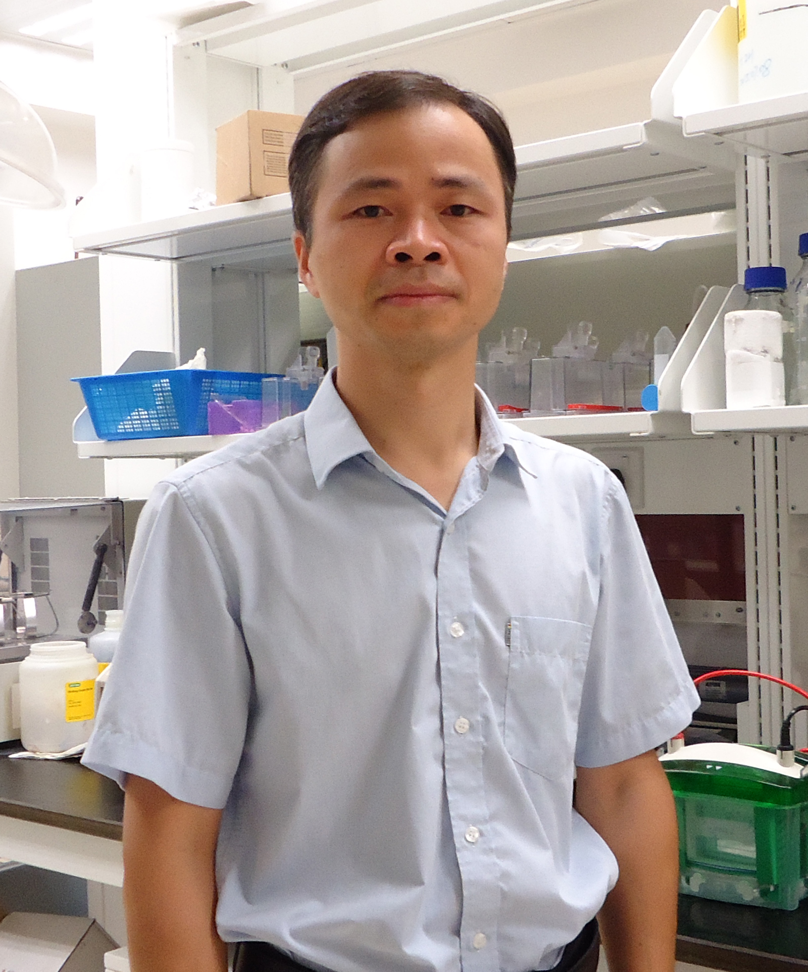 Cheng-Wei He (何承伟)合作论文数Institute of Chinese Medical Sciences,University of Macau23
 Hsiang-Wen Chien合作论文数22
 Shun-Fa Yang合作论文数Institute of Medicine, Chung Shan Medical University;Chung Shan Medical University Hospital22
 Jianbo Wan (万建波)合作论文数Institute of Chinese Medical Sciences,University of Macau17
 Jia Xuejing合作论文数16
 Jiaolin Bao (鲍娇琳)合作论文数School of Pharmacy, Hainan University13
 Mei Wan Chen (陈美婉)合作论文数Institute of Chinese Medical Sciences, University of Macau13
 Chia-Yi Lee合作论文数11
 Yanbei Tu合作论文数11
 Haiyong Wang合作论文数11
 Peng Li (李鵬)合作论文数Institute of Chinese Medical Sciences,University of Macau11
 Huanxing Su (蘇煥興)合作论文数Institute of Chinese Medical Sciences,University of Macau11
 Yanqi Dang合作论文数8
 Wenjun Zhou合作论文数8Chenyue Zhang合作论文数7Yeer Liang合作论文数7
 Guang Ji (季光)合作论文数Shanghai University of Traditional Chinese Medicine7
 Liping Wang (王丽萍)合作论文数College of Life Science, Jilin University7Zhou Yu合作论文数6FengHua Li (李风华)合作论文数6
 Chao Zhang (张超)合作论文数中国药科大学中药学院6
 Baoqiang Song (宋保强)合作论文数第四军医大学西京医院整形外科6
 Jing-Yang Huang (黃景揚)合作论文数Institute of Medicine, Chung Shan Medical University;Department of Medical Research, Chung Shan Medical University Hospital6
 Yitao Wang (王一濤)合作论文数Institute of Chinese Medical Sciences, University of Macau6Yong-Syuan Chen合作论文数5Bing Han合作论文数5
 Zhiqiang Mei合作论文数5
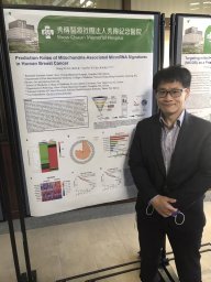 Hung-Yu Lin合作论文数College of Medicine, Chang Gung University5Binyu Song合作论文数4Yuhan Zhu合作论文数4Juanli Dang合作论文数4Lihua Tan合作论文数4Xiao-Yan Liu合作论文数4
 Mingzhe Zhu (朱明哲)合作论文数Shanghai University of Traditional Chinese Medicine4
 Fang Liu (刘芳)合作论文数School of Pharmacy, Chengdu University of Traditional Chinese Medicine4
 Hui Li (李卉)合作论文数School of Public Health, Jilin University4
 Li Zhang (张力)合作论文数Sun Yat-sen University Cancer Center4Hui-Ling Chiou合作论文数4Yi-Hsien Hsieh合作论文数4Nuo-Yi Yu合作论文数3H-W Lin合作论文数3Yixuan Peng合作论文数3Shenghui Chen合作论文数3Xiaotong Wang合作论文数3Jing Zheng合作论文数3Tong Wang合作论文数3Yuting Liang合作论文数3Sijia LI (李思佳)合作论文数3




