OBJECTIVE:A clinically nonfunctioning pituitary adenoma (CNFPA) is the likely diagnosis in patients presenting with a sellar mass consistent with an adenoma on imaging and without clinical or laboratory evidence of hormone excess. A better understanding of the outcome of observation alone for CNFPAs not requiring surgery at diagnosis is needed. METHODS:We conducted a prospective, observational study of apparent CNFPAs ≥6 mm in diameter following the Endocrine Society guidelines for clinical, endocrine, and imaging follow-ups. RESULTS:A total of 118 patients aged 56 years (range: 24.7-81)-of which 62% were female, and 72% had macroadenomas-were followed for a median of 3.9 years (range: 0.42-16). On follow-up, 49% of tumors increased in size (56% of macroadenomas and 31% of microadenomas), 12% decreased, and 39% remained unchanged. The median time to increase in size was 2.2 years (range: 0.43-12.8) for macroadenomas and 3.44 years (range: 0.41-7.4) for microadenomas (P = .16). Twenty-nine (24.6%) patients (26 who had macroadenomas and 3 who had microadenomas at diagnosis) underwent pituitary surgery after 2.58 years of follow-up (range: 0.4-7.6). Multivariable analysis found male sex to be a significant predictor of tumor growth, and macroadenoma to be a significant predictor of surgery. Most surgically removed CNFPAs were typical, hormone-immunonegative, or gonadotropin-staining tumors. CONCLUSION:In this prospective study of 6 to 9 mm micro-CNFPAs and macro-CNFPAs followed conservatively, growth and surgery can occur within the first year or after many years of observation. Risks of enlargement and surgery are increased for certain patient groups. These factors should be considered in designing a plan for follow-up.
BACKGROUND:Patients with functioning pituitary adenomas (FPA) present a diagnostic challenge with identification of microadenomas and/or invasion of the cavernous sinus. OBJECTIVE:The aim of this study was to provide evidence-based recommendations on the use of imaging to facilitate an accurate diagnosis. METHODS:PubMed and Embase were searched from the inception of the database to June 8, 2021, using search terms and search strategies to identify pertinent abstracts. These were then screened using published exclusion/inclusion criteria to identify full-text review articles. Evidence tables were constructed using data from full-text reviews, and recommendations were made. RESULTS:Of the total 8685 identified abstracts pertinent to this topic, 138 full articles met the eligibility criteria. Of these, 18 met the inclusion criteria and were included in the evidence tables. Class III evidence supported 4 Level III recommendations for adult patients with FPA. CONCLUSION:This systematic review provides evidence-based recommendations to guide providers caring for adult patients with FPA when making decisions pertinent to imaging. The Congress of Neurological Surgeons will continue to pursue timely updates and to further improve the care of patients with diagnosis.
BACKGROUND AND OBJECTIVES:Pituitary adenomas (PAs) are the most common intrasellar tumor. Clinically relevant adenomas have a prevalence of 1 per 1000 in the general population. Transsphenoidal surgery (TSS) is the most common surgical treatment and is the first-line management for most PAs. Most patients fare well postoperatively, but a subset of patients experience a prolonged length of stay (PLOS). In this article, we aim to identify demographic and clinical factors associated with PLOS after TSS for PA. METHODS:Patients with sellar pathologies surgically treated at a single tertiary center from March 1, 2009, to May 31, 2020, were retrospectively reviewed. All patients older than 18 years receiving nonemergent endoscopic TSS for pituitary adenoma were included. Clinical and demographic characteristics were analyzed using χ 2 -tests and student t -tests. For those factors with a P -value less than .01, multivariate logistic regression and negative binomial regression models were constructed to estimate the adjusted odds of PLOS across predictive factors. RESULTS:A total of 301 patients were included in the study. This cohort had an average age of 54.65 ± 15.06 years and an average body mass index of 29.47 ± 6.69. The median length of stay was 54.9 hours [25th-75th percentiles: 43.5-72.9]. Postoperative cerebrospinal fluid leak ( P < .01), postoperative diabetes insipidus (DI) ( P < .01), increased surgery duration ( P = .01), and elevated maximal tumor dimension ( P = .01) were predictive of PLOS in logistic regression. Increased surgery duration, previous pituitary radiation, intraoperative complications, and postoperative DI (all P < .01) were associated with increased rate of PLOS in negative binomial regression. CONCLUSION:Patients undergoing endoscopic TSS for PA resection demonstrate prolonged lengths of stay if they have higher tumor burden, have lengthier surgeries with intraoperative complications, or develop postoperative complications such as cerebrospinal fluid leak or DI. Careful monitoring of these factors will allow for better resource optimization, reducing costs to both the hospital and the patient.
CONTEXT:Long-term growth hormone/insulin-like growth factor-1 (GH/IGF-1) excess could increase the risk of cancer in acromegaly, but individual levels of these hormones do not relate to this risk. OBJECTIVE:We newly investigated longitudinally-measured IGF-1 levels as a potential predictor of cancer in a large New York City acromegaly cohort. METHODS:We conducted a prospective, longitudinal study of 598 acromegaly (309 men, 289 women) and 292 clinically nonfunctioning pituitary adenoma (CNFPA) (140 women, 152 men) patients from the same underlying population. GH and IGF-1 levels were measured longitudinally and outcomes were observed during long-term follow-up. Cumulative exposure to IGF-1 excess was tested as a predictor of cancer. We compared cancer prevalence in acromegaly and CNFPA cohorts and incidence in each to that expected from Surveillance, Epidemiology, and End Results (SEER) data. RESULTS:Cancer prevalence by last follow-up was 22.6% in acromegaly and 12.7% in CNFPAs (odds ratio [OR] = 1.99 [95% CI, 1.34-2.97]) (P = .0005). Overall standardized incidence ratio for cancer was 1.78 (1.51-1.81) in the acromegaly and 1.26 (0.89-1.70) in the CNFPA cohorts. Cumulative exposure to IGF-1 excess, OR = 1.278 (1.060-1.541) (P = .01), years from acromegaly diagnosis to cancer or last follow-up, OR = 1.03 (1.004-1.057) (P = .024), and age at follow up, OR = 1.064 (1.047-1.082) (P < .001), were predictors of cancer. CONCLUSION:Cancer risk is increased in acromegaly, but not in CNFPA patients. Cumulative exposure to IGF-1 excess is a predictor of cancer in acromegaly. Our data suggest that cancer risk in acromegaly relates to the degree and duration of IGF-1 excess and that full appreciation of this risk requires long-term follow up.
Introduction: Pituitary adenomas (PA) are the most common intrasellar tumor and have a prevalence of a prevalence of 17%, and transsphenoidal surgical approach (TSA) is the most common surgical treatment. Most patients fare well postoperatively, but a subset of patients experience a prolonged length of stay (PLOS). The aim of this study was to identify demographic and clinical factors associated with PLOS following TSA for PA.
In the 2022 fifth edition of the WHO Classification of Endocrine Tumours and of Central Nervous System Tumours, pituitary adenomas are reclassified as neuroendocrine tumours (NETs). This change confers an oncology label to neoplasms that are overwhelmingly benign. A comprehensive clinical classification schema is required to guide prognosis, therapy and outcomes for all patients with pituitary adenomas. Pituitary adenomas and NETs exhibit some morphological and ultrastructural similarities. However, unlike NETs, pituitary adenomas are highly prevalent, yet indolent and rarely become malignant. This Perspective presents the outcomes of an interdisciplinary international workshop that addressed the merit and clinical implications of the classification change of pituitary adenoma to NET. Many non-histological factors provide mechanistic insight and influence the prognosis and treatment of pituitary adenoma. We recommend the development of a comprehensive classification that integrates clinical, genetic, biochemical, radiological, pathological and molecular information for all anterior pituitary neoplasms.
The Department of Neurosurgery's residency program at The Mount Sinai Hospital was founded in 1946. The department has its origins in 1914 as a division of general surgery, with Charles Elsberg at the helm. Neurosurgery then became a separate department in 1932 under the leadership of Ira Cohen. Dr. Cohen oversaw the creation of the neurosurgery residency training program 75 years ago. Since its inception, the residency program has graduated 120 residents. For more than 100 years, The Mount Sinai Hospital has been a site of clinical excellence, groundbreaking research, and technological innovation in neurosurgery. Currently, the Department of Neurosurgery has 39 clinical faculty members, performs more than 5300 surgeries and endovascular procedures annually, and is in the top 25 neurosurgical departments for NIH funding.
Purpose Vision loss following surgery for pituitary adenoma is poorly described in the literature and cannot be reliably predicted with current prognostic models. Detailed characterization of this population is warranted to further understand the factors that predispose a minority of patients to post-operative vision loss. Materials and methods The medical records of 587 patients who underwent endoscopic transsphenoidal surgery at the Mount Sinai Medical Centre between January 2013 and August 2018 were reviewed. Patients who experienced post-operative vision deterioration, defined by reduced visual acuity, worsened VFDs, or new onset of blurry vision, were identified and analysed. Results Eleven out of 587 patients who received endoscopic surgery for pituitary adenoma exhibited post-operative vision deterioration. All eleven patients presented with preoperative visual impairment (average duration of 13.1 months) and pre-operative optic chiasm compression. Seven patients experienced visual deterioration within 24 h of surgery. The remaining four patients experienced delayed vision loss within one month of surgery. Six patients had complete blindness in at least one eye, one patient had complete bilateral blindness. Four patients had reduced visual acuity compared with preoperative testing, and four patients reported new-onset blurriness that was not present before surgery. High rates of graft placement (10/11 patients) and opening of the diaphragma sellae (9/11 patients) were found in this series. Four patients had hematomas and four patients had another significant post-operative complication. Conclusions While most patients with pituitary adenoma experience favourable ophthalmological outcomes following endoscopic transsphenoidal surgery, a subset of patients exhibit post-operative vision deterioration. The present study reports surgical and disease features of this population to further our understanding of factors that may underlie vision loss following pituitary adenoma surgery. Graft placement and opening of the diaphragma sellae may be important risk factors in vision loss following ETS and should be an area of future investigation.
Abstract Introduction Dopamine-agonist (DA) resistant prolactinomas are characterized by failure to achieve normal prolactin levels or reduce tumor size by at least 50% on maximal conventional DA dosages. This occurs in about 10% of patients on cabergoline and is more common in larger macroadenomas, and in men compared with women. The underlying cause of resistance may be a lack of D2 receptor (D2R) tumor expression. As high dose DA therapy may have serious cardiac side effects, it is important to determine the D2R expression. Further, the sexual dimorphism observed in resistant prolactinomas may be due to estrogen receptor alpha (ERα) expression in female tumors, as ERα signaling has been shown to enhance sensitivity to DAs. We investigated the possibility that androgen receptor (AR) signaling in males promotes resistance via induction of transmembrane serine protease 2 (TMPRSS2), a cell surface serine protease, a known promoter of tumor progression in prostate and lung cancer. Methods This is a retrospective, single-center study conducted at an academic quaternary-care center. Immunohistochemical (IHC) analysis was performed on 16 specimens from surgically removed lactotroph cell type tumors, including D2R, growth hormone (GH), prolactin (PRL), TMPRSS2, ERα, AR, and the stem cell marker, CD133. We simultaneously conducted a chart review of the Electronic Medical Record to determine age at diagnosis, tumor size, initial PRL level, medical treatment, and reason for surgical resection. Results A total of 14 patients were included in our study. Two patients had multiple surgeries for resistant prolactinomas such that the sample size included 16 specimens- 7 resistant prolactinomas and 9 controls (resected for another reason other than treatment resistance). There were 2 men and 3 women in the resistant group, and 5 women and 4 men in the control group. Baseline prolactin level was higher in the resistant group (2413ng/mL) than the control group (676.4ng/mL) and tumor size was larger—2.05cm vs. 1.57cm in the largest diameter in the resistant vs. control groups, respectively. D2R expression was positive in 8 out of 9 controls vs. 1 out of 7 in resistant tumors. ERα positivity did not correlate with DA resistance. AR expression was negative in all specimens although the majority of tumors in both groups did express TMPRSS2. CD133 was negative in all but one of the resistant tumors. Conclusion We found that 80% of specimens from patients with DA-resistant prolactinomas had negative D2R expression. The one resistant tumor that had positive D2R and negative TMPRSS2 expression was from an 11 year old girl who also had negative ERα expression that may explain the lack of response to dopamine agonist therapy. D2R IHC can identify lack of D2R expression in DA-resistant tumors and provide a rationale for discontinuation of high dose DA therapy to avoid toxicity. Presentation: Monday, June 13, 2022 12:30 p.m. - 2:30 p.m.
PURPOSE:Resection of pituitary adenomas presents a number of unique challenges in neuro-oncology. The proximity of these lesions to key vascular and endocrine structures as well as the need to interpret neuronavigation in the context of shifting tumor position increases the complexity of the operation. More recently, substantial advances in fluorescence-guided surgery have been demonstrated to facilitate the identification of numerous tumor types and result in increased rates of complete resection and overall survival.METHODS:A review of the literature was performed, and data regarding the mechanism of the fluorescence agents, their administration, and intraoperative tumor visualization were extracted. Both in vitro and in vivo studies were assessed. The application of these agents to pituitary tumors, their advantages and limitations, as well as future directions are presented here.RESULTS:Numerous laboratory and clinical studies have described the use of 5-ALA, fluorescein, indocyanine green, and OTL38 in pituitary lesions. All of these drugs have been demonstrated to accumulate in tumor cells. Several studies have reported the successful use of the majority of the agents in inducing intraoperative tumor fluorescence. However, their sensitivity and specificity varies across the literature and between functioning and non-functioning adenomas.CONCLUSIONS:At present, numerous studies have shown the feasibility and safety of these agents for pituitary adenomas. However, further research is needed to assess the applicability of fluorescence-guided surgery across different tumor subtypes as well as explore the relationship between their use and postoperative clinical outcomes.
OBJECTIVE:To describe an unusual presentation of a patient with recurrent pituitary apoplexy of an adenoma that switched phenotypes from a nonfunctioning, or silent gonadotroph adenoma (SGA), to a silent corticotroph adenoma (SCA). We discuss the potential etiologies of both recurrent pituitary apoplexy and phenotype switching of pituitary tumors.METHODS:The presented case includes clinical and biochemical findings, surgical outcomes, and pathologic reports related to the treatment of our patient who presented with recurrent pituitary apoplexy.RESULTS:A 56-year-old man presented for evaluation of decreased libido and was found to have a low testosterone level. A pituitary magnetic resonance image demonstrated an 8-mm pituitary adenoma. He underwent transsphenoidal surgery (TSS) to remove the tumor and pathology demonstrated an SGA immunopositive for luteinizing hormone and follicle-stimulating hormone with evidence of apoplexy. Eight years later, the patient underwent another TSS after developing acute-onset headache, vomiting, and a cranial nerve palsy. Pathology at this time showed a necrotic tumor consistent with apoplexy with negative immunostains for all pituitary tumors. Three years after this, the tumor recurred and after another TSS the tumor stained positive for adrenocorticotropic hormone but was negative for luteinizing hormone and follicle-stimulating hormone with hemorrhage consistent with apoplexy. A few years afterward, he again developed acute-onset headache and cranial nerve palsies and had another TSS. On pathology, the tumor demonstrated extensive necrosis consistent with apoplexy and again stained positive for adrenocorticotropic hormone. The patient was then referred for radiation therapy and was subsequently lost to follow up.CONCLUSION:Recurrent pituitary apoplexy in the same patient has only been described 3 times in the literature. There have been no case reports of a pituitary adenoma that switched phenotypes from an SGA to SCA. We suggest that pituitary apoplexy may recur multiple times due to a tumor with particularly fragile vessel walls and increased vascularization. We review the literature that suggests clinical and molecular similarities between SGAs and SCAs. Further studies are needed to determine the etiologies of recurrent apoplexy and pituitary adenomas with switching phenotypes.
Abstract Silent corticotroph adenomas (SCAs) are tumors of the TPIT anterior pituitary cell lineage that do not lead to biochemical or clinical Cushings syndrome. Thus, they present as clinically nonfunctioning pituitary adenomas (CNFPAs) and are only diagnosed pathologically. Since they are often aggressive tumors, identification of a peripheral blood marker of SCA activity would be useful for diagnosis and monitoring. Some data suggest that aberrant processing of POMC, the precursor of ACTH, underlies the lack of elevated biologically active ACTH and thus cortisol excess in SCAs. We hypothesized that these tumors could secrete POMC, resulting in elevated plasma levels of POMC in patients with SCAs. Therefore, we investigated plasma POMC levels as a potential marker of this tumor type and correlated with tumor ACTH immunoreactivity (IR), which may be detecting unprocessed POMC. We studied 267 patients (134M, 133F, age 56.3±13.6yr) with CNFPAs (Cushings excluded) prior to surgery and at enrollment in a prospective, observational study. We also studied 9 patients with known SCAs with residual macroadenomas after surgery. Peripheral blood was sampled for POMC, ACTH and cortisol levels. POMC was measured by in-house two-site ELISA (detects POMC and 22kD pro-ACTH) and ACTH and cortisol by Immulite(Siemens). POMC levels were compared to the 95%CI of the mean of 70 healthy subjects and considered elevated if ≥ the 97.5 percentile of 39 fmol/mL. Of the CNFPA cohort, 12/267 had elevated POMC levels (range 39-166 fmol/mL) and 4 of the 12 had elevated ACTH levels (> 50 pg/ml)(range 53.6-76 pg/ml). Of the 12 with elevated POMC, 9 underwent surgery and 6 of them had positive ACTH IR in their tumors. An additional 13 patients in the CNFPA cohort had weak ACTH IR tumors, but POMC <39 fmol/mL and normal ACTH. POMC levels were elevated in all 9 known SCA patients (range 40-996 fmol/mL), being highest in those with the most aggressive tumors. ACTH levels were elevated in 6 of them (range 50.6-397 pg/ml). POMC levels were lowered with surgery in 4 of 4 SCA patients followed longitudinally. Two SCAs were treated with monthly pasireotide LAR (40 mg escalated to 60 mg) for 8 months. Plasma POMC levels fell from 104 to 21 fmol/mL in 1 patient, but did not change in the 2nd, 124 to 112 fmol/mL. Both patients had no change in the size of their macroadenomas during treatment. In summary, patients with elevated plasma POMC levels were correctly identified as SCAs in most surgically treated patients and POMC levels were lowered by tumor removal in all who were tested longitudinally. Although POMC levels were not elevated in some other patients with weakly positive ACTH IR tumors, further characterization of these tumors by lineage specific transcription factors is underway to confirm them to be of corticotroph cell origin. These data suggest that plasma POMC measurements may have clinical utility in the evaluation of SCAs and this warrants further study.
CONTEXT:Clinically nonfunctioning pituitary adenomas (CNFPAs) typically remain undetected until mass effect symptoms develop. However, currently, head imaging is performed commonly for many other indications, which may increase incidental discovery of CNFPAs. Since current presentation and outcome data are based on older, retrospective series, a prospective characterization of a contemporary CNFPA cohort was needed.OBJECTIVE:To determine the prevalence of incidental presentation and hypopituitarism and its predictors in a CNFPA cohort that spanned 6 to 9 mm micro- to macroadenoma included observational and surgical therapy.METHODS:At enrollment in a prospective, observational study, 269 patients with CNFPAs were studied by history, examination, blood sampling, and pituitary imaging analysis and categorized into incidental or symptoms presentation groups that were compared.RESULTS:Presentation was incidental in 48.7% of patients and due to tumor symptoms in 51.3%. In the symptoms and incidental groups, 58.7% and 27.4% of patients had hypopituitarism, respectively, and 25% of patients with microadenomas had hypopituitarism. Many had unappreciated signs and symptoms of pituitary disease. Most tumors were macroadenomas (87%) and were larger in the symptoms than incidental and hypopituitary groups than in the eupituitary groups. The patients in the incidental group were older, and males were older and had larger tumors in both the incidental and symptoms groups.CONCLUSIONS:Patients with CNFPAs commonly present incidentally and with previously unrecognized hypopituitarism and symptoms that could have prompted earlier diagnosis. Our data support screening all large micro and macro-CNFPAs for hypopituitarism. Most patients with CNFPAs still have mass effect signs at presentation, suggesting the need for more awareness of pituitary disease. Our ongoing, prospective observation of this cohort will assess outcomes of these CNFPA groups.
Outcome of acromegaly surgery is assessed by IGF-1 and glucose-suppressed GH, but whether the latter provides additional clinically relevant information when IGF-1 is normal is unclear. The role of GH suppression testing after surgery requires clarification. We studied 97 acromegaly patients with normal IGF-1 after surgery by measuring GH after oral glucose longitudinally, initially at ≥ 3 months after surgery and repeated one or more times ≥ 1 year later. Nadir GH was categorized as normal or abnormal relative to the 97.5th percentile of nadir GH in 100 healthy subjects, which were ≤ 0.14 µg/L (DSL IRMA) or ≤ 0.15 µg/L(IDS iSYS). Signs and symptoms scores and insulin resistance were followed longitudinally. Of 68 patients with initial normal GH suppression 63 (93%) remained in remission and of 29 with initial abnormal GH suppression, 9 (31%) recurred. Recurrence was more common in patients with abnormal suppression (P < 0.001). A total of 14 patients recurred, including 5 with normal GH suppression progressing to abnormal and then recurrence. Overall, serial signs and symptoms and insulin resistance assessments did not identify patients with abnormal suppression or recurrence. Risk of recurrence after surgery is increased for patients with a normal IGF-1 level, but abnormal GH suppression. We newly find, using both our and others’ cut-offs, that while normal suppression predicts long-term remission in most patients, some can progress from normal to abnormal suppression and then recurrence after many years of follow up. Nadir GH levels are of prognostic value in acromegaly patients with normal IGF-1 levels after surgery.
The 13th Acromegaly Consensus Conference was held in November 2019 in Fort Lauderdale, Florida, and comprised acromegaly experts including endocrinologists and neurosurgeons who considered optimal approaches for multidisciplinary acromegaly management. Focused discussions reviewed techniques, results, and side effects of surgery, radiotherapy, and medical therapy, and how advances in technology and novel techniques have changed the way these modalities are used alone or in combination. Effects of treatment on patient outcomes were considered, along with strategies for optimizing and personalizing therapeutic approaches. Expert consensus recommendations emphasize how best to implement available treatment options as part of a multidisciplinary approach at Pituitary Tumor Centers of Excellence.
Context: Pituitary corticotroph adenomas are rare tumors that can be associated with excess adrenocorticotropin (ACTH) and adrenal cortisol production, resulting in the clinically debilitating endocrine condition Cushing disease. A subset of corticotroph tumors behave aggressively, and genomic drivers behind the development of these tumors are largely unknown. Objective: To investigate genomic drivers of corticotroph tumors at risk for aggressive behavior. Design: Whole-exome sequencing of patient-matched corticotroph tumor and normal deoxyribonucleic acid (DNA) from a patient cohort enriched for tumors at risk for aggressive behavior. Setting: Tertiary care center Patients: Twenty-seven corticotroph tumors from 22 patients were analyzed. Twelve tumors were macroadenomas, of which 6 were silent ACTH tumors, 2 were Crooke's cell tumors, and 1 was a corticotroph carcinoma. Intervention: Whole-exome sequencing. Main outcome measure: Somatic mutation genomic biomarkers. Results: We found recurrent somatic mutations in USP8 and TP53 genes, both with higher allelic fractions than other somatic mutations. These mutations were mutually exclusive, with TP53 mutations occurring only in USP8 wildtype (WT) tumors, indicating they may be independent driver genes. USP8-WT tumors were characterized by extensive somatic copy number variation compared with USP8-mutated tumors. Independent of molecular driver status, we found an association between invasiveness, macroadenomas, and aneuploidy. Conclusions: Our data suggest that corticotroph tumors may be categorized into a USP8-mutated, genome-stable subtype versus a USP8-WT, genome-disrupted subtype, the latter of which has a TP53-mutated subtype with high level of chromosome instability. These findings could help identify high risk corticotroph tumors, namely those with widespread CNV, that may need closer monitoring and more aggressive treatment.
Abstract Surgical removal of the GH-secreting tumor is the initial treatment of choice for acromegaly. Outcome of surgery is assessed by measuring IGF-1 and glucose-suppressed GH levels. IGF-1 normalization is an essential biochemical criterion for remission. The cut-off for nadir GH after oral glucose that signifies remission, however, is debated. It also remains unclear whether GH levels provide additional prognostic or clinically relevant information when IGF-1 results are definitive. To address this question, we examined how initial postoperative glucose-suppressed GH levels change over time on serial testing in patients who achieve initial remission as defined by IGF-1 normalization. We studied 87 acromegaly patients (48M, 39F) who achieved a normal IGF-1 level after surgery alone longitudinally from 1996 to 2019. All had GH measured before and 60, 90 and 120 minutes after 75 or 100 mg oral glucose (OGTT) at ≥ 3 months after surgery and GH and IGF-1 repeated ≥ 1 year later. GH was by measured by sensitive, 22KDa GH specific assays, either a IRMA (DSL, International Reference Standard (IRS) 88/624) or a chemiluminescence immunoassay (IDS-iSYS, IRS 98/574). OGTT Nadir GH levels were also measured in healthy subjects; n=46 (26 M, 20 F, ages 19-71 yr.) by DSL and n=46 (29 M, 17 F; ages 20-66 yr.) by IDS-iSYS. Nadir GH levels in acromegaly patients were compared to the 95%CI of healthy subjects’ mean and categorized relative to healthy subjects’ 97.5 percentile, which was 0.14 µg/L for both assays. IGF-1 levels were compared to age and gender adjusted normal ranges. Subjects were grouped based on initial nadir GH ≤ or > 0.14 µg/L and the patterns of change in nadir GH and IGF-1 at last follow up or until IGF-1 became elevated (i.e. recurrence). Follow up durations are given as median(range). In follow up, 73 patients remained in remission (normal IGF-1) and 14 had a recurrence (elevated IGF-1). Of the 73 in remission, 55 had initial nadir GH ≤ 0.14 µg/L that persisted to 10 yr.(1-22yr.) of follow up, 5 had initial GH ≤ 0.14 µg/L that rose to > 0.14 µg/L by 9(3-21)yr., 10 had GH > 0.14 µg/L that persisted at 5.5(2-22)yr., and 3 had GH > 0.14 µg/L that fell to ≤ 0.14 µg/L at 5(4-7)yr. of follow up. Of the 14 that recurred, 11 had an initial and persistent GH > 0.14 µg/L and developed an elevated IGF-1 level after 6(1-23) yr.. The 3 other patients that recurred had an initial GH ≤ 0.14 µg/L that rose to > 0.14 µg/L by 1-6 years later and subsequently developed an elevated IGF-1 level by 14-16 years of follow up. In summary, we found that the pattern of normal IGF-1 along with nadir GH > 0.14 µg/L on initial testing or developing with time, was associated with recurrence in 14/32 patients. We also found that initial nadir GH ≤ 0.14 µg/L was highly predictive of long-term persistent remission: 60/63 such patients remained in remission. In conclusion, glucose-suppressed GH levels are of prognostic value in acromegaly patients with normal IGF-1 after surgery.
Introduction: Pituitary apoplexy (PA) is a neurosurgical and endocrine emergency. Prompt recognition and treatment can significantly improve outcomes. Although there are several case series and retrospective reviews of patients with pituitary apoplexy, the vast majority of these include less than 50 patients. Furthermore, endocrine follow-up data is lacking from these studies. We aimed to retrospectively review recovery of neuroendocrine function in early vs. late surgery and to determine whether there was a lower incidence of recurrence of pituitary tumors after apoplexy. Methods: We reviewed the charts of 72 patients managed surgically for PA at our hospital between 1986 and 2018. We analyzed presentation patterns, imaging, and pathology findings, and pre and post-operative endocrine data. Patients were included in the study if they had surgical or pathologic confirmation of PA. There were no exclusion criteria. Early intervention was defined as within 8 days of presentation. Post-operative hormone replacement was evaluated by review of patients’ medications. Results: Of the 72 patients reviewed, data regarding early vs. late surgery was available for 69 patients. 47.8% (n=33) of patients received early intervention and 52.2% (n=36) received surgery 8 or more days after symptom onset. No association between timing of surgery and recovery of neuroendocrine function was observed. 51.5% (n=17) of patients who received early surgery vs. 52.8% (n=19) who received late surgery required post-operative steroid replacement (p= 0.92). 42.4% (n=14) vs. 40.0% (n=14) required post-operative thyroid replacement (p=0.84). 24.2% (n=8) vs. 17.1% (n=6) required post-operative testosterone replacement (p=0.47). 12.1% (n=4) vs. 8.8% (n=3) required post-operative DDAVP replacement (p=0.71). 17% (n=12) of our patients had tumors that recurred. Of these, 8 were non-functioning adenomas, 2 were Rathke cleft cysts, 1 was a prolactinoma, and 1 was a chordoma. 4 of our patients were lost to follow up. Neurological outcomes resolved in 45% (n=15) vs. 50% (n=18) of patients, and improved in 45% (n=15) vs. 33% (n=12) in the early vs late intervention group respectively. Conclusion: Whether to perform early surgery in patients with PA is a topic of debate. We report no significant difference in neuroendocrine outcomes of patients who were treated with early vs. late surgery. Our recurrence incidence after PA is consistent with that of non-functioning pituitary adenomas that do not undergo apoplexy, which is quoted to be about 16%. Future studies are needed to corroborate our findings. Unless otherwise noted, all abstracts presented at ENDO are embargoed until the date and time of presentation. For oral presentations, the abstracts are embargoed until the session begins. Abstracts presented at a news conference are embargoed until the date and time of the news conference. The Endocrine Society reserves the right to lift the embargo on specific abstracts that are selected for promotion prior to or during ENDO.
Pituitary apoplexy is a neurosurgical entity in which rapid diagnosis and prompt surgical treatment enhance the probability of good endocrinologic and neurologic outcomes. The difficulty lies in the fact that patients present with myriad signs and symptoms. Meningeal signs, visual and oculomotor disturbances, as well as endocrine deficits are all possible. Computed tomography (CT) and magnetic resonance imaging (MRI) are important in defining the pituitary tumor, the hemorrhage, and their relationship to other nearby anatomical structures. Angiography or magnetic resonance angiography (MRA) may be necessary to exclude an aneurysm. Transsphenoidal resection of the pituitary tumor and hemorrhage is the procedure of choice. It offers definitive treatment for the pituitary apoplexy as well as its underlying neoplastic pathology. It also carries with it a low morbidity and mortality, even in seriously ill patients. Intensive hormonal support is a necessary adjunct in the perioperative period, and endocrine evaluation is necessary postoperatively to establish need for long-term replacement therapy.
 Pamela U. Freda合作论文数29
 Joshua B Bederson合作论文数Department of Neurosurgery, Icahn School of Medicine, Mount Sinai Medical Center18
 Eliza B. Geer合作论文数14
 Raj K Shrivastava合作论文数The Center for Cranial Base Surgery, St. Luke's-Roosevelt Medical Center11Reyes-Vidal Carlos M合作论文数9Sharon L. Wardlaw合作论文数9
 Edward R Laws合作论文数Neurosciences Center, Brigham and Women's Hospital;Harvard Medical School9
 Jeffrey Bruce合作论文数Princess Margaret Cancer Centre, University Health Network8
 Mark E Molitch合作论文数Division of Endocrinology, Metabolism and Molecular Medicine, Department of Medicine, Feinberg School of Medicine, Northwestern University7
 Zhezhen Jin合作论文数Department of Biostatistics, Mailman School of Public Health, Columbia University7
 Chirag D. Gandhi合作论文数Department of Neurosurgery, School of Medicine, New York Medical College7
 Marc R. Mayberg合作论文数Department of Neurological Surgery, School of Medicine, University of Washington7Jeffrey N. Bruce合作论文数7
 Alexander Khandji合作论文数7Ciric Ivan S合作论文数6Kostadinov Jane合作论文数6
 Paolo Cappabianca合作论文数6Jane Walsh合作论文数5K Hague合作论文数5Giorgio Frank合作论文数Department of Neurosurgery, Villa Maria Group5
 Mônica Gadelha合作论文数Departamento de Clínica Médica - Endocrinologia, Faculdade de Medicina, Universidade Federal do Rio de Janeiro;Ambulatório de Adenomas Hipofisários, Hospital Universitário Clementino Fraga Filho, Universidade Federal do Rio de Janeiro4Seymour Reichlin合作论文数Department of Medicine, School of Medicine, Tufts University4
 Wei Shen合作论文数Institute of Human Nutrition, Columbia University College of Physicians and Surgeons4Ivor M. D. Jackson合作论文数4
 Adam Mamelak合作论文数Laboratory of Neurophysiology, Harvard Medical School4Chandranath Sen合作论文数4
 Rudolf Fahlbusch合作论文数Center for Endocrine Neurosurgery at the International Neuroscience Institute (INI) in Hannover4Khadeen C Cheesman合作论文数3
 Tirissa Reid合作论文数3Scott A Meyer合作论文数3
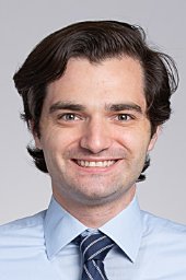 Jonathan T Dullea合作论文数Icahn Sch Med Mt Sinai, Dept Neurosurg, 1 Gustave Levy Pl, New York, NY 10029 USA3
 John W Rutland合作论文数Icahn Sch Med Mt Sinai, Dept Neurosurg, New York, NY 10029 USA3
 Laligam N. Sekhar合作论文数Department of Neurological Surgery, UW Medicine, University of Washington3
 Maria Chiara Zatelli合作论文数Dipartimento di Scienze Mediche, Università degli Studi di Ferrara;Section of Endocrinology and Metabolic Diseases, Azienda Ospedaliero Universitaria di Ferrara3Brisman M H合作论文数3
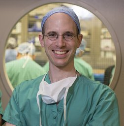 Oermann Eric K合作论文数Icahn Sch Med Mt Sinai, Dept Neurosurg, New York, NY 10029 USA3Ben-Shlomo Anat合作论文数3Pain Margaret合作论文数3
 Susan Morgello合作论文数Friedman Brain Institute, Icahn School of Medicine at Mount Sinai3Fernandez Jean Carlos合作论文数3
 Philippe Chanson合作论文数University Paris-Saclay;Bicêtre’s Hospital3William F. Chandler合作论文数3
 Paul C Mccormick合作论文数Irving Medical Center, Columbia University;NewYork-Presbyterian Allen Hospital;College of Physicians and Surgeons, Columbia University;The Och Spine Hospital, The Neurological Institute of New York3
 Isabelle M Germano合作论文数3
 Michael Buchfelder合作论文数Department of Neurosurgery, University Hospital Erlangen3
 Nienke Biermasz合作论文数Leiden University Medical Center;Faculteit Geneeskunde, Leiden University3
 Serge Cremers合作论文数Department of Pathology and Cell Biology, Columbia University Irving Medical Center3Samuel M. Wolpert合作论文数3
 Kevin Yuen合作论文数3




