It is shown here that microwave sensors can be used to monitor glucose in serum concentration with minimum detectable as well as resolution of 1 mMol . L-1 (approximate to 18 mg . dL(-1)). The ultrasensitive detection technique relies on a split ring resonator, operating at the frequency of 1.156 GHz, as the core of the sensor where its loss is compensated to enhance the quality factor from similar to 190 (passive mode)to similar to 3850 (active mode) to enable high resolution (mod ified frequency detection error from +/- 12 kHz down to +/- 2.5 kHz) frequency-shift sensing. Initially, glucose concentrations of 100-1000 mMol . L-1 (1800-18000 mg . dL(-1)) in water were detected within 250 kHz of dynamic range (between two spectrum ends). Selectivity of the sensor to glucose is verified with respect to common interstitial fluid ingredients with biological levels. Finally, to enhance the resolution of the proposed sensor, its loss-compensation is further improved leading to increased accuracy of measuring glucose samples in a 0.9 % NaCl solution containing 10 % horse serum that closely resembles blood plasma and interstitial fluid. This allows exploration of lower concentrations in the physiological range 1-30 mMol . L-1 (18-540 mg . dL(-1)) with improved frequency detection error down to +/- 0.75 kHz for two cases of with/without serum solutions with dynamic range of 30 kHz/38 kHz. The highly accurate glucose monitoring technique could be utilized for developing noninvasive glucose sensors for biomedical applications in real-time glucose monitoring.
Age-related macular degeneration (AMD) is a condition affecting the retina and is the leading cause of vision loss. Dry AMD is caused by the accumulation of lipid deposits called drusen, which form under the retina. This work demonstrates, for the first time, the removal of drusen-like deposits underneath ARPE-19 cell layers using femtosecond laser pulses. A novel cell culture model was created in response to the limited access to primary cell lines and the absence of animal models that recapitulate all aspects of AMD. In the cell culture model, deposits were identified with fluorescent stains specific to known deposit constituents. Trains of sub-10 femtosecond laser pulses from a Ti:Sapphire laser were used to successfully ablate the deposits without causing damage to surrounding cells. This drusen removal method can be used as a potential treatment for dry-stage AMD.
Retinoblastoma is a retinal cancerous disease that primarily affects young children. To date, preservation of the eye and its functionality is secondary to saving the child's life. EpCAM+ Y79 retinoblastoma cells behave like cancer stem cells that are recognized as cells that are resistant to treatment. Additionally, reoccurrence of tumours is attributed to the persistence of cancer stem cells. An effective technique to treat retinoblastoma cancer cells is demonstrated using femtosecond laser pulses and EpCAM targeting gold nanorods (Au-NRs). Both fluorescence viability assay and MTS cellular metabolism assay confirm an astonishing cellular viability drop, to similar to 10%. It is shown that right after laser irradiation the cellular membrane ruptures. FESEM imaging shows that Au-NRs reach melting temperature after laser pulse exposure. The medium of the eye is transparent to NIR laser irradiation, making this treatment ideal for this type of cancer. This treatment methodology would also be an invaluable tool for treatment of chemotherapy-resistant and radiation-resistant cancers.
Retinoblastoma is a cancerous disease that affects the retina, and primarily affects young children. To date, the primary treatment goal of retinoblastoma is to save the child's life, while the preservation of the eye and its functionality are the secondary goals. Reoccurrence of tumors is mainly attributed to the persistence of cancer stem cells. EpCAM+ Y79 retinoblastoma cells behave like cancer stem cells and are recognized as cells that are resistant to treatment. We demonstrate an effective technique to treat retinoblastoma cancer cells, using femtosecond laser pulses and epithelial cell adhesion molecule (EpCAM)-targeting gold nanorods (Au-NRs). Complete assessment of the optimal laser parameters required for the development of a translational retinoblastoma cancer treatment is provided. Both an MTS cellular metabolism assay and a fluorescence viability assay demonstrate an astonishing cellular viability drop, to approximate to 10%. Right after laser irradiation the cellular membrane ruptures. Calculations and field-emission scanning electron microscopy (FESEM) imaging show that Au-NRs reach melting temperature after laser pulse exposure. Delivering femtosecond laser pulses directly onto the retina to treat retinoblastoma through the medium of the eye is possible without interacting with its compartments-making this treatment ideal for this type of cancer. This treatment methodology would be an invaluable tool for treatment of chemotherapy-resistant and radiation-resistant cancers.
The mechanism of femtosecond laser nanosurgical attachment is investigated in the following article. Using sub-10 femtosecond laser pulses with 800 nm central wavelength were used to attach retinoblastoma cells. During the attachment process the cell membrane phospholipid bilayers hemifuse into one shared phospholipid bilayer, at the location of attachment. Transmission electron microscopy was used in order to verify the above hypothesis. Based on the imaging results, it was concluded that the two cell membrane coalesce to form one single shared membrane. The technique of cell-cell attachment via femtosecond laser pulses could potentially serve as a platform for precise cell membrane manipulation. Manipulation of the cellular membrane is valuable for studying diseases such as cancer; where the expression level of plasma proteins on the cell membrane is altered.
This article provides insight into the mechanism of femtosecond laser nanosurgical attachment of cells. We have demonstrated that during the attachment of two retinoblastoma cells using sub-10 femtosecond laser pulses, with 800 nm central wavelength, the phospholipid molecules of both cells hemifuse and form one shared phospholipid bilayer, at the attachment location. In order to verify the hypothesis that hemifusion takes place, transmission electron microscope images of the cell membranes of retinoblastoma cells were taken. It is shown that at the attachment interface, the two cell membranes coalesce and form one single membrane shared by both cells. Thus, further evidence is provided to support the hypothesis that laser-induced ionization process led to an ultrafast reversible destabilization of the phospholipid layer of the cellular membrane, which resulted in cross-linking of the phospholipid molecules in each membrane. This process of hemifusion occurs throughout the entire penetration depth of the femtosecond laser pulse train. Thus, the attachment between the cells takes place across a large surface area, which affirms our findings of strong physical attachment between the cells. The femtosecond laser pulse hemifusion technique can potentially provide a platform for precise molecular manipulation of cellular membranes. Manipulation of the cellular membrane is an important procedure that could aid in studying diseases such as cancer; where the expression level of plasma proteins on the cell membrane is altered.
Neuronal injury may cause an irreversible damage to cellular, organ and organism function. While preventing neural injury is ideal, it is not always possible. There are multiple etiologies for neuronal injury including trauma, infection, inflammation, immune mediated disorders, toxins and hereditary conditions. We describe a novel laser application, utilizing femtosecond laser pulses, in order to connect neuronal axon to neuronal soma. We were able to maintain cellular viability, and demonstrate that this technique is universal as it is applicable to multiple cell types and media.
Attachment of single cells via hemifusion of cellular membranes using femtosecond laser pulses is reported in this manuscript. This is a method to attach single cells using sub-10 femtosecond laser pulses, with 800 nm central wavelength delivered from a Ti:Sapphire laser is described. A fluorescent dye, Calcein AM, was used to verify that the cell’s cytoplasm did not migrate from a dyed cell to a non-dyed cell, in order to ascertain that the cells did not go through cell-fusion process. An optical tweezer was used in order to assess the mechanical integrity of the attached joint membranes. Hemifusion of cellular membranes was successful without initiating full cell fusion. Attachment efficiency of 95% was achieved, while the cells’ viability was preserved. The attachment was performed via the delivery of one to two trains of sub-10 femtosecond laser pulses lasting 15 milliseconds each. An ultrafast reversible destabilization of the phospholipid molecules in the cellular membranes was induced due to a laser-induced ionization process. The inner phospholipid cell membrane remained intact during the attachment procedure, and cells’ cytoplasm remained isolated from the surrounding medium. The unbounded inner phospholipid molecules bonded to the nearest free phospholipid molecule, forming a joint cellular membrane at the connection point. The cellular membrane hemifusion technique can potentially provide a platform for the creation of engineered tissue and cell cultures.
Background and ObjectiveLaser‐induced cell‐cell surgical attachment using femtosecond laser pulses is reported.Study Design/Materials and MethodsWe have demonstrated the ability to attach single cells using sub‐10 femtosecond laser pulses, with 800 nm central wavelength delivered from a Ti:Sapphire laser. To check that the cells did not go through a cell‐fusion process, a fluorescent dye Calcein AM was used to verify that the fluorescent dye did not migrate from a dyed cell to a non‐dyed cell. The mechanical integrity of the attached joint was assessed using an optical tweezer.ResultsAttachment of cells was performed without the induction of cell‐cell fusion, with attachment efficiency of 95%, and while preserving the cells' viability. Cell‐cell attachment was achieved by delivery of one to two trains of femtosecond laser pulses lasting 15 ms each.ConclusionsLaser‐induced ionization process led to an ultrafast reversible destabilization of the phospholipid layer of the cellular membrane. The inner cell membrane remained intact during the attachment procedure, and isolation of the cells' cytoplasm from the surrounding medium was obtained. A strong physical attachment between the cells was obtained due to the bonding of the membranes' ionized phospholipid molecules and the formation of a joint cellular membrane at the connection point. The cellular attachment technique, femtosecond laser‐induced cell‐cell surgical attachment, can potentially provide a platform for the creation of engineered tissue and cell cultures. Lasers Surg. Med. 46:335–341, 2014. © 2014 Wiley Periodicals, Inc.
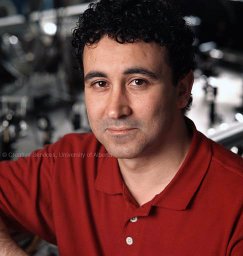 A. Y. Elezzabi合作论文数Department of Electrical and Computer Engineering, Faculty of Engineering, University of Alberta9
 Roseline Godbout合作论文数Department of Oncology;University of Alberta;Cross Cancer Institute;Cross Cancer Institute, University of Alberta7Goez Helly R合作论文数Division of Pediatric Neurology, University of Alberta3
 Ali Hatef合作论文数Department of Physics and Astronomy, University of Western Ontario2
 Mohammad Abdolrazzaghi合作论文数Department of Electrical and Computer Engineering, University of Alberta, Edmonton, AB, Canada1
 Indrani Dutta合作论文数1
 Peter E Light合作论文数1Mrinal K. Mandal合作论文数Department of Electrical and Computer Engineering, Multimedia Computing and Communications Laboratory, University of Alberta1
 Mahmoud Daneshmand合作论文数1




