The contractile apparatus of striated muscle, including both skeletal and cardiac muscle, intrinsically exhibits self-sustained oscillatory behavior under intermediate activation states between contraction and relaxation. This phenomenon, termed SPOC (spontaneous oscillatory contraction), provides unique insights into the dynamic regulation of force generation. In this review, we first highlight experimental findings that characterize SPOC and then focus on theoretical frameworks developed to capture its essential features. A key feature of these models is the assumption that the lattice spacing, that is, the distance between the thick and thin filaments, varies dynamically during muscle contraction and that actomyosin activity depends on this spacing. This assumption naturally explains the emergence of SPOC. Particular attention is given to models constructed for single sarcomeres, isolated myofibrils, and small bundles of myofibrils, through which the underlying mechanisms of SPOC can be systematically understood. Finally, by extending the mathematical structure of these models, we propose that SPOC may exhibit chaotic properties, thereby providing a comprehensive understanding of its mechanisms and physiological significance.
Transport networks spanning the entire body of an organism are key infrastructures for achieving a functional system and facilitating the distribution of nutrients and signals. The large amoeba-like organism Physarum polycephalum has gained attention as a useful model for studying biological transport networks owing to its visible and rapidly adapting vein structure. Using particle-tracking velocimetry, we measured the flow velocity of protoplasmic streaming over the entire body of Physarum plasmodia during the development of its intricate vein network. Based on these measurements, we estimated how the protoplasm is transported and mixed throughout the body. Our findings suggest that the vein network significantly enhances effective mixing of the protoplasm throughout the organism, which may have important physiological implications for nutrient distribution and signaling.
Interactions between different animal species are a critical determinant of each species' evolution and range expansion. Chemical, visual, and mechanical interactions have been abundantly reported, but the importance of electric interactions is not well understood. Here, we report the discovery that the nematode Caenorhabditis elegans transfers across electric fields to achieve phoretic attachment to insects. First, we found that dauer larvae of C. elegans nictating on a substrate in a Petri dish moved directly to the lid through the air due to the electrostatic force from the lid. To more systematically investigate the transfer behavior, we constructed an assay system with well-controlled electric fields: the worms flew up regardless of whether a positive or negative electric field was applied, suggesting that an induced charge within the worm is related to this transfer. The mean take-off speed is 0.86 m/s, and the worm flies up under an electric field exceeding 200 kV/m. This worm transfer occurs even when the worms form a nictation column composed of up to 100 worms; we term this behavior "multiworm transfer."These observations led us to conclude that C. elegans can transfer and attach to the bumblebee Bombus terrestris, which was charged by rubbing with flower pollen in the lab. The charge on the bumblebee was measured with a coulomb-meter to be 806 pC, which was within the range of bumblebee charges and of the same order of flying insect charges observed in nature, suggesting that electrical interactions occur among different species.
For organisms that respond to environmental stimuli using taxes, reversal of the tactic sign should be tightly regulated for survival. The biciliate green alga Chlamydomonas reinhardtii is an excellent model for studying reversal between positive and negative phototaxis. C. reinhardtii cells change swimming direction by modulating the balance of beating forces between their two cilia after photoreception at the eyespot; however, it remains unknown how they reverse phototactic sign. In this study, we observed cells undergoing phototactic turns with a high-speed camera and found that two key factors determine the phototactic sign: which of the two cilia beats more strongly for phototactic turning and when the strong beating starts. The timing of the strong ciliary beating is suggested to be regulated by ROS-regulated switching between the light-on and light-off responses at the eyespot, which leads to the switching between positive and negative phototaxis. This idea is supported by a mathematical model that introduces the timing of the strong ciliary beating after photoreception. ### Competing Interest Statement The authors have declared no competing interest.
Many fundamental biological processes are dependent on cellular migration. Although the mechanical mechanisms of single-cell migration are relatively well understood, those underlying migration of multiple cells adhered to each other in a cluster, referred to as cluster migration, are poorly understood. A key reason for this knowledge gap is that many forces-including contraction forces from actomyosin networks, hydrostatic pressure from the cytosol, frictional forces from the substrate, and forces from adjacent cells-contribute to cell cluster movement, making it challenging to model, and ultimately elucidate, the final result of these forces. This paper describes a two-dimensional cell membrane model that represents cells on a substrate with polygons and expresses various mechanical forces on the cell surface, keeping these forces balanced at all times by neglecting cell inertia. The model is discrete but equivalent to a continuous model if appropriate replacement rules for cell surface segments are chosen. When cells are given a polarity, expressed by a direction-dependent surface tension reflecting the location dependence of contraction and adhesion on a cell boundary, the cell surface begins to flow from front to rear as a result of force balance. This flow produces unidirectional cell movement, not only for a single cell but also for multiple cells in a cluster, with migration speeds that coincide with analytical results from a continuous model. Further, if the direction of cell polarity is tilted with respect to the cluster center, surface flow induces cell cluster rotation. The reason why this model moves while keeping force balance on cell surface (i.e., under no net forces from outside) is because of the implicit inflow and outflow of cell surface components through the inside of the cell. An analytical formula connecting cell migration speed and turnover rate of cell surface components is presented.
How smart can cells behave under complicated environmental conditions in their natural habitat? Further, what physical mechanism brings about such smart behaviors? These two questions are closely related to each other and are to be studied in both cell biology and physics. Here, we focus on behavioral smartness and variability observed mainly in unicellular eukaryotic organisms. We review a few typical cases in which cell behavior seemed smart in a complex experimental condition and=or variable in a similar experimental condition. On each of these behaviors, a possible mechanism was considered by formulating and analyzing the model equations of cell motion. By revisiting the thought that cells might be simple machines that could repeat the same response to the same stimuli and conditions, we threw some light on the plausible mechanisms for behavioral smartness and variability in cellular movements.
Protists ubiquitously live in nature and play key roles in the food web chain. Their habitats consist of various geometrical structures, such as porous media and rigid surfaces, affecting their motilities. A kind of protist, Stentor coeruleus, exhibits free swimming and adhering for feeding. Under environmental and culture conditions, these organisms are often found in sediments with complex geometries. The determination of anchoring location is essential for their lives. However, the factors that induce the behavioral transition from swimming to adhering are still unknown. In this study, we quantitatively characterized the behavioral transitions in S. coeruleus and observed the behavior in a chamber with dead ends made by a simple structure mimicking the environmental structures. As a result, the cell adheres and feeds in narrow spaces between the structure and the chamber wall. It may be reasonable for the organism to hide itself from predators and capture prey in these spaces. The behavioral strategy for the exploration and exploitation of spaces with a wide variety of geometries in their habitats is discussed.
Living cells actively deform and move by their force generations in three-dimensional (3D) space. These 3D cell dynamics occur over a long-term time scale, ranging from tens of minutes to days. On such a time scale, turnover of cell membrane constituents due to endocytosis and exocytosis cannot be ignored, i.e., the surface membrane dynamically deforms without mass conservation. Although membrane turnover is essential for large deformation of cells, there is no computational framework yet to simulate long-term cell dynamics with a non-conservative fluidic membrane. In this paper, we proposed a computational framework for simulating the long-term dynamics of a cell membrane in 3D space. For this purpose, in the proposed framework, the cell surface membrane is treated as a viscous fluid membrane without mass conservation. Cell shape is discretized by a triangular mesh, and its dynamics are expressed by effective energy and dissipation function. The mesh structure, distorted by membrane motion, is dynamically optimized by introducing a modified dynamic remeshing method. To validate the proposed framework, numerical simulations were performed, showing that the membrane flow is reproduced in a physically consistent manner and that the artificial effects of the remeshing method were negligible. To further demonstrate the applicability of the proposed framework, numerical simulations of cell migration induced by a mechanism similar to the Marangoni effect, i.e., the polarized surface tension actively generated by the cell, were performed. The observed cell behaviors agreed with existing analytical solutions, indicating that the proposed computational framework can quantitatively reproduce long-term active cell dynamics with membrane turnover. Based on the simple description of cell membrane dynamics, this framework provides a useful basis for analyzing various cell shaping and movement.
In embryogenesis and cancer invasion, cells collectively migrate as a cluster in 3D tissues. Many studies have elucidated mechanisms of either individual or collective cell migration on 2D substrates; however, it remains unclear how cells collectively migrate as a cluster through 3D tissues. To address this issue, we considered the interfacial tension at cell-cell boundaries expressing cortical actomyosin contractions and cell-cell adhesive interactions. The strength of this tension is polarized; i.e., spatially biased within each cell according to a chemoattractant gradient. Using a 3D vertex model, we performed numerical simulations of multicellular dynamics in 3D space. The simulations revealed that the polarized interfacial tension enables cells to migrate collectively as a cluster through a 3D tissue. In this mechanism, interfacial tension induces unidirectional flow of each cell surface from the front to the rear along the cluster surface. Importantly, this mechanism does not necessarily require convection of cells, i.e., cell rearrangement, within the cluster. Moreover, several migratory modes were induced, depending on the strengths of polarity, adhesion, and noise; i.e., cells migrate either as single cells, as a cluster, or aligned like beads on a string, as occurs in embryogenesis and cancer invasion. These results indicate that the simple expansion and contraction of cell-cell boundaries enables cells to move directionally forward and to produce the variety of collective migratory movements observed in living systems.
The vertex model is a useful mathematical model to describe the dynamics of epithelial cell sheets. However, existing vertex models do not distinguish contraction forces on the cell boundary from adhesion between cells, employing a single parameter to express both. In this paper, we introduce the rest length of the cell boundary and its dynamics into the existing vertex model, giving a novel formulation of the model that treats separately the contraction force and the strength of adhesion between cells. We apply this vertex model to the phenomenon of compartment boundary in the fruit fly pupa, recapturing the observation that increasing the strength of adhesion between cells straightens the compartment boundary, even though contraction forces at cell boundaries remain unchanged. We also discuss possibilities of the novel vertex models by considering the stretching of a cell sheet by external forces.
Maintaining lineage restriction boundaries in proliferating tissues is vital to animal development. A long-standing thermodynamics theory, the differential adhesion hypothesis, attributes cell sorting phenomena to differentially expressed adhesion molecules. However, the contribution of the differential adhesion system during tissue morphogenesis has been unsubstantiated despite substantial theoretical support. Here, we report that Toll-1, a transmembrane receptor protein, acts as a differentially expressed adhesion molecule that straightens the fluctuating anteroposterior compartment boundary in the abdominal epidermal epithelium of the Drosophila pupa. Toll-1 is expressed across the entire posterior compartment under the control of the selector gene engrailed and displays a sharp expression boundary that coincides with the compartment boundary. Toll-1 corrects local distortions of the boundary in the absence of cable-like Myosin II enrichment along the boundary. The reinforced adhesion of homotypic cell contacts, together with pulsed cell contraction, achieves a biased vertex sliding action by resisting the separation of homotypic cell contacts in boundary cells. This work reveals a self-organizing system that integrates a differential adhesion system with pulsed contraction of cells to maintain lineage restriction boundaries.
ABSTRACT For organisms that respond to environmental stimuli using taxes, reversal of the tactic sign should be tightly regulated for survival. The biciliate green alga Chlamydomonas reinhardtii is an excellent model for studying reversal between positive and negative phototaxis. C. reinhardtii cells change swimming direction by modulating the balance of beating forces between their two cilia after photoreception at the eyespot; however, it remains unknown how they reverse phototactic sign. In this study, we observed cells undergoing phototactic turns with a high-speed camera and found that two key factors determine the phototactic sign: which of the two cilia beats more strongly for phototactic turning and when the strong beating starts. The timing of the strong ciliary beating is suggested to be regulated by ROS-regulated switching between the light-on and light-off responses at the eyespot, which leads to the switching between positive and negative phototaxis. This idea is supported by a mathematical model that introduces the timing of the strong ciliary beating after photoreception.
Epithelial sheet integrity is robustly maintained during morphogenesis, which is essential to shape organs and embryos. While maintaining the planar monolayer in three-dimensional space, cells dynamically flow via rearranging their connections between each other. However, little is known about how cells maintain the plane sheet integrity in three-dimensional space and provide cell flow in the in-plane sheet. In this study, using a three-dimensional vertex model, we demonstrate that apical junctional fluctuations allow stable cell rearrangements while ensuring monolayer integrity. In addition to the fluctuations, direction-dependent contraction on the apical cell boundaries, which corresponds to forces from adherens junctions, induces cell flow in a definite direction. We compared the kinematic behaviors of this apical-force-driven cell flow with those of typical cell flow that is driven by forces generated on basal regions and revealed the characteristic differences between them. These differences can be used to distinguish the mechanism of epithelial cell flow observed in experiments, i.e., whether it is apical- or basal-force-driven. Our numerical simulations suggest that cells actively generate fluctuations and use them to regulate both epithelial integrity and plasticity during morphogenesis.
Single-celled organisms show a fascinating faculty for integrating spatial information and adapting their behaviour accordingly. As such they are of potential interest for elucidating fundamental mechanisms of developmental biology. In this mini-review we highlight current research on two organisms, the true slime mould Physarum polycephalum and the ciliates Paramecium and Tetrahymena. For each of these, we present a case study how applying physical principles to explain behaviour can lead to the understanding of general principles possibly relevant to developmental biology.
Flow instability under shear flow is commonly observed in many soft-bio materials. This instability causes the formation of macroscopic band referred to as "shear bands", which can be regarded as non-equilibrium phase transition. This shear band formation has been reported in a variety of systems, including DNA, polymer and surfactant systems. Very recently, we found that biological materials such as actin filament and microtubule systems show the shear band formation. These biological materials are a component of the cytoskeleton. It is interesting to know their mechanical instability under shear deformation because their structure formations are very important in a number of cellular processes. In this study, we directly visualize the shear deformation of a microtubule solution systems, and discuss the physical origin of the shear banding. Microtubule movement is characterized by dynamic instability and treadmilling. This non-equilibrium phenomena can be suppressed by adding microtubule stabilizing agent "Paclitaxel". In the absence of paclitaxel, the shear band formation is not observed. In microtubule solution with paclitaxel, on the other hand, we observed the shear band formation at shear rate of 0.01-0.1 s−1, which roughly corresponds to cell migration speed. As the paclitaxel concentration is increased, the shear band formation is observed in wide shear rates. Rheological property of the microtubule may play a role in mechanical propagation of the force for the cell migration.
Transport networks are ubiquitous in multicellular organisms and include leaf veins, fungal mycelia and blood vessels. While transport of materials and signals through the network plays a crucial role in maintaining the living system, the transport capacity of the network can best be understood in terms of hydrodynamics. We report here that plasmodium from the large, single-celled amoeboid Physarum was able to construct a hydrodynamically optimized veinnetwork when evacuating biomass from confined arenas of various shapes through a narrow exit. Increasingly thick veins developed towards the exit, and the network spanned the arena via repetitive bifurcations to give a branching tree. The Hausdorff distance from all parts of the plasmodium to the vein network was kept low, whilst the hydrodynamic conductivity from distal parts of the network to the exit was equivalent, irrespective of the arena shape. This combination of spatial patterning and differential vein thickening served to evacuate biomass at an equivalent rate across the entire arena. The scaling relationship at the vein branches was determined experimentally to be 2.53-3.29, consistent with predictions from Murray's law. Furthermore, we show that mathematical models for self-organised, adaptive transport in Physarum simulate the experimental network organisation well if the scaling coefficient of the current-reinforcement rule is set to 3. In simulations, this resulted in rapid development of an optimal network that minimised the combined volume and frictional energy in comparison with other scaling coefficients. This would predict that the boundary shear forces within each vein are constant throughout the network, and would be consistent with a feedback mechanism based on a sensing a threshold shear at the vein wall.
Vein networks span the whole body of the amoeboid organism in the plasmodial slime mould Physarum polycephalum, and the network topology is rearranged within an hour in response to spatio-temporal variations of the environment. It has been reported that this tube morphogenesis is capable of solving mazes, and a mathematical model, named the 'current reinforcement rule', was proposed based on the adaptability of the veins. Although it is known that this model works well for reproducing some key characters of the organism's maze-solving behaviour, one important issue is still open: In the real organism, the thick veins tend to trace the shortest possible route by cutting the corners at the turn of corridors, following a center-in-center trajectory, but it has not yet been examined whether this feature also appears in the mathematical model, using corridors of finite width. In this report, we confirm that the mathematical model reproduces the center-in-center trajectory of veins around corners observed in the maze-solving experiment.
During early embryonic development, epithelial cells form a monolayer sheet and migrate in a definite direction. This phenomenon, called epithelial cell migration, is an important topic in developmental biology. A characteristic feature of this process is attachment to adjacent cells during migration, which is necessary for maintaining the integrity of the sheet. However, it is unclear how these cohesive cells migrate without breaking their attachments. A mechanism for this phenomenon was recently proposed, in which direction-dependent contraction forces acting on cell boundaries induce unidirectional epithelial migration. In this review, we examine this proposed mechanism from various aspects and provide theoretical background for the collective migration of epithelial cells. This information may be helpful for investigators to realize the basic principles underlying collective epithelial migration and devise new mechanisms for it.
 Shoichi Yamada (山田章一)合作论文数School of Advanced Science and Engineering, Faculty of Science and Engineering, Waseda University20
 Hideo Kodama合作论文数Yukawa Institute for Theoretical Physics, Kyoto University18
 S. Nagataki合作论文数16
 Shinichi Ishiwata (石渡信一)合作论文数Department of Physics, School of Advanced Science and Engineering, Waseda University16
 Gentaro Watanabe (渡边元太郎)合作论文数School of Physics, Zhejiang University16
 Yuta Shimamoto合作论文数The Rockefeller University14Ohtaki Masako合作论文数13Kei Kotake合作论文数13Hajime Takami合作论文数13
 Kenji Yasuoka (泰岡顕治)合作论文数Department of Mechanical Engineering, Faculty of Science and Technology, Keio University;School of Science for Open and Environmental Systems, Keio University;Center for Applied and Computational Mechanics, Keio University;RIKEN12
 Tomoya Takiwaki合作论文数Department of Physics;School of Science;University of Tokyo;School of Science, University of Tokyo12
 Toshiyuki Nakagaki合作论文数Hokkaido University Research Institute for Electronic Science12Mariko Takahara合作论文数11Hiroyuki Yoshiguchi合作论文数11Toshikazu Ebisuzaki合作论文数11K Kotake合作论文数10masaaki hashimoto合作论文数9Shin’Ichiro Ando合作论文数Caltech9Kunita Itsuki合作论文数8
 Kei Iida合作论文数Department of Physics;University of Tokyo;Department of Physics, University of Tokyo8Hideyuki Suzuki合作论文数8
 Yudai Suwa合作论文数Department of Physics;School of Science;University of Tokyo;School of Science, University of Tokyo8Nishigami Yukinori合作论文数Graduate School of Life Science, University of Hyogo7
 Yasushi Suto (須藤靖)合作论文数Research Institute, Kochi University of Technology7
 Misao Sasaki (佐佐木節)合作论文数Yukawa Institute for Theoretical Physics, Kyoto University7
 Kazunori Kohri (郡和範)合作论文数Kavli Institute for the Physics and Mathematics of the Universe, The University of Tokyo;Division of Science, National Astronomical Observatory of Japan;The Graduate University for Advanced Studies;Institute of Particle and Nuclear Studies, High Energy Accelerator Research Organization7
 Kunihiko Kaneko合作论文数Center for Models of Life, Niels Bohr Institute, Faculty of Science, University of Copenhagen7
 Shigehiro Nagataki合作论文数6Masahiro Kawasaki (川崎雅裕)合作论文数Graduate School of Science Research Center for The Early Universe, University of Tokyo6Hiroshi Orihara合作论文数6
 Tomonori Totani (戸谷友則)合作论文数Department of Astronomy, School of Science, The University of Tokyo6Koutaro Nakagome合作论文数5S Ando合作论文数5Mizuo Izawa合作论文数5Hidetaka Sonoda合作论文数5N Terasawa合作论文数4Shin-ichiro Fujimoto合作论文数4
 Junichi Yokoyama (横山順一)合作论文数School of Science, The University of Tokyo4
 Nobuya Nishimura合作论文数Department of Neuropsychiatry, Osaka University Medical School4Yoichiro Ito合作论文数4Yoshimi Tanaka合作论文数3Dai Akita合作论文数3Takikawa Yoshinori合作论文数3Okuda Satoru合作论文数Organogenesis and Neurogenesis Group, Center for Developmental Biology, RIKEN, 2-2-3 Minatojima-Minamimachi, Chuo-ku, Kobe, Hyogo, 650-0047, Japan, okuda@cdb.riken.jp.3Yuzuru Yoshii合作论文数3
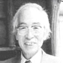 Humitaka Sato合作论文数3
 Akihiko Toda (戸田昭彦)合作论文数Graduate School of Advanced Science and Engineering, Hiroshima University3Shunji Matsuura合作论文数3
 Riou Nakamura合作论文数3




