Over the past two decades, genetic and proteomic screens have identified the Hippo pathway as a complex signaling network that controls tissue growth and human cancer. Despite these advances, our understanding of how Hippo signaling regulates transcription is less clear. To address this, we used live microscopy to study the nuclear behavior of the major Hippo pathway transcription effectors, YAP and TEADs. We reveal that TEADs are a major determinant of YAP DNA binding and nuclear mobility, while YAP minorly influences TEADs. YAP and TEAD1 associate with DNA for longer periods in cells with intrinsically low Hippo pathway activity and upon acute Hippo pathway perturbation. TEAD1 binds the genome on a broad range of timescales, and this is extended substantially in nuclear condensates. Last, a cancer-associated YAP fusion protein exhibits substantially different biophysical behavior than either YAP or TEAD1. Thus, we reveal that Hippo signaling regulates transcription, in part, by influencing the DNA binding times of YAP and TEADs.
The Hippo pathway is a highly conserved signalling network that controls tissue growth and cell fate, responding to physical properties of the tissue microenvironment and cell biological features such as adhesion and polarity. Hippo signalling perturbation is associated with several human diseases, particularly various solid cancers. Hippo pathway-targeted therapies are beginning to emerge for the treatment of cancer, most of which are focused on disrupting the ability of the YAP and TAZ transcription co-activator proteins to promote transcription of genes with their cognate TEAD1-4 DNA binding proteins. Recently, TEAD inhibitors have shown promise in a phase I clinical trial in cancers that are enriched for Hippo pathway mutations, such as mesothelioma. Moreover, Hippo pathway-targeted therapies have great potential to be combined with RAS-MAPK pathway inhibitors, given the close functional relationship that these signalling pathways share in development and disease.
Epithelial organs maintain their integrity and prevent tumor initiation by actively removing defective cells, such as those that have lost apicobasal polarity. Here, we identify how transcription factors of two key signaling pathways—Jun-N-terminal kinase (JNK) and Hippo—regulate epithelial integrity by controlling transcription of an overlapping set of target genes. Targeted DamID experiments reveal that, in proliferating cells of the Drosophila melanogaster eye, the AP-1 transcription factor Jun and the Hippo pathway transcription regulators Yorkie and Scalloped bind to a common suite of target genes that promote organ growth. In defective neoplastic cells, AP-1 transcription factors repress transcription of growth genes together with the C-terminal binding protein (CtBP) co-repressor. If gene repression by AP-1/CtBP fails, neoplastic tumor growth ensues, driven by Yorkie/Scalloped. Thus, AP-1/CtBP eliminates defective cells and prevents tumor initiation by acting in parallel to Yorkie/Scalloped to repress expression of a shared transcriptome. These findings shed new light on the maintenance of epithelial integrity and tumor suppression.
The Hippo pathway is an important regulator of organ growth and cell fate. The major mechanism by which Hippo is known to control transcription is by dictating the nucleo-cytoplasmic shuttling rate of Yorkie, a transcription co-activator, which promotes transcription with the DNA binding protein Scalloped. The nuclear biophysical behavior of Yorkie and Scalloped, and whether this is regulated by the Hippo pathway, remains unexplored. Using multiple live-imaging modalities on Drosophila tissues, we found that Scalloped interacts with DNA on a broad range of timescales, and enrichment of Scalloped at sites of active transcription is mediated by longer DNA dwell times. Further, Yorkie increased Scalloped’s DNA dwell time, whereas the repressors Nervous fingers 1 (Nerfin-1) and Tondu-domain-containing growth inhibitor (Tgi) decreased it. Therefore, the Hippo pathway influences transcription not only by controlling nuclear abundance of Yorkie but also by modifying the DNA binding kinetics of the transcription factor Scalloped.
The Hippo tumor suppressor pathway controls transcription by regulating nuclear abundance of YAP and TAZ, which activate transcription with the TEAD1-TEAD4 DNA-binding proteins. Recently, several small-molecule inhibitors of YAP and TEADs have been reported, with some entering clinical trials for different cancers with Hippo pathway deregulation, most notably, mesothelioma. Using genome-wide CRISPR/Cas9 screens we reveal that mutations in genes from the Hippo, MAPK, and JAK-STAT signaling pathways all modulate the response of mesothelioma cell lines to TEAD palmitoylation inhibitors. By exploring gene expression programs of mutant cells, we find that MAPK pathway hyperactivation confers resistance to TEAD inhibition by reinstating expression of a subset of YAP/TAZ target genes. Consistent with this, combined inhibition of TEAD and the MAPK kinase MEK, synergistically blocks proliferation of multiple mesothelioma and lung cancer cell lines and more potently reduces the growth of patient-derived lung cancer xenografts in vivo. Collectively, we reveal mechanisms by which cells can overcome small-molecule inhibition of TEAD palmitoylation and potential strategies to enhance the anti-tumor activity of emerging Hippo pathway targeted therapies.
Our understanding of heterochromatin nanostructure and its capacity to mediate gene silencing in a living cell has been prevented by the diffraction limit of optical microscopy. Thus, here to overcome this technical hurdle, and directly measure the nucleosome arrangement that underpins this dense chromatin state, we coupled fluorescence lifetime imaging microscopy (FLIM) of Förster resonance energy transfer (FRET) between histones core to the nucleosome, with molecular editing of heterochromatin protein 1 alpha (HP1α). Intriguingly, this super-resolved readout of nanoscale chromatin structure, alongside fluorescence fluctuation spectroscopy (FFS) and FLIM-FRET analysis of HP1α protein-protein interaction, revealed nucleosome arrangement to be differentially regulated by HP1α oligomeric state. Specifically, we found HP1α monomers to impart a previously undescribed global nucleosome spacing throughout genome architecture that is mediated by trimethylation on lysine 9 of histone H3 (H3K9me3) and locally reduced upon HP1α dimerisation. Collectively, these results demonstrate HP1α to impart a dual action on chromatin that increases the dynamic range of nucleosome proximity. We anticipate that this finding will have important implications for our understanding of how live cell heterochromatin structure regulates genome function.
SUMMARY:DamID sequencing is a technique to map the genome-wide interaction of a protein with DNA. Damsel is the first Bioconductor package to provide an end to end analysis for DamID sequencing data within R. Damsel performs quantification and testing of significant binding sites along with exploratory and visual analysis. Damsel produces results consistent with previous analysis approaches. AVAILABILITY AND IMPLEMENTATION:The R package Damsel is available for install through the Bioconductor project https://bioconductor.org/packages/release/bioc/html/Damsel.html and the code is available on GitHub https://github.com/Oshlack/Damsel/.
Organ size is controlled by numerous factors including mechanical forces, which are mediated in part by the Hippo pathway. In growing Drosophila epithelial tissues, cytoskeletal tension influences Hippo signaling by modulating the localization of key pathway proteins to different apical domains. Here, we discovered a Hippo signaling hub at basal spot junctions, which form at the basal-most point of the lateral membranes and resemble adherens junctions in protein composition. Basal spot junctions recruit the central kinase Warts via Ajuba and E-cadherin, which prevent Warts activation by segregating it from upstream Hippo pathway proteins. Basal spot junctions are prominent when tissues undergo morphogenesis and are highly sensitive to fluctuations in cytoskeletal tension. They are distinct from focal adhesions, but the latter profoundly influences basal spot junction abundance by modulating the basal-medial actomyosin network and tension experienced by spot junctions. Thus, basal spot junctions couple morphogenetic forces to Hippo pathway activity and organ growth.
Supplementary Materials and Methods from Transcriptional Output of the Salvador/Warts/Hippo Pathway Is Controlled in Distinct Fashions in <i>Drosophila melanogaster</i> and Mammalian Cell Lines
Deregulation of the Hippo pathway is a driver for cancer progression and treatment resistance. In the context of gastric cancer, YAP1 is a biomarker for poor patient prognosis. Although genomic tumor profiling provides information of Hippo pathway activation, the present study demonstrates that inhibition of Yap1 activity has anti-tumor effects in gastric tumors driven by oncogenic mutations and inflammatory cytokines. We show that Yap1 is a key regulator of cell metabolism, proliferation, and immune responses in normal and neoplastic gastric epithelium. We propose that the Hippo pathway is targetable across gastric cancer subtypes and its therapeutic benefits are likely to be mediated by both cancer cell-intrinsic and -extrinsic mechanisms.
Correlation between mRNA expression and copy number (CN) in the 10 most commonly amplified Hippo pathway genes
The Hippo signaling pathway regulates developmental organ growth, regeneration, and cell fate decisions. Although the role of the Hippo pathway, and its transcriptional effectors YAP and TAZ, has been well documented in many cell types and species, only recently have the roles for this pathway come to light in vascular development and disease. Experiments in mice, zebrafish, and in vitro have uncovered roles for the Hippo pathway, YAP, and TAZ in vasculogenesis, angiogenesis, and lymphangiogenesis. In addition, the Hippo pathway has been implicated in vascular cancers and cardiovascular diseases, thus identifying it as a potential therapeutic target for the treatment of these conditions. However, despite recent advances, Hippo's role in the vasculature is still underappreciated compared with its role in epithelial tissues. In this review, we appraise our current understanding of the Hippo pathway in blood and lymphatic vessel development and highlight the current knowledge gaps and opportunities for further research.
Dedifferentiation is the reversion of mature cells to a stem cell-like fate, whereby gene expression programs are altered and genes associated with multipotency are (re)expressed. Misexpression of multipotency factors and pathways causes the formation of ectopic neural stem cells (NSCs). Whether dedifferentiated NSCs faithfully produce the correct number and types of progeny, or undergo timely terminal differentiation, has not been assessed. Here, we show that ectopic NSCs induced via bHLH transcription factor Deadpan (Dpn) expression fail to undergo appropriate temporal progression by constantly expressing mid-temporal transcription factor(tTF), Sloppy-paired 1/2 (Slp). Consequently, this resulted in impaired terminal differenation and generated an excess of Twin of eyeless (Toy)-positive neurons at the expense of Reversed polarity (Repo)-positive glial cells. Preference for a mid-temporal fate in these ectopic NSCs is concordant with an enriched binding of Dpn at mid-tTF loci and a depletion of Dpn binding at early- and late-tTF loci. Retriggering the temporal series via manipulation of the temporal series or cell cycle is sufficient to reinstate neuronal diversity and timely termination.
Correlation amongst melanoma cell lines of viability following YAP siRNA- and verteporfin-treatment
The Germinal Center Kinase III (GckIII) pathway is a Hippo-like kinase module defined by sequential activation of Ste20 kinases Thousand and One (Tao) and GckIII, followed by nuclear dbf2-related (NDR) kinase Tricornered (Trc). We previously uncovered a role for the GckIII pathway in Drosophila melanogaster tracheal (respiratory) tube morphology. The trachea form a network of branched epithelial tubes essential for oxygen transport, and are structurally analogous to branched tubular organs in vertebrates, such as the vascular system. In the absence of GckIII pathway function, aberrant dilations form in tracheal tubes characterized by mislocalized junctional and apical proteins, suggesting that the pathway is important in maintaining tube integrity in development. Here, we observed a genetic interaction between trc and Cerebral cavernous malformations 3 (Ccm3), the Drosophila ortholog of a human vascular disease gene, supporting our hypothesis that the GckIII pathway functions downstream of Ccm3 in trachea, and potentially in the vertebrate cerebral vasculature. However, how GckIII pathway signaling is regulated and the mechanisms that underpin its function in tracheal development are unknown. We undertook biochemical and genetic approaches to identify proteins that interact with Trc, the most downstream GckIII pathway kinase. We found that known GckIII and NDR scaffold proteins are likely to control GckIII pathway signaling in tracheal development, consistent with their conserved roles in Hippo-like modules. Furthermore, we show genetic interactions between trc and multiple enzymes in glycolysis and oxidative phosphorylation, suggesting a potential function of the GckIII pathway in integrating cellular energy requirements with maintenance of tube integrity.
Supplementary Figure 2 from Transcriptional Output of the Salvador/Warts/Hippo Pathway Is Controlled in Distinct Fashions in Drosophila melanogaster and Mammalian Cell Lines
Supplementary Figure 1 from Transcriptional Output of the Salvador/Warts/Hippo Pathway Is Controlled in Distinct Fashions in <i>Drosophila melanogaster</i> and Mammalian Cell Lines
Correlation between mRNA expression and copy number (CN) in the 10 most commonly deleted Hippo pathway genes
Our understanding of heterochromatin nanostructure and its capacity to mediate gene silencing in a living cell has been prevented by the diffraction limit of optical microscopy. Thus, here to get around this technical hurdle, and directly measure the nucleosome arrangement that underpins this dense chromatin state in a living cell, we coupled fluorescence lifetime imaging microscopy of Förster resonance energy transfer (FRET) between histones core to the nucleosome, with genome editing of heterochromatin protein 1 alpha (HP1α). Intriguingly, this super-resolved readout of nanoscale chromatin structure revealed nucleosome arrangement to be differentially regulated by HP1α oligomeric state. Specifically, we found HP1α monomers to impart a previously undescribed global nucleosome spacing throughout genome architecture that is locally reduced upon HP1α dimerisation. Further, this dynamic scaffold is impervious to sub-micron higher order chromatin structure and HP1α foci formation. Collectively these findings demonstrate that the macroscale features of live cell genome architecture are dictated by different concentrations, but not configurations, of spaced and bridged nucleosomes. This result has key implications for our understanding of transcription, replication, and DNA repair.
 Xiaomeng Zhang合作论文数26
 Sharad Kumar合作论文数Centre for Cancer Biology, University of South Australia13Katrina Mitchell合作论文数11
 Anthony Papenfuss合作论文数Division of Bioinformatics, The Walter and Eliza Hall Institute of Medical Research11Poon Carole L C合作论文数10Joseph Ha Vissers合作论文数10Vergara Ismael A合作论文数GenomeDx Biosci Inc9
 Shackleton Mark合作论文数9
 Manning Samuel A合作论文数9You-Fang Zhang合作论文数8Pacman Szeto合作论文数Peter MacCallum Canc Ctr, Melbourne, Vic, Australia8
 Lie Yang (杨烈)合作论文数West China Hospital of Sichuan University8
 Shu Kondo合作论文数7Claire C. Milton合作论文数7DI Cook合作论文数6A Dinudom合作论文数6Benjamin Kroeger合作论文数6
 Iswar Hariharan合作论文数Department of Molecular and Cell Biology, University of California, Berkeley6
 Helena Richardson合作论文数Department of Biochemistry and Chemistry, La Trobe University6Lachlan McIntosh合作论文数Peter MacCallum Cancer Centre5Chris Mintoff合作论文数5
 Aishwarya Kulkarni合作论文数Division of Biomedical Informatics, Cincinnati Children's Hospital Medical Center, Cincinnati, OH, 45229, USA.5
 Georgina Long合作论文数Faculty of Medicine and Health, The University of Sydney;Melanoma Institute Australia5Permsak Komwatana合作论文数5
 Grant Mcarthur合作论文数Peter MacCallum Cancer Centre;Victorian Comprehesive Cancer Centre;University of Melbourne5
 Lucas Dent合作论文数Cell Growth and Proliferation Laboratory, Peter MacCallum Cancer Centre;Department of Pathology, University of Melbourne;Peter MacCallum Cancer Centre, University of Melbourne5
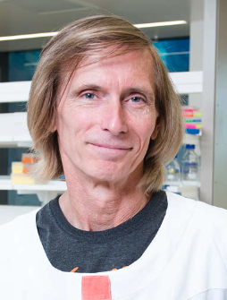 Nicholas Hayward合作论文数Oncogenomics Laboratory, QIMR Berghofer Medical Research Institute5Colebatch Andrew J合作论文数5
 Helen Rizos合作论文数School of Biochemistry;University of New South Wales;School of Biochemistry, University of New South Wales5
 Patrick O. Humbert合作论文数Cell Cycle and Cancer Genetics Laboratory, Peter MacCallum Cancer Center5
 Louise Cheng合作论文数Cancer Research Division, Peter MacCallum Cancer Centre;The Sir Peter MacCallum Department of Oncology, Faculty of Medicine, Dentistry and Health Sciences, University of Melbourne5
 Alexey Veraksa合作论文数Department of Biology, University of Massachusetts Boston5Jane I Lin合作论文数4Degoutin Joffrey L合作论文数4Froldi Francesca合作论文数4Felix A. Grusche合作论文数4
 Nicolas Tapon合作论文数Medical Research Council Laboratory for Molecular Cell Biology, and Department of Biochemistry, University College London4Munir R. Tanas合作论文数4E A Takano合作论文数3Dushyandi Rajendran合作论文数3Abdul Jabbar Saiful Hilmi合作论文数3Jonathan M Pojer合作论文数3Nicole Merritt合作论文数3J A Young合作论文数3
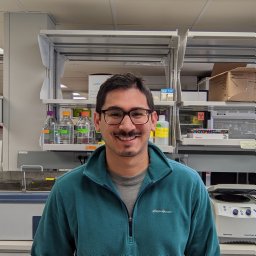 Keith Garcia合作论文数Department of Pathology, University of Iowa3Zhen-Yuan Lin合作论文数3Jieqiong Lou合作论文数School of Physics, Faculty of Science, The University of Melbourne3
 Liz Hinde合作论文数School of Physics, Faculty of Science, The University of Melbourne3
 Paul Gregorevic合作论文数Department of Anatomy and Physiology, School of Biomedical Sciences, Faculty of Medicine, Dentistry and Health Sciences, The University of Melbourne3




