Development of an effective HCV vaccine requires the induction of both broadly neutralizing antibodies (bnAbs) and a robust cellular response. One issue that has arisen is that HCV subunit vaccines have limited immunogenicity, thus requiring multivalent formats in order to elicit a robust anti-HCV immune response. Toward that end, nanoparticle vaccines possess the ability to facilitate a controlled multivalent presentation and trafficking to lymph nodes, where they can interact with both arms of the immune system. Here, we used a soluble, secreted form of E1E2 (sE1E2) to assemble native E1E2 into a nanoparticle platform using a post-purification coupling assembly system. Nanoparticles were assembled by purifying sE1E2 containing a C-terminal SpyTag and an mi3-SpyCatcher fusion separately and covalently coupling the components via incubation. Free sE1E2-SpyTag was removed from nanoparticle preparations via gel filtration. The sE1E2-mi3 nanoparticles are fully competent to bind conformation-dependent bnAbs, indicating retention of a native assembly in the nanoparticle format. Electron microscopy analysis showed a clear incorporation of sE1E2 on the surface of the nanoparticle. Immunogenicity of sE1E2-mi3 nanoparticles was examined relative to sE1E2 alone and membrane-bound E1E2 (mbE1E2) following inoculation of groups of CD1 mice. Assessment of the immunogenicity of the sE1E2-mi3 nanoparticles showed that the nanoparticle assembly has a similar immunogenicity profile to that of mbE1E2 after only a prime and one boost, and overall superior to sE1E2. This proof-of-principle study sets the stage for further exploration of nanoparticles and other multivalent platforms for the development of E1E2-based vaccines. Importance:Hepatitis C virus infects approximately 50 million people, and at present no effective HCV vaccine exists. Due to the high sequence variability of HCV and the resulting difficulty in developing a vaccine that elicits a broadly neutralizing response, multiple efforts are underway to enhance the immunogenicity of HCV vaccine candidates. In this study, we incorporated native soluble, secreted E1E2 (sE1E2) into a 60-mer nanoparticle via the SpyTag-SpyCatcher system and covalent isopeptide bond attachment using the purified components. These nanoparticles are antigenically intact and elicit a neutralizing antibody response at an earlier time point in the immunization regimen than the corresponding subunit vaccine. These studies show that a well-characterized sE1E2 platform compatible with multiple genotypes can be coupled to nanoparticles for use as a vaccine candidate.
An effective vaccine against hepatitis C virus (HCV) must elicit the production of broadly neutralizing antibodies (bnAbs) reproducibly against the E1E2 glycoprotein complex. Little is known about how glycan content affects this process. Ideally, glycans would maximize epitope exposure without compromising antigen stability or exposing new epitopes. However, typical recombinant vaccines contain considerable heterogeneity in glycan content, which can affect the antibody response and neutralization potency. Here we employed glycoengineered Chinese hamster ovary (geCHO) cell lines that impart nearly homogeneous glycosylation as a means to test how specific glycan features influence antigenicity and immunogenicity for the secreted HCV E2 ectodomain (sE2). Specific geCHO antigens exhibited a modest but reproducible increase in affinity for some mAbs relative to CHO- and HEK293-produced sE2. Surprisingly, one geCHO sE2 antigen failed to bind the CD81 receptor, indicating the potential for significant glycan effects on biochemical properties. We immunized mice with the four antigens and found the total antibody response to be the same for all groups. However, sera from one geCHO group exhibited a 7-fold improvement in neutralization against the homologous HCV pseudovirus (HCVpp) and had the most mice whose sera exhibited neutralization activity against genotypes 1b, 2a, 2b, and 3. Further analysis identified beneficial and deleterious glycan features, and the glycan that correlated the most with decreased potency was relatively small. However, size was not the sole determinant of glycan-driven effects on the antibody response. In summary, glycan content impacts biochemical properties of antigens to varying degrees and such effects can influence immune response quality and uniformity.
The rise of multi-drug-resistant bacteria that cannot be treated with traditional antibiotics has prompted the search for alternatives to combat bacterial infections. Endolysins, which are bacteriophage-derived peptidoglycan hydrolases, are attractive tools in this fight. Several studies have already demonstrated the efficacy of endolysins in targeting bacterial infections. Endolysins encoded by bacteriophages that infect Gram-positive bacteria typically possess an N-terminal catalytic domain and a C-terminal cell-wall binding domain (CWBD). In this study, we have uncovered the molecular mechanisms that underlie formation of a homodimer of Cpl-1, an endolysin that targets Streptococcus pneumoniae. Here, we use site-directed mutagenesis, analytical size exclusion chromatography, and analytical ultracentrifugation to disprove a previous suggestion that three residues at the N-terminus of the CWBD are involved in the formation of a Cpl-1 dimer in the presence of choline in solution. We conclusively show that the C-terminal tail region of Cpl-1 is involved in formation of the dimer. Alanine scanning mutagenesis generated various tail mutant constructs that allowed identification of key residues that mediate Cpl-1 dimer formation. Finally, our results allowed identification of a consensus sequence (FxxEPDGLIT) required for choline-dependent dimer formation─a sequence that occurs frequently in pneumococcal autolysins and endolysins. These findings shed light on the mechanisms of Cpl-1 and related enzymes and can be used to inform future engineering efforts for their therapeutic development against S. pneumoniae.
The monoclonal antibody (mAb) protein class has become a primary therapeutic platform for the production of new life saving drug products. MAbs are comprised of two domains: the antigen-binding fragment (Fab) and crystallizable fragment (Fc). Despite the success in the clinic, NMR assignments of the complete Fab domain have been elusive, in part due to problems in production of properly folded, triply-labeled 2H,13C,15N Fab domain. Here, we report the successful recombinant expression of a triply-labeled Fab domain, derived from the standard IgG1κ known as NISTmAb, in yeast. Using the 2H,13C,15N Fab domain, we assigned 94% of the 1H, 13C, and 15N backbone atoms.
Four tailspike proteins (TSP1-4) of Escherichia coli O157:H7 bacteriophage CBA120 enable infection of multiple hosts. They form a branched complex that attaches to the tail baseplate. Each TSP recognizes a different lipopolysaccharide on the membrane of a different bacterial host. The 335 N-terminal residues of TSP4 promote the assembly of the TSP complex and anchor it to the tail baseplate. The crystal structure of TSP4-N 335 reveals a trimeric protein comprising four domains. The baseplate anchor domain (AD) contains an intertwined triple-stranded β-helix. The ensuing XD1, XD2 and XD3 β-sheet containing domains mediate the binding of TSP1-3 to TSP4. Each of the XD domains adopts the same fold as the respective XD domains of bacteriophage T4 gp10 baseplate protein, known to engage in protein–protein interactions via its XD2 and XD3 domains. The structural similarity suggests that XD2 and XD3 of TSP4 also function in protein–protein interactions. Analytical ultracentrifugation analyses of TSP4-N 335 and of domain deletion proteins showed how TSP4-N 335 promotes the formation of the TSP quaternary complex. TSP1 and TSP2 bind directly to TSP4 whereas TSP3 binding requires a pre-formed TSP4-N 335 :TSP2 complex. A 3-dimensional model of the bacteriophage CBA120 TSP complex has been developed based on the structural and ultracentrifuge information.
Critical assessment of structure prediction (CASP) conducts community experiments to determine the state of the art in computing protein structure from amino acid sequence. The process relies on the experimental community providing information about not yet public or about to be solved structures, for use as targets. For some targets, the experimental structure is not solved in time for use in CASP. Calculated structure accuracy improved dramatically in this round, implying that models should now be much more useful for resolving many sorts of experimental difficulties. To test this, selected models for seven unsolved targets were provided to the experimental groups. These models were from the AlphaFold2 group, who overall submitted the most accurate predictions in CASP14. Four targets were solved with the aid of the models, and, additionally, the structure of an already solved target was improved. An a posteriori analysis showed that, in some cases, models from other groups would also be effective. This paper provides accounts of the successful application of models to structure determination, including molecular replacement for X-ray crystallography, backbone tracing and sequence positioning in a cryo-electron microscopy structure, and correction of local features. The results suggest that, in future, there will be greatly increased synergy between computational and experimental approaches to structure determination.
Hepatitis C virus (HCV) is a major worldwide health burden, and a preventive vaccine is needed for global control or eradication of this virus. A substantial hurdle to an effective HCV vaccine is the high variability of the virus, leading to immune escape. The E1E2 glycoprotein complex contains conserved epitopes and elicits neutralizing antibody responses, making it a primary target for HCV vaccine development. However, the E1E2 transmembrane domains that are critical for native assembly make it challenging to produce this complex in a homogenous soluble form that is reflective of its state on the viral envelope. To enable rational design of an E1E2 vaccine, as well as structural characterization efforts, we have designed a soluble, secreted form of E1E2 (sE1E2). As with soluble glycoprotein designs for other viruses, it incorporates a scaffold to enforce assembly in the absence of the transmembrane domains, along with a furin cleavage site to permit native-like heterodimerization. This sE1E2 was found to assemble into a form closer to its expected size than full-length E1E2. Preservation of native structural elements was confirmed by high-affinity binding to a panel of conformationally specific monoclonal antibodies, including two neutralizing antibodies specific to native E1E2 and to its primary receptor, CD81. Finally, sE1E2 was found to elicit robust neutralizing antibodies in vivo. This designed sE1E2 can both provide insights into the determinants of native E1E2 assembly and serve as a platform for production of E1E2 for future structural and vaccine studies, enabling rational optimization of an E1E2-based antigen.
Abstract Purpose: Gasdermin B (GSDMB) overexpression/amplification occurs in about 60% of HER2 breast cancers, where it promotes cell migration, resistance to anti-HER2 therapies, and poor clinical outcome. Thus, we tackle GSDMB cytoplasmic overexpression as a new therapeutic target in HER2 breast cancers. Experimental Design: We have developed a new targeted nanomedicine based on hyaluronic acid–biocompatible nanocapsules, which allow the intracellular delivery of a specific anti-GSDMB antibody into HER2 breast cancer cells both in vitro and in vivo. Results: Using different models of HER2 breast cancer cells, we show that anti-GSDMB antibody loaded to nanocapsules has significant and specific effects on GSDMB-overexpressing cancer cells' behavior in ways such as (i) lowering the in vitro cell migration induced by GSDMB; (ii) enhancing the sensitivity to trastuzumab; (iii) reducing tumor growth by increasing apoptotic rate in orthotopic breast cancer xenografts; and (iv) diminishing lung metastasis in MDA-MB-231-HER2 cells in vivo. Moreover, at a mechanistic level, we have shown that AbGB increases GSDMB binding to sulfatides and consequently decreases migratory cell behavior and may upregulate the potential intrinsic procell death activity of GSDMB. Conclusions: Our findings portray the first evidence of the effectiveness and specificity of an antibody-based nanomedicine that targets an intracellular oncoprotein. We have proved that intracellular-delivered anti-GSDMB reduces diverse protumor GSDMB functions (migration, metastasis, and resistance to therapy) in an efficient and specific way, thus providing a new targeted therapeutic strategy in aggressive HER2 cancers with poor prognosis.
The functional and biological significance of the selected CASP12 targets are described by the authors of the structures. The crystallographers discuss the most interesting structural features of the target proteins and assess whether these features were correctly reproduced in the predictions submitted to the CASP12 experiment.
Two Borrelia burgdorferi interacting proteins, BB0238 and BB0323, play distinct roles in pathogen biology and infectivity although a significance of their interaction remained enigmatic. Here we identified the polypeptide segment essential for BB0238-BB0323 interaction and examined how it supports spirochete infectivity. We show that the interaction region in BB0323 requires amino acid residues 22–200, suggesting that the binding encompasses discontinuous protein segments. In contrast, the interaction region in BB0238 spans only 11 amino acids, residues 120–130. A deletion of these 11 amino acids neither alters the overall secondary structure of the protein, nor affects its stability or oligomerization property, however, it reduces the post-translational stability of the binding partner, BB0323. Mutant B. burgdorferi isolates producing BB0238 lacking the 11-amino acid interaction region were able to persist in ticks but failed to transmit to mice or to establish infection. These results suggest that BB0238-BB0323 interaction is critical for post-translational stability of BB0323, and that this interaction is important for mammalian infectivity and transmission of B. burgdorferi. We show that saturation or inhibition of BB0238-BB0323 interaction could be studied in a luciferase assay, which could be amenable for future identification of small molecule inhibitors to combat B. burgdorferi infection.
Oxford Glycobiology Institute, Department of Biochemistry, University of Oxford, South Parks Road, Oxford OX1 3QU, England, United Kingdom UCIBIO, REQUIMTE, Departamento de Química, Faculdade de Cien̂cias e Tecnologia, Universidade Nova de Lisboa, Caparica, 2829-516, Portugal Institute for Bioscience and Biotechnology Research, University of Maryland, Rockville, Maryland 20850 Department of Structural Biology, Weizmann Institute of Science, Rehovot, Israel Department of Chemistry, University of Copenhagen, Universitetsparken 5, 2100 Copenhagen Ø, Denmark Department of Molecular Biology and Biochemistry/Pharmaceutical Sciences, University of California Irvine, Irvine, California 92697 Department of Molecular, Cellular and Developmental Biology/Biomolecular Science and Engineering Program, University of California, Santa Barbara, Santa Barbara, California 93106
The exact function of human gasdermin-B (GSDMB), which regulates differentiation and growth of epithelial cells, is yet to be elucidated. In human epidermal growth factor receptor 2 (HER2)-positive breast cancer, GSDMB gene amplification and protein overexpression indicate a poor response to HER2-targeted therapy. Genome-wide association studies revealed a correlation between GSDMB SNPs and an increased susceptibility to Crohn's disease, ulcerative colitis, and asthma. The N- and C-terminal domains of all gasdermins possess lipid-binding and regulatory activities, respectively. Inflammatory caspases cleave gasdermin-D in the interdomain linker but not GSDMB. The cleaved N- terminal domain binds phosphoinositides and cardiolipin, forms membrane-disrupting pores, and executes pyroptosis. We show that both full-length GSDMB and the N- terminal domain bind to nitrocellulose membranes immobilized with phosphoinositides or sulfatide, but not with cardiolipin. In addition, the GSDMB N- terminal domain binds liposomes containing sulfatide. The crystal structure of the GSDMB C-terminal domain reveals the structural impact of the amino acids encoded by SNPs that are linked to asthma and inflammatory bowel disease (IBD). A loop that carries the polymorphism amino acids corresponding to healthy individuals (Gly299: Pro306) exhibits high conformational flexibility, whereas the loop carrying amino acids found in individuals with increased disease risk (Arg299: Ser306) exhibits a well-defined conformation and higher positive surface charge. Apoptotic executioner caspase-3, - 6, and - 7, but not the inflammatory caspases, cleave GSDMB at 88DNVD91 within the N- terminal domain. Selective sulfatide binding may indicate possible function for GSDMB in the cellular sulfatide transport.
We recently published structural and biochemical studies of gasdermin-B (GSDMB) (1), a protein that regulates the maintenance of the epithelial cell barrier as well as cell proliferation and differentiation processes (2, 3). GSDMB amplification and GSDMB overexpression lead to a poor response to HER2-targeted therapy in HER2-positive breast cancer (4). We showed that GSDMB binds to sulfatide and phosphoinositides, components of the epithelial cell membrane. Because sulfatide promotes cell migration and metastasis (5), we speculated that GSDMB might be directly or indirectly involved in the transport of sulfatide to the cell membrane. Genome-wide association studies show that the presence of two GSDMB missense SNPs (dbSNP:rs2305479 and dbSNP:rs2305480) correlate with an increased … [↵][1]1To whom correspondence may be addressed. Email: osnat{at}umd.edu or jmoult{at}umd.edu. [1]: #xref-corresp-1-1
Background: RON and MET receptors bind their ligands MSP and HGF selectively and activate different signaling pathways. Results: Crystallographic and analytical ultracentrifugation studies provide important information about RON-MSP interaction. Conclusion: RON-MSP and MET-HGF exhibit 2:2 complex stoichiometry, but differences within the respective interfaces explain the strict ligand-receptor specificity. Significance: Signaling pathways must be exquisitely regulated with no cross-reactivity between related systems.Recepteur d'origine nantais (RON) receptor tyrosine kinase and its ligand, serum macrophage-stimulating protein (MSP), play important roles in inflammation, cell growth, migration, and epithelial to mesenchymal transition during tumor development. The binding of mature MSP (disulfide-linked - and -chains) to RON ectodomain modulates receptor dimerization, followed by autophosphorylation of tyrosines in the cytoplasmic receptor kinase domains. Receptor recognition is mediated by binding of MSP -chain (MSP) to the RON Sema. Here we report the structure of RON Sema-PSI-IPT1 (SPI1) domains in complex with MSP at 3.0 resolution. The MSP serine protease-like -barrel uses the degenerate serine protease active site to recognize blades 2, 3, and 4 of the -propeller fold of RON Sema. Despite the sequence homology between RON and MET receptor tyrosine kinase and between MSP and hepatocyte growth factor, it is well established that there is no cross-reactivity between the two receptor-ligand systems. Comparison of the structure of RON SPI1 in complex with MSP and that of MET receptor tyrosine kinase Sema-PSI in complex with hepatocyte growth factor -chain reveals the receptor-ligand selectivity determinants. Analytical ultracentrifugation studies of the SPI1-MSP interaction confirm the formation of a 1:1 complex. SPI1 and MSP also associate primarily as a 1:1 complex with a binding affinity similar to that of SPI1-MSP. In addition, the SPI1-MSP ultracentrifuge studies reveal a low abundance 2:2 complex with approximate to 10-fold lower binding affinity compared with the 1:1 species. These results support the hypothesis that the -chain of MSP mediates RON dimerization.
Human RON (Recepteur d’Origine Nantais) receptor tyrosine kinase is a cell surface receptor for Macrophage Stimulating Protein (MSP). RON mediates signal transduction pathways that regulate cell adhesion, invasion, motility and apoptosis processes. Elevated levels of RON and its alternatively spliced variants are implicated in the progression and metastasis of tumor cells. The binding of MSP α/β heterodimer to the extracellular region of RON receptor induces receptor dimerization and activation by autophosphorylation of the intracellular kinase domains. The ectodomain of RON, containing the ligand recognition and dimerization domains, is composed of a semaphorin (Sema), Plexins-Semaphorins-Integrins domain (PSI), and four Immunoglobulins-Plexins-Transcription factor (IPT) domains. High affinity association between MSP and RON is mediated by the interaction between MSP β-chain and RON Sema, although RON activation requires intact RON and MSP proteins. Here, we report the structure of RON Sema-PSI domains at 1.85 Å resolution. RON Sema domain adopts a seven-bladed β-propeller fold, followed by disulfide bond rich, cysteine-knot PSI motif. Comparison with the homologous Met receptor tyrosine kinase reveals that RON Sema-PSI contains distinguishing secondary structural features. These define the receptors’ exclusive selectivity towards their respective ligands, RON for MSP and Met for HGF. The RON Sema-PSI crystal packing generates a homodimer with interface formed by the Sema domain. Mapping of the dimer interface using the RON homology to Met, MSP homology to Hepatocyte Growth Factor (HGF), and the structure of the Met/HGF complex shows the dimer interface overlapping with the putative MSPβ binding site. The crystallographically determined RON Sema-PSI homodimer may represent the dimer assembly that occurs during ligand-independent receptor activation and/or the inhibition of the constitutive activity of RONΔ160 splice variant by the soluble RON splice variant, RONΔ85.
High throughput genome wide associations studies (GWAS) are now identifying a large number of genome loci related to risk of common human disease. Each such locus presents a challenge in identifying the relevant underlying mechanism. Here we report the experimental characterization of a proposed causal single nucleotide polymorphism (SNP) in a locus related to risk of Crohn's disease and ulcerative colitis. The SNP lies in the MST1 gene encoding Macrophage Stimulating Protein (MSP), and results in an R689C amino acid substitution within the β-chain of MSP (MSPβ). MSP binding to the RON receptor tyrosine kinase activates signaling pathways involved in the inflammatory response. We have purified wild-type and mutant MSPβ proteins and compared biochemical and biophysical properties that might impact the MSP/RON signaling pathway. Surface plasmon resonance (SPR) binding studies showed that MSPβ R689C affinity to RON is approximately 10-fold lower than that of the wild-type MSPβ and differential scanning fluorimetry (DSF) showed that the thermal stability of the mutant MSPβ was slightly lower than that of wild-type MSPβ, by 1.6 K. The substitution was found not to impair the specific Arg483-Val484 peptide bond cleavage by matriptase-1, required for MSP activation, and mass spectrometry of tryptic fragments of the mutated protein showed that the free thiol introduced by the R689C mutation did not form an aberrant disulfide bond. Together, the studies indicate that the missense SNP impairs MSP function by reducing its affinity to RON and perhaps through a secondary effect on in vivo concentration arising from reduced thermodynamic stability, resulting in down-regulation of the MSP/RON signaling pathway.
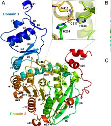
The ydcF gene from E. coli encodes a conserved 266 amino acid residue protein of unknown function. At the time of this writing, a sequence search of the nonredundant GenBank database using PSI-BLAST yielded 831 sequences related to YdcF.1 YdcF belongs to the PF02698 family in PfamA (containing 762 members) proteins that share the domain of unknown function, DUF218.2 DUF218 domains are found mostly in the four phyla of bacteria but also in fungi, plants, and archaea. The genomes of many organisms encode multiple proteins possessing this domain. Despite low sequence identity, both Pfam and PSI-BLAST identify four E. coli genes that contain DUF218 domains, ydcF, ycbC, ygjQ, and sanA. The amino acid sequence identity between YdcF and its three paralogs is very low (10%–18% over less than half the polypeptide chain), indicating different function for each of the paralogs. SanA participates in the barrier function of the cell envelope3 whereas its ortholog from Salmonella typhimurium, SifX, may be involved in murein synthesis.4 However, these are rather vague definitions of function, which shed no light on the biochemical function of SanA or SifX. Both biological and biochemical functions of ycbC and ygjQ are unknown. GdmH from Gram-positive bacteria is also a DUF218 protein domain with broad biological function. The gene is located on an operon that encodes proteins involved in the biosynthesis of the antimicrobial peptides Gallidermin and Epidermin. GdmH is one of the accessory factors of the ABC transporters that secrete these peptides.5 The crystal structure of YdcF reported here reveals a fold of the adenine nucleotide α hydrolase-like family. Following the structural clues, a nucleotide binding screen wascarried out and showed that YdcF binds S-adenosyl-L-methionine (AdoMet) with a Kd value of 25 μM. The ydcF gene was amplified from the E. coli K-12 genomic DNA (ATCC700926), cloned into a pET100/D-TOPO (Invitrogen) and sequenced. E. coli BL21*(DE3) cells transformed with the plasmid were grown in the auto-induction media (ZYP5052) containing 0.2% (v/v) α-lactose inducer and 0.1 mg/mL ampicillin for 16 h at 27°C.6 Cells suspended in 20 mM Tris-HCl (pH 8.0), 50mM NaCl, 1 mM phenylmethanesulfonyl fluoride, 1 mM ethylene-diamine-tetra-acetic acid (EDTA), 5 mM dithiothreitol (DTT), 10% (v/v) glycerol, 1% (v/v) Triton X-100, 2.5 mM benzimidine, 0.17 mg/mL lysozyme, and 3.3 units/mL Benzonase (Novagen) were lysed by sonication and centrifuged for 1 h at 23,700g. The soluble fraction was applied onto a Q Sepharose column (GE Health Sciences) in Buffer A [20 mM Tris-HCl (pH 8.0), 50 mM NaCl, 1 mM PMSF, 1 mM EDTA, 5 mM DTT, 10% (v/v) glycerol]. YdcF in the flow thru was precipitated with 55% saturated ammonium sulfate and centrifuged for 30 min at 14,000g. The pellet was re-suspended in Buffer A to a final conductivity of ∼1.0 M ammonium sulfate and loaded onto a Phenyl Sepharose column (GE Health Sciences). YdcF was eluted with a linear gradient of 1.0–0.0 M ammonium sulfate in Buffer A, and applied onto a Sephacryl S100 column (GE Health Sciences) in Storage buffer (20 mM Tris-HCl (pH 7.5) 0.10 M NaCl, 0.5 mM EDTA). The protein concentration was determined at 280 nm (calculated extinction coefficient: 41,940 M−1 cm−1). All purification steps were conducted at 4°C. The molecular weight was confirmed by MALDI-TOF mass spectrometry (measured and calculated molecular masses of 29,718 and 29,702 Da, respectively). Dynamic light scattering indicated that the protein was monomeric. YdcF containing selenomethionine (SeMet) was produced in E. coli B834(DE3). Transformed cells were grown in the Studier's PASM-5052 media supplemented with 0.125 mg/mL L-(+)-selenomethionine (Anatrace), 0.2% α-lactose inducer and 0.10 mg/mL ampicillin at 27°C for 24 h.6 The purification protocol was identical to that of the wild-type protein, and incorporation of the SeMet was verified by MALDI-TOF mass spectrometry. YdcF crystals were obtained at room temperature using the vapor diffusion method in hanging drops. Equal volumes of wild-type YdcF (10 mg/mL in storage buffer and 10 mM DTT) and mother liquor (0.1 M 4-(2-hydroxyethyl)piperazine-1-ethanesulfonic acid (pH 7.5), 1.7 M ammonium sulfate, and 4% (v/v) (±)-2-methyl-(phenyl)-2,3-pentanediol (MPD)) were mixed and equilibrated against the mother liquor reservoir. The resulting crystals belong to space group P21. Crystals were also obtained from wild-type (5 mg/mL) or SeMet-containing YdcF (3.5 mg/mL) at 1:1 volumes of protein and 0.10 M Tris-HCl (pH 8.0), 0.7 M sodium citrate and 0.02% (v/v) polyethylene glycol 20,000. These crystals belong to space group R3. For diffraction data collection, the crystals were transferred to a cryogenic solution composed of mother liquor containing 30% (v/v) glycerol and flash-cooled at 100 K in liquid propane. Multiple wavelength diffraction data, exploiting the absorption edge of Se, were collected at the industrial macromolecular crystallography association-collaborative access team (IMCA-CAT) 17-ID beamline at the advanced photon source (Argonne National Laboratory, Argonne, IL). The beamline was equipped with a MAR CCD detector. Data for the wild-type enzyme were also collected at the IMCA-CAT 17-ID beamline. The data were processed with the HKL suite of programs7 (Table I). The positions of five Se atoms were determined using the ShelxD program.8 Phases were calculated at 2.0 Å resolution with Solve9 and improved by solvent flattening techniques with Resolve.10 Nearly a third of the polypeptide chain was traced automatically by Resolve and the remaining model was built with use of the program O.11 This initial structure served as a search model for Molecular Replacement executed with BEAST12 and using the diffraction data of the P21 crystal. Refinement of the structure was performed with CNS13 followed by REFMAC.14 Ligand binding was measured by Isothermal Titration Calorimetry using a VP Microcal, Inc. isothermal titration calorimeter. The solution vessel contained 70–80 μM YdcF in storage buffer and 5 mM MgCl2. Aliquots (5 μL) of 2 mM ligand solutions were titrated into the YdcF solution. Duplicate measurements were performed. The following ligands were tested: β-nicotinamide adenine dinucleotide, β-nicotinamide adenine dinucleotide phosphate (NADP+), flavin adenine dinucleotide, adenosine triphosphate (ATP), deoxyadenosine monophosphate (dAMP), and AdoMet. The 1.8 Å resolution crystal structure of YdcF defines the coordinates of residues 5–265 and 5–263 of the 266 amino acids of molecules A and B in the asymmetric unit, respectively (Table I). The root-mean-square deviation (rmsd) between the Cα atoms of the two protomers in the asymmetric unit is 0.2 Å. The crystal packing indicates that YdcF is monomeric, consistent with the dynamic light scattering data. YdcF contains two closely associated domains and the chain crosses twice from one domain to another [Fig. 1(A)]. One domain adopts an α/β open-sheet fold (residues 38–180) with five-stranded parallel β-sheet (β-strands order 32145) flanked by α2 and α4 on one face and α5 and α6 on the opposing face. The large loop connecting β2 to α4 contains helix α3. The second, helical domain (residues 5–37, 181–263), encompasses α1 and α7-α9 and contains a β-hairpin (β6-β7). The large cleft at the domains' interface is flanked by residues that are located at the C-termini of β1 and β4 (the classical α/β-fold switch region where enzyme active sites are located), and on α3, α6, and α7. Crystal structure of E. coli YdcF. (A) The overall fold with α-helices colored in cyan and β-strands in yellow. (B) Superposition of E. coli YdcF (cyan) and Methanococcus jannaschii. MJ0577 (yellow) with bound ATP. (C) Putative active site of E. coli YdcF. The ligand binding cavity and the surrounding residues are shown. Atomic colors used: carbon-gray; oxygen-red; nitrogen-blue; phosphate-magenta. A search for structure relatives using the program DALI15 failed to identify PDB entries that span the entire YdcF molecule. However, there are many structure homologs of the α/β domain. The closest structure is that of the Methanococcus jannaschii universal stress protein-like MJ0577 (1MJH) (Z = 7.3, rmsd = 2.8 Å for 103 common Cα atoms, 5% sequence identity)16 [Fig. 1(B)]. MJ0577 contains only the α/β domain and assembles into dimers, in contrast to the monomeric YdcF that contains an additional helical domain. MJ0577 belongs to the adenine nucleotide α hydrolases-like superfamily in the SCOP database.17 Most of the structural homologs in the DALI list bind adenosine-containing compounds. A DALI search of the YdcF's helical domain with or without the β-hairpin segment yielded a few homologs with low Z-scores, the closest of which is a fragment of the RNA helicase from Pyrococcus furiosus Hef helicase/nuclease (1WP9, Z = 3.3, rmsd = 3.3 Å for 78 Cα atoms, 13% sequence identity).18 Because helical domains occur in many function contexts, the structure homology does not provide clues about the function of YdcF. The superimposed structures of YdcF and MJ0577/ATP complex support the hypothesis that the YdcF putative ligand binds at the C-termini of β1 and β4 [Fig. 1(B)]. A sulfate molecule is positioned in the deep cleft, forming an ion pair with Arg158, an invariant residue in the YdcF sequence family, and also interacts with the invariant His73 [Fig. 1(C)]. The sulfate may occupy the site of a phosphate group of a nucleotide or other negatively charged group of a substrate. A second invariant arginine residue in the cleft, Arg218, may also contribute to substrate binding. The structural homology to nucleotide-binding proteins, in particular adenosine-containing compounds, inspired the binding screening. Hence various adenosine-containing compounds were examined by isothermal titration calorimetry. Only AdoMet binds to YdcF and the binding isotherms fit well to a single site binding model, which yields a dissociation constant of 25 ± 3 μM and a binding enthalpy of −1.5 ± 0.3 kJ/mol. Indeed, the DALI results include several methyltransferases, although YdcF does not span the entire canonical methyltransferase fold. The dissociation constant is physiologically significant as a survey of the BRENDA database shows that this value is well within the Km values of methyltransferases. The YdcF fold is different from any of the known α/β-folds of methyltransferases, AdoMet-dependent oxidoreductases and AdoMet decarboxylase. Presently, the exact biochemical activity of YdcF remains unknown. Information about the biological function of YdcF is scarce. A whole genome transcriptional profiling study detected an increased expression of the ydcF transcript under anaerobic growth condition in fnr− and ArcA-deficient E. coli strains when compared with expression in wild-type E. coli (4.3- and 4.7-fold, respectively). This was interpreted as indicative of YdcF expression that is regulated by both the ferric nitrate reductase global regulatory protein FNR and the two-component system ArcAB.19, 20 A whole E. coli genome protein pull-down study identified YdcF interactions with the 50S ribosomal protein L2 (a protein that interacts with extraordinarily large number of proteins) and with dTDP-glucose-4,6-dehydrogenase associated with the lipopolysaccharide biosynthesis pathway.21 Both interactions are yet to be verified by accurate biophysical methods. The E. coli ydcF gene is not located on an operon, and its gene neighbors or those of homologs from other organisms do not provide insight into YdcF's function. Thus, the structure insight, the ligand screening, and the expression profiling suggest that YdcF is an AdoMet-dependent enzyme that plays a role in an anaerobic respiratory pathway. We greatly appreciate John Moult's leadership of the Structural Genomics program at CARB. We thank the staff of IMCA-CAT at the Advanced Photon Source for help during data collection. The IMCA-CAT facility is supported by the companies of the Industrial Macromolecular Crystallographic Association, through a contract with IIT. Use of the Advanced Photon Source was supported by the U.S. Department of Energy, Basic Energy Sciences, Office of Science, under contract W-31-109-Eng-38. Protein Data Bank coordinates entry code: 3CA8.
 Osnat Herzberg合作论文数Department of Chemistry and Biochemistry, University of Maryland;Institute for Bioscience and Biotechnology Research, National Institute of Standards and Technology, University of Maryland17
 John Moult合作论文数Institute for Bioscience and Biotechnology Research, University of Maryland6
 Sara B. Linden合作论文数3
 Daniel Nelson合作论文数Institute for Bioscience & Biotechnology Research, University of Maryland3
 Thomas R Fuerst合作论文数3Kulakova Liudmila合作论文数3
 Lipika R. Pal合作论文数3Reinhard Albrecht合作论文数3
 Eric A Toth合作论文数3
 Alexander K. Andrianov合作论文数Apogee3
 Alexander Marin合作论文数Center for Biopolymers on Interfaces, University of Utah3
 Andriy Kryshtafovych合作论文数Protein Structure Prediction Center, University of California, Davis3
 Marcus D. Hartmann合作论文数Max Planck Unit for Structural Molecular Biology, MPG-ASMB c/o DESY3José Palacios合作论文数2
 Andrea Gonella合作论文数2Laura Garcia Bermejo合作论文数2Magnus Besev合作论文数2
 Marco Nardini合作论文数2Folmer Fredslund合作论文数2
 Marcos Garcia-Fuentes合作论文数2Thanh-Hong Nguyen合作论文数2Adit B Alreja合作论文数2Gema Moreno-Bueno合作论文数2
 Magda Fontes合作论文数2
 Xiaoran Shang合作论文数2
 Natalia Gorlatova合作论文数2Manuel Gámez-Chiachio合作论文数2Johan C Hill合作论文数2Macarena Rodriguez-Serrano合作论文数2
 Ana Olivera合作论文数Dept Farm & Tecnol Farmaceut, Univ Santiago de Compostela2
 Marco Mangiagalli合作论文数Univ Milano Bicocca, Dept Biotechnol & Biosci, Piazza Sci 2, I-20126 Milan, Italy2
 Lidia Martínez合作论文数ESISNA Res Grp, Inst Mat Sci Madrid CSIC2
 Mark J. van Raaij合作论文数Gorlaeus Laboratoria, Leiden University, Einsteinweg 55 NL-2300 RA Leiden Netherlands2David Sarrió合作论文数2
 Ana Luísa Carvalho合作论文数2
 Guido Pintacuda合作论文数Institut des Sciences Analytiques, Centre National de la Recherche Scientifique2
 Kaspars Tars合作论文数2
 Ndeh Didier A合作论文数Inst Cell & Mol Biosci, Newcastle Univ2
 Dolores Torres合作论文数2
 Ana Cadete Pires合作论文数2
 Singh Abhimanyu K合作论文数Departamento de Estructura de Macromoleculas, Centro Nacional de Biotecnologia (CNB-CSIC), Madrid, Spain;NanoBiomedicine Initiative, Centro Nacional de Biotecnologia (CNB-CSIC),;Centro Nacional de Biotecnologia (CNB-CSIC),, Centro Nacional de Biotecnologia (CNB-CSIC),2Postel Sandra合作论文数2
 Pedro Bule合作论文数Faculdade de Medicina Veterinária, Universidade de Lisboa2
 Juan Carlos Trivino合作论文数2
 Valentina Nardone合作论文数Center for Nano Science and Technology @PoliMi, Istituto Italiano di Tecnologia2Molina-Crespo Ángela合作论文数2Greenfield Julia合作论文数2
 Nicole Zitzmann合作论文数2Amparo Cano合作论文数2




