Epithelial mesenchymal transition (EMT) is considered to be correlated with malignancy of cancer cells and responsible for cancer invasion and metastasis. We previously reported that distant metastasis was associated with hypoxia in gastric cancer. We therefore investigated the effect of hypoxic condition on EMT of gastric cancer cells. Gastric cancer cells were cultured in normoxia (21% O2) or hypoxia (1% O2) for 24 h. EMT was evaluated as the percentage of spindle-shaped cells in total cells. Effect of transforming growth factor β1 (TGFβ1) or tyrosine kinase inhibitors on the EMT was evaluated. The expression level of TGFβ1 and TGFβR was evaluated by real time RT-PCR. The TGFβ1 production from cancer cells was measured by ELISA. Hypoxia stimulated EMT of OCUM-2MD3 and OCUM-12 cells, but not that of OCUM-2M cells. The expression level of TGFβ1 mRNA under hypoxia was significantly higher than that under normoxia in all of three cell lines. The expression level of TGFβR mRNA was significantly increased by hypoxia in OCUM-2MD3 cells, but not in OCUM-2M cells. TGFβR inhibitor, SB431542 or Ki26894, significantly suppressed EMT of OCUM-2MD3 and OCUM-12. TGFβ1 production from OCUM-2MD3 and OCUM-12 cells was significantly increased under hypoxia in comparison with that under normoxia. These findings might suggest that hypoxia stimulates the EMT of gastric cancer cells via autocrine TGFβ/TGFβR signaling.
Cancer-associated fibroblasts (CAFs) have recently been implicated in tumor growth and metastasis in gastric cancer. Cancer stem cells (CSCs) have been proposed to have an important role in cancer progression. The aim of this study was to clarify the effect of CAFs on CSCs characteristics in gastric carcinoma. Scirrhous gastric cancer cell lines, OCUM-12 and OCUM-2MD3, and non-scirrhous gastric cancer cell lines, MKN-45 and MKN-74, were used. OCUM-12/side population (SP) cells and OCUM-2MD3/SP cells were sorted by flow cytometry as CSC-rich cells from the parent cells. CaF-37 was established from the tumoral gastric specimens as CAFs. Flow cytometric analysis of SP fraction, spheroid colony assay, and RT-PCR analysis of CSC markers were performed to identify CSCs properties. Effect of CAFs on the tumorigenicity by OCUM-12/SP cells was examined using nude mice. CAF CM significantly increased the percentages of the SP fraction of OCUM-12/SP and OCUM-2MD3/SP cells, but not that of MKN-45/SP and MKN-74/SP cells. Taken together, CM from CaF-37 significantly increased the number of spheroid colonies and the expression level of CSC markers of OCUM-12/SP and OCUM-2MD3/SP cells. These stimulating-activities by CM were significantly decreased by TGFβ inhibitors, but not FGFR and cMet inhibitor. Tumorigenicity by subcutaneous coinoculation of OCUM-12/SP cells with CAFs was significantly high in comparison with that by OCUM-12/SP cells alone. Phospho-Smad2 expression level was significantly increased by co-inoculation with CAFs. These findings suggested that CAFs might regulate the stemness of CSCs in scirrhous gastric cancer by TGFβ signaling.
Skeletal muscle expressing Pro104Leu mutant caveolin 3 (CAV3(P104L)) in mouse becomes atrophied and serves as a model of autosomal dominant limb-girdle muscular dystrophy 1C. We previously found that caveolin 3-deficient muscles showed activated intramuscular transforming growth factor beta (TGF-β) signals. However, the cellular mechanism by which loss of caveolin 3 leads to muscle atrophy is unknown. Recently, several small-molecule inhibitors of TGF-β type I receptor (TβRI) kinase have been developed as molecular-targeting drugs for cancer therapy by suppressing intracellular TGF-β1, -β2, and -β3 signaling. Here, we show that a TβRI kinase inhibitor, Ki26894, restores impaired myoblast differentiation in vitro caused by activin, myostatin, and TGF-β1, as well as CAV3(P104L). Oral administration of Ki26894 increased muscle mass and strength in vivo in wild-type mice, and improved muscle atrophy and weakness in the CAV3(P104L) mice. The inhibitor restored the number of satellite cells, the resident stem cells of adult skeletal muscle, with suppression of the increased phosphorylation of Smad2, an effector, and the upregulation of p21 (also known as Cdkn1a), a target gene of the TGF-β family members in muscle. These data indicate that both TGF-β-dependent reduction in satellite cells and impairment of myoblast differentiation contribute to the cellular mechanism underlying caveolin 3-deficient muscle atrophy. TβRI kinase inhibitors could antagonize the activation of intramuscular anti-myogenic TGF-β signals, thereby providing a novel therapeutic rationale for the alternative use of this type of anticancer drug in reversing muscle atrophy in various clinical settings.
Transforming growth factor‐β (TGF‐β) signals are closely associated with the distant metastases of gastric cancer. The aim of this study was to clarify the effect of a TGF‐β receptor I (TβR‐I) phosphorylation inhibitor, Ki26894, in combination with anticancer drugs, on the lymph node (LN) metastasis of scirrhous gastric cancer. A novel TβR‐I kinase inhibitor, Ki26894, inhibits the phosphorylation of Smad2 at the ATP binding site of TβR‐I. S1 is a 5‐fluorouracil analog. The human scirrhous gastric cancer cell line OCUM‐2MLN and the human gastric fibroblasts NF‐33 were used. OCUM‐2MLM cells in the upper well and NF‐33 cells in the lower well were co‐incubated with or without Ki26894. The proliferation of OCUM‐2MLN cells was significantly stimulated by co‐culture with NF‐33 cells. Ki26894 significantly suppressed the growth interactions between OCUM‐2MLN cells and NF‐33 cells. Gastric cancer models established by orthotopic inoculation of OCUM‐2MLN cells showed diffusely infiltrating gastric adenocarcinoma accompanied by LN metastases. We divided these mice into four groups, (control vehicle, Ki26894, S1, Ki26894 plus S1), and examined the effect of Ki26894 and/or S1 on phosphorylation of Smad2, tumor size, LN metastases, and lymphatic involvements. Ki26894 inhibited the Smad2 phosphorylation of cancer cells and decreased the extent of lymphatic involvement, compared with the control or S1 only group. The Ki26894 plus S1 administration group significantly suppressed tumor growth and decreased LN metastasis more effectively than either alone. These findings suggested that the TβR‐I kinase inhibitor with S1 is useful for the treatment of scirrhous gastric carcinoma with LN metastasis. ( Cancer Sci 2010)
Transforming growth factor (TGF)-beta signaling has been shown to promote tumor growth and metastasis in advanced cancer. Use of inhibitors of TGF-beta signaling may thus be a novel strategy for treatment of patients with such cancers. In this study, we investigated the effects of a novel TGF-beta type I receptor (TbetaR-I) kinase inhibitor, Ki26894, on bone metastasis of a highly bone-metastatic variant of human breast cancer MDA-MB-231 cells, termed MDA-MB-231-5a-D (MDA-231-D). Ki26894 blocked TGF-beta signaling in MDA-231-D cells, as detected by suppression of phosphorylation of Smad2 and inhibition of TGF-beta-responsive reporter activity. Moreover, Ki26894 decreased the motility and the invasion of MDA-231-D cells induced by TGF-beta in vitro. Ki26894 also suppressed transcription of plasminogen activator inhibitor-1 (PAI-1), parathyroid hormone-related protein (PTHrP), and interleukin-11 (IL-11) mRNA of MDA-231-D cells, which were stimulated by TGF-beta. X-ray radiography revealed that systemic Ki26894 treatment initiated 1 day before the inoculation of MDA-231-D cells into the left ventricle of BALB/cnu/nu female mice resulted in decreased bone metastasis of breast cancer cells. Moreover, Ki26894 prolonged the survival of mice inoculated with MDA-231-D cells compared to vehicle-treated mice. These findings suggest that TbetaR-I kinase inhibitors such as Ki26894 may be useful for blocking the progression of advanced cancers.
The Notch proteins constitute a family of transmembrane receptors that play a pivotal role in cellular differentiation, proliferation and apoptosis. Although it has been recognized that excess Notch signaling is potentially tumorigenic, little is known about precise mechanisms through which dysregulated Notch signaling induces neoplastic transformation. Here we demonstrate that Notch signaling has a transcriptional cross-talk with transforming growth factor-beta (TGF-beta) signaling, which is well characterized by its anti proliferative effects. TGF-beta-mediated transcriptional responses are suppressed by constitutively active Notch1, and this inhibitory effect is canceled by introduction of transcriptional coactivator p300. We further show that this blockade of TGF-beta signaling is executed by the sequestration of p300 from Smad3. Moreover, in a human cervical carcinoma cell line, CaSki, in which Notch1 is spontaneously activated, suppression of Notch1 expression with small interfering RNA significantly restores the responsiveness to TGF-beta. Taken together, we propose that Notch oncoproteins promote cell growth and cancer development partly by suppressing the growth inhibitory effects of TGF-beta through sequestrating p300 from Smad3.
The transforming growth factor-beta (TGF-beta)-Smad signaling pathway inhibits the growth of human epithelial cells and plays a role in tumor suppression. The Smad4 gene is mutated or deleted in 50% of pancreatic cancers. In this study, the Smad4-null pancreatic cancer cell line BxPC-3 was transfected with either the Smad4 expression vector or the empty vector and incubated in the presence or absence of TGF-beta. The cells were analysed using a cDNA microarray, which included 2280 named genes to screen for target genes regulated by TGF-beta in either a Smad4-dependent or -independent manner. The microarray and subsequent quantitative RT-PCR analysis demonstrated that the Smad4-independent and -dependent signaling pathways driven by TGF-beta upregulated only one of the 2280 genes, respectively, suggesting that Smad4-independent signaling downstream of TGF-beta might be as widespread as Smad4-dependent signaling. In this study, we demonstrated that the cyclin-dependent kinase inhibitor p21/WAF1, which has been considered the major effector of the Smad-dependent growth inhibitory signal of TGF-beta, is upregulated in a Smad4-independent manner. The upregulation occurs through Smad2/3-dependent transcriptional activation of the p21/WAF1 promoter region. These results suggest a novel mechanism of gene regulation, that is, a novel signal mediator other than Smad4.
Transforming growth factor-beta (TGF-beta) has growth-stimulating effects on mesenchymal cells and several tumor cell lines. The signaling pathway for this effect is, however, not well understood. We examined how TGF-beta stimulates proliferation of MG63 human osteosarcoma cells. Two distinct type I receptors for TGF-beta, ALK-1 and ALK-5, were expressed and functional in MG63 cells. Of these two receptors, ALK-5 appears to be responsible for the growth stimulation because expression of constitutively active ALK-5, but not ALK-1, stimulated proliferation of MG63 cells. SB-431542 (0.3 microM), a novel inhibitor of ALK4/5/7 kinase, suppressed TGF-beta-induced growth stimulation. DNA microarray analysis as well as quantitative real-time PCR analysis of RNAs from TGF-beta-treated cells demonstrated that several growth factors, including platelet-derived growth factor AA, were induced in response to TGF-beta in MG63 cells. Gleevec (1 microM) as well as AG1296 (5 microM) inhibited TGF-beta-induced growth stimulation of MG63 cells, suggesting that platelet-derived growth factor AA was mainly responsible for the growth-stimulatory effect of TGF-beta. We also examined the mechanisms of perturbation of growth-suppressing signaling in MG63 cells. We found that expression of c-Myc, which is down-regulated by TGF-beta in many other cells, was up-regulated in MG63 cells, suggesting that up-regulation of c-Myc expression may be the mechanism canceling growth-suppressing signaling of TGF-beta in MG63 cells.
In the transforming growth factor β (TGF-β) superfamily, activin A, TGF-β1, and bone morphogenic protein 4 (BMP-4) have various effects on hematopoiesis, including early mesodermo-hematogenesis. After these cytokines bind to their respective receptor, a regulatory Smad is phosphorylated and becomes associated with Smad4, the common Smad, and the resulting complex translocates to the nucleus to regulate transcription. DLX1 is the product of a member of the distal-less homeobox gene family, which is known to have important roles in embryogenesis, particularly in craniofacial development, and in GABAergic neurogenesis. DLX1 has been reported to be temporally and spatially coexpressed with BMP-4 during embryogenesis in selected contexts. We report here that, in addition to the previously reported regions/cells, DLX1 is expressed in hematopoietic cells in a lineage-dependent manner and that DLX1 interacts with Smad4 through its homeodomain. We show that it blocks multiple signals from TGF-β superfamily cytokines such as activin A, TGF-β1, and BMP-4, including differentiation of a hematopoietic cell line by activin A. Taken together, these data suggest that DLX1 may function as a regulator of multiple signals from TGF-β superfamily members in broad biological contexts during blood production.
We report the cDNA sequence and expression of a mouse homeobox gene, Dmbx1, from the PRD class and comparison to its human orthologue. The gene defines a new homeobox gene family, Dmbx, phylogenetically distinct from the Ptx, Alx, Prx Otx, Gsc, Otp and Pax gene families. The Dmbx1 gene is expressed in the developing mouse diencephalon, midbrain and hindbrain, and has dynamic expression during forelimb and hindlimb development. Unusually for homeobox genes, there is no orthologue in the Drosophila or Caenorhabditis genomes; we argue this reflects secondary loss.
 Toshiyuki Shimizu合作论文数5
 Kosei Hirakawa合作论文数3
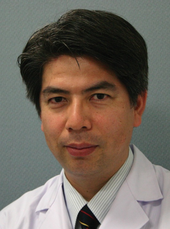 Masakazu Yashiro (八代正和)合作论文数Department of Clinical Medical Science, Graduate School of Medicine, Osaka Metropolitan University;Department of Medical Science, School of Medicine, Osaka Metropolitan University;Osaka City University Graduate School of Medicine3
 Hisamaru Hirai合作论文数Department of Hematology and Oncology, Graduate School of Medicine, University of Tokyo3
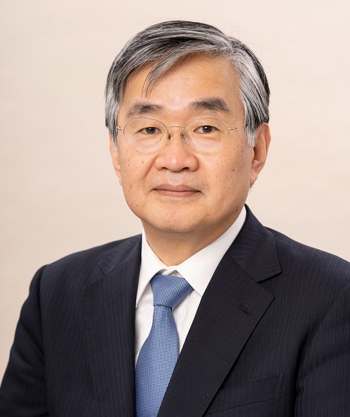 Kohei Miyazono (宮園浩平)合作论文数Graduate School of Medicine, The University of Tokyo;RIKEN3
 Mineo Kurokawa (黒川峰夫)合作论文数Department of Hematology and Oncology, The University of Tokyo Hospital;Department of Cell Therapy and Transplantation Medicine, The University of Tokyo Hospital3
 Atsushi Miwa合作论文数3S. Chiba合作论文数2K. Kumano合作论文数2Hasegawa Tsuyoshi合作论文数2Osamu Shinto合作论文数2Yuhiko Fuyuhiro合作论文数2Yoichi Imai合作论文数Graduate School of Medicine, University of Tokyo2Junko Matsuoka合作论文数2Shigeo Masuda合作论文数Center for Molecular Medicine, Jichi Medical University2Aki Hanyu合作论文数2S Matsuyama合作论文数1M Miyagishi合作论文数1Takeshi Imamura合作论文数1F Kanai合作论文数1Kouichiro Goto合作论文数1Hk Mishima合作论文数1Yutaka Ohsawa合作论文数1Tatsunari Fukuoka合作论文数1T Kawabe合作论文数1Yukihiro Kato合作论文数1Takafumi Nishii合作论文数1Tadashi Okada合作论文数1
 Toki Takahashi合作论文数Division of Developmental Biology and Medicine, School of Medical Sciences, Faculty of Biology, Medicine and Health, The University of Manchester1Ehata Shogo合作论文数Dept Mol Pathol, Univ Tokyo1
 Kunihiro Tsuchida (土田邦博)合作论文数Institute for Comprehensice Medical Sciences, Fujita Health University;Center for Medical Science, Fujita Health University1Aomatsu Naoki合作论文数1Morisaki Tamami合作论文数1
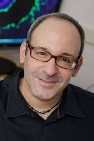 Martin J Cohn合作论文数Department of Molecular Genetics & Microbiology, College of Medicine, University of Florida1Iwadate Manabu合作论文数1Hirakawa Toshiki合作论文数1
 Masao Omata合作论文数Department of Gastroenterology, University of Tokyo1Tetsuji Sawada合作论文数1Kazunari Taira合作论文数1
 Makoto Uchino合作论文数1
 Yoshihiro Arakawa合作论文数1
 Toru Imamura合作论文数Signaling Molecules Research Laboratory, National Institute of Advanced Industrial Science and Technology (AIST),1
 Hiroyuki Aburatani (油谷浩幸)合作论文数Laboratory for Systems Biology and Medicine, Research Center for Advanced Science and Technology, University of Tokyo1
 Yoshihide Sunada合作论文数1Tsuneo Ikenoue合作论文数1Shuji Nakamura合作论文数1
 Keiji Miyazawa合作论文数Department of Biochemistry, University of Yamanashi1
 Sumihare Noji合作论文数Tokushima University1
 Frank Ruddle合作论文数Yale University1




