INTRODUCTION:Operating room (OR) noise during holmium laser enucleation of the prostate (HoLEP), particularly from laser activation, may impair surgical workflow and staff performance. METHODS:84 HoLEP procedures were recorded at three academic institutions between June 2024 and January 2026. Laser platforms included Lumenis Moses 2.0, Quanta Litho 150, Quanta Magneto, and Dornier Thulio. Sound measurements were obtained using a standardized smartphone-based system during three phases: laser-off (baseline), idle (not firing), and active lasing. Acoustic metrics included average sound level (LAeq), peak sound level (Lcpeak), and sound levels exceeded for 10%, 50%, and 90% of the recording time (L10, L50, and L90). Background noise correction was performed using baseline measurements to account for ambient noise and isolate laser-related sound. OR staff completed a validated survey assessing perceived noise burden (0-10 scale). RESULTS:Background-corrected noise during active lasing differed significantly by platform. Litho demonstrated lower mean noise levels compared to Thulio for LAeq (60.6 vs. 68.2 dB, p = 0.023) and L10 (61.5 vs. 70.6 dB, p = 0.023), while Moses had higher LAeq compared to Litho (63.1 vs. 60.6 dB, p = 0.043). Overall, Litho had the lowest noise levels, whereas Thulio had the highest. Survey responses paralleled these findings. Litho was associated with lower perceived overall effort (2.1 ± 0.9), while Moses demonstrated higher overall effort (3.6 ± 1.2) for communication and concentration (Figure 2). Significant pairwise differences were observed between Moses and Litho (all p < 0.05). Trends were consistent across sites. CONCLUSIONS:Noise levels during HoLEP varied by laser technology and correlated with differences in perceived communication and cognitive effort. Litho demonstrated the lowest acoustic burden, while Moses and Thulio had higher noise exposure. These findings highlight the impact of laser selection on the OR environment and intraoperative team performance.
Introduction: We aimed to assess the impact of discharge instruction (DCI) readability on 30-day postoperative contact with the healthcare system. Materials and methods: Utilizing a multidisciplinary team, DCI were modified for patients undergoing cystoscopy, retrograde pyelogram, ureteroscopy, laser lithotripsy, and stent placement (CRULLS) from a 13(th) grade to a 7(th) grade reading level. We retrospectively reviewed 100 patients including 50 consecutive patients with original DCI (oDCI) and 50 consecutive patients with improved readability DCI (irDCI). Clinical and demographic data collected including healthcare system contact (communications [phone or electronic message], emergency department [ED], and unplanned clinic visits) within 30 days of surgery. Uni/multivariate logistic regression analyses used to identify factors, including DCI-type, associated with increased healthcare system contact. Findings reported as odds ratios with 95% confidence intervals and p values (< 0.05 significant). Results: There were 105 contacts to the healthcare system within 30 days of surgery: 78 communications, 14 ED visits and 13 clinic visits. There were no significant differences between cohorts in the proportion of patients with communications (p = 0.16), ED visits (p =1.0) or clinic visits (p = 0.37). On multivariable analysis, older age and psychiatric diagnosis were associated with significantly increased odds of overall healthcare contact (p = 0.03 and p = 0.04) and communications (p = 0.02 and p = 0.03). Prior psychiatric diagnosis was also associated with significantly increased odds of unplanned clinic visits (p = 0.003). Overall, irDCI were not significantly associated with the endpoints of interest. Conclusions: Increasing age and prior psychiatric diagnosis, but not irDCI, were significantly associated with an increased rate of healthcare system contact following CRULLS.
ABSTRACT Introduction and Objective: Upper tract urothelial carcinoma (UTUC) represents 5% of all urothelial malignancies ( 1 – 3 ). Accurate pathologic diagnosis is key and may direct treatment decisions. Current ureteroscopic biopsy techniques include cold-cup, backloaded cold-cup and stone basket ( 4 – 6 ). The study objective was to compare a standard cold-cup biopsy technique to a novel cold-cup biopsy technique and evaluate histopathologic results. Materials and Methods: We developed a novel UTUC biopsy technique termed the “form tackle” biopsy. Ureteroscope is passed into ureter/renal collecting system. Cold-cup forceps are opened and pressed into the lesion base (to engage the urothelial wall/submucosal tissue) then closed. Ureteroscope/forceps are advanced forward 3-10mm and then extracted from the patient. We compared standard versus novel upper tract biopsy techniques in a series of patients with lesions ≥1cm. In each procedure, two standard and two novel biopsies were obtained from the same lesion. The primary study aim was diagnosis of malignancy. IRB approved: 21-006907. Results: Fourteen procedures performed on 12 patients between June 2020 and March 2021. Twenty-eight specimens sent (14 standard, 14 novel) (Two biopsies per specimen). Ten procedures with concordant pathology. In 4 procedures the novel biopsy technique resulted in a diagnosis of UTUC (2 high-grade, 2 low-grade) in the setting of a benign standard biopsy. Significant difference in pathologic diagnoses was detected between standard and novel upper tract biopsy techniques (p=0.008). Conclusions: The “form tackle” upper tract ureteroscopic biopsy technique provides higher tissue yield which may increase diagnostic accuracy. Further study on additional patients required. Early results are encouraging.
Introduction:Retrograde ureteroscopy with holmium laser lithotripsy (HLL) is a standard treatment for urolithiasis. Moses technology has been shown to improve fragmentation efficiency in vitro; however, it is still unclear how it performs clinically compared to standard HLL. We performed a systematic review and meta-analysis evaluating the differences in efficiency and outcomes between Moses mode and standard HLL.Material and methods:We searched the MEDLINE, EMBASE, and CENTRAL databases for randomized clinical trials and cohort studies comparing Moses mode and standard HLL in adults with urolithiasis. Outcomes of interest included operative (operation, fragmentation, and lasing times; total energy used; and ablation speed) and perioperative parameters (stone-free rate and overall complication rate).Results:The search identified six studies eligible for analysis. Compared to standard HLL, Moses was associated with significantly shorter average lasing time (mean difference [MD] -0.95, 95% confidence interval [CI] -1.22 to -0.69 minutes), faster stone ablation speed (MD 30.45, 95% CI 11.56-49.33 mm3/min), and higher energy used (MD 1.04, 95% CI 0.33-1.76 kJ). Moses and standard HLL were not significantly different in terms of operation (MD -9.89, 95% CI -25.14 to 5.37 minutes) and fragmentation times (MD -1.71, 95% CI -11.81 to 8.38 minutes), as well as stone-free (odds ratio [OR] 1.04, 95% CI 0.73-1.49) and overall complication rates (OR 0.68, 95% CI 0.39-1.17).Conclusions:While perioperative outcomes were equivalent between Moses and standard HLL, Moses was associated with faster lasing time and stone ablation speeds at the expense of higher energy usage.
OBJECTIVE To perform a cost-effectiveness evaluation comparing the management options for mid-size (1-2cm) renal stones including percutaneous nephrolithotomy (PCNL), retrograde intrarenal surgery (RIRS), and shockwave lithotripsy (SWL). METHODS A Markov model was created to compare cost-effectiveness of PCNL, mini-PCNL, RIRS, and SWL for 1-2cm lower pole (index patient 1) and PCNL, RIRS, and SWL for 1-2 cm non-lower pole (index patient 2) renal stones. A literature review provided stone free, complication, retreatment, secondary procedure rates, and quality adjusted life years (QALYs). Medicare costs were used. The incremental cost-effectiveness ratio (ICER) was compared with a willingness-to-pay (WTP) threshold of $100,000/QALY. One-way and probabilistic sensitivity analyses were performed. RESULTS At 3 years, costs for index patient 1 were $10,290(PCNL), $10,109(mini-PCNL), $5,930(RIRS), and $10,916(SWL). Mini-PCNL resulted in the highest QALYs(2.953) followed by PCNL (2.951), RIRS(2.946), and SWL(2.943). This translated to RIRS being most cost-effective followed by mini-PCNL(ICER $624,075/QALY) and PCNL(ICER $946,464/QALY). SWL was dominated with higher costs and lower effectiveness. For index patient 2, RIRS dominated both PCNL and SWL. For index patient 1: mini-PCNL and PCNL became cost effective if cost <=$5,940 and <=$5,390, respectively. SWL became cost-effective with SFR >= 75% or cost <=$1,236. On probabilistic sensitivity analysis, the most cost-effective strategy was RIRS in 97%, miniPCNL in 2%, PCNL in 1%, and SWL in 0% of simulations. CONCLUSION For 1-2cm renal stones, RIRS is most cost-effective. However, mini and standard PCNL could become cost-effective at lower costs, particularly for lower pole stones. Published by Elsevier Inc.
PURPOSE:Previously published studies have shown small prostate size, capsular perforation and intraoperative bladder distension are associated with failed trial without a catheter (TWOC) after HoLEP. The study objective was to determine the relationship between MOSES pulse modulation versus standard laser technology and short-term catheter reinsertion following failed TWOC. MATERIALS AND METHODS:The study included 487 patients who underwent HoLEP, using standard holmium laser settings (180 patients) or MOSES pulse modulation (255 patients), between August 2018 and February 2021. Catheter reinsertion defined as reinsertion following failed TWOC within 30 days of surgery. Association of pulse modulation with catheter reinsertion was examined using single and multivariable logistic regression models. Comparisons of pre and intraoperative characteristics between patients treated without and with pulse modulation were made using a Wilcoxon rank sum test for numeric characteristics or Fisher's exact test for categorical characteristics. RESULTS:Short-term catheter reinsertion occurred in 14% (26/180) of the standard laser setting group as compared with 10% (24/252) of the pulse modulation group. There was no statistically significant association with short-term catheter reinsertion in single (unadjusted OR [standard settings vs. pulse modulation], 1.60; 95% CI, 0.80-2.91; p=0.12) or multivariable analysis adjusting for specimen weight and operative time (adjusted OR [standard settings vs. pulse modulation], 1.44; 95% CI, 0.77-2.68; p=0.25). CONCLUSIONS:In this study, we found no association between post-HoLEP short-term catheter reinsertion following failed TWOC and MOSES pulse modulation. Although MOSES pulse modulation offers several well-documented advantages, catheter reinsertion events appear to be attributable to other factors.
You have accessJournal of UrologyImaging/Radiology: Uroradiology II (MP22)1 Sep 2021MP22-07 IN VIVO ASSESSMENT OF STONE FRAGILITY USING COMPUTED TOMOGRAPHY Dane Klett, Aaron Potretzke, Andrea Ferrero, Tristan Juvet, Marcelino Rivera, John Knoedler, Jayse Weaver, John Lieske, Felicity Enders, and Cynthia McCollough Dane KlettDane Klett More articles by this author , Aaron PotretzkeAaron Potretzke More articles by this author , Andrea FerreroAndrea Ferrero More articles by this author , Tristan JuvetTristan Juvet More articles by this author , Marcelino RiveraMarcelino Rivera More articles by this author , John KnoedlerJohn Knoedler More articles by this author , Jayse WeaverJayse Weaver More articles by this author , John LieskeJohn Lieske More articles by this author , Felicity EndersFelicity Enders More articles by this author , and Cynthia McColloughCynthia McCollough More articles by this author View All Author Informationhttps://doi.org/10.1097/JU.0000000000002013.07AboutPDF ToolsAdd to favoritesDownload CitationsTrack CitationsPermissionsReprints ShareFacebookLinked InTwitterEmail Abstract INTRODUCTION AND OBJECTIVE: Computed tomography (CT) is recommended for non-invasively imaging symptomatic urinary stones as it provides accurate submillimeter details of stone size and location within the urinary system. Previous ex vivo studies demonstrated routine dual-energy CT (DECT) stone protocols provide an accurate estimate of stone fragility by quantifying morphological features. The objective of this study was to validate the accuracy of a stone fragility model based on preoperative DECT to predict comminution time in patients undergoing percutaneous nephrolithotripsy (PCNL). METHODS: Patients received a DECT followed by PCNL using the ShockPulse SE ultrasonic lithotripter (Olympus). A quantitative model consisting of 7 DECT metrics, including stone volume, internal and surface morphology and stone composition, was developed using stones ex vivo stones and then applied in vivo to stone procedures. During the procedure, the time the ultrasonic lithotripter was actively engaged in stone breaking was measured. The observed and predicted comminution times for each stone were individually ranked from most to least fragile. Stone fragility classifications based on comminution time were then created: soft stones in the first quartile, hard stones in the last quartile and average stones in between. Postprocedure, the performing urologist subjectively assessed stone fragility, and was blinded to measured and estimated comminution times. RESULTS: A total of 11 stones from 10 patients were studied. With stones ranked by volume, the average ranking difference (predicted vs actual comminution time) was 1.8 with 4/11 having a ranking difference greater than 2. Using the stone fragility model, the average ranking difference was 0.7, with only one stone having a ranking difference greater than 2. The 3-class classification showed only 2 stones erroneously labeled using the predictive stone fragility model. The urologist's impression of stone fragility correlated well with comminution time-based classification. CONCLUSIONS: A predictive stone fragility model based on morphological and mineral characteristics of renal stones, as quantified on DECT, may accurately predict in vivo comminution time. Source of Funding: Grants DK100227 & EB028591 from the National Institute of Health © 2021 by American Urological Association Education and Research, Inc.FiguresReferencesRelatedDetails Volume 206Issue Supplement 3September 2021Page: e393-e393 Advertisement Copyright & Permissions© 2021 by American Urological Association Education and Research, Inc.MetricsAuthor Information Dane Klett More articles by this author Aaron Potretzke More articles by this author Andrea Ferrero More articles by this author Tristan Juvet More articles by this author Marcelino Rivera More articles by this author John Knoedler More articles by this author Jayse Weaver More articles by this author John Lieske More articles by this author Felicity Enders More articles by this author Cynthia McCollough More articles by this author Expand All Advertisement PDF downloadLoading ...
Background: Decidual reaction bladder endometriosis (DRBE) is exceedingly rare with few reported cases in the literature. It presents as a bladder mass during pregnancy, and may be accompanied by lower urinary tract symptoms. Histologic diagnosis is necessary to rule out primary bladder malignancy. We present a case of a bladder tumor identified during pregnancy. The mass was managed endoscopically and found to be DRBE, a rare benign entity. Case Presentation: We present a 31 year old 15 weeks pregnant nonsmoker woman with a rapidly enlarging bladder mass concerning for primary bladder malignancy. Mass confirmed on formal renal/bladder ultrasound and in-office cystoscopy. After informed consent was obtained, the patient was taken to the operating room. A 5.5 cm bladder mass, with an atypical nodular appearance and minor calcifications, was identified. Transurethral resection of the mass was performed. Final pathology report showed florid endometriosis with stromal decidualization. Final diagnosis: pregnancy induced vesical decidualized endometriosis simulating a bladder tumor. Patient continued routine obstetrics follow-up, and has experienced no pregnancy-related complications. Three months after delivery the patient will follow up with outside urology provider for cystoscopy, and subsequent surgical management should it be necessary. Conclusion: DRBE is a rare benign bladder mass that presents in pregnancy. It can grow rapidly raising concern for an aggressive primary bladder malignancy. Any bladder mass identified in pregnancy should undergo early, appropriate work-up given the potential risk for bladder cancer. After diagnosis, DRBE is most often managed conservatively. After delivery, should the patient experience ongoing urinary symptoms, medical and surgical treatment options are available. Overall, DRBE is considered rare, but should be considered in the differential diagnosis for any bladder mass presenting during pregnancy.
Background: Iatrogenic ureteral injury represents an uncommon, but significant, complication of gynecologic surgery. Endoscopy has typically played little to no role in the treatment of these injuries, which are traditionally managed with re-exploration or delayed repair. Delayed repair with temporary urinary diversion exposes the patient to significant morbidity. We present a case in which iatrogenic ureteral injury is managed definitively with endoscopy alone. Case Presentation: We present a 32-year-old female who developed a delayed postpartum hemorrhage following cesarean section, necessitating emergent hysterectomy. Postoperatively, there was concern for right ureteral injury. A computed tomography (CT) urogram was obtained showing right-sided hydronephrosis, but no obvious ureteral injury. After developing right flank pain, the patient was taken to the operating room for further evaluation. On semirigid ureteroscopy, a suture was identified within the lumen of the ureter and incised with the holmium laser, effectively treating the obstruction. At a 10-week follow-up, a renal ultrasound showed no hydronephrosis. At 8 months, the patient reports she is doing well with no flank pain. Conclusion: We present, to the best of our knowledge, the first published report in the United States of an iatrogenic ureteral ligation managed effectively in an acute postoperative setting with endoscopic holmium laser release, without balloon dilation, sparing the patient from delayed surgical intervention and the potentially associated morbidity. It is our belief that an initial retrograde pyelogram followed by a ureteroscopic evaluation should be performed as this allows for proper characterization of the injury, and may allow one to attempt definitive endoscopic management.
Purpose: To describe a novel modification to robot-assisted partial cystectomy (RAPC) that allows for intraoperative surgical margin assessment by bimanual-examination and frozen-section analysis. Materials and Methods: A total of 7 patients underwent RAPC at a single tertiary-care institution between 2008 and 2013. The technique evolved over the study-period and permitted real-time intraoperative surgical margin evaluation in the last 5 patients via bimanual-examination and frozen-section analysis, utilizing the GelPOINT platform (a hand-assist device). The GelPOINT platform was placed through a 4- to 5-cm vertical supraumbilical incision and allowed for rapid retrieval of the bladder specimen without compromising the pneumoperitoneum or prolonging the operative time. Perioperative, oncological and functional outcomes were evaluated; all patients had a minimum 12-month follow-up. At the time of last follow-up, a cross-sectional survey of patients was performed to evaluate regret/satisfaction utilizing validated questionnaires. Results: The mean age was 72.5 years; 71.4% of the patients were men (n=5). All patients underwent RAPC for a malignant indication. The mean operative and console times were 291 and 217 minutes, respectively. No patient had a positive surgical margin. Mean length-of-stay was 1.7 days. At a median follow-up of 38.9 months, 1 patient experienced a local recurrence 6 months postsurgery. The only mortality was secondary to Lewy-body disease, in the same patient, 1 year postoperatively. Patient assessment of regret and satisfaction indicated 0% regret and 0% dissatisfaction. Conclusions: The ‘modified’ technique of RAPC is technically feasible, safe, and reproducible; further, RAPC leads to favorable oncological, functional and quality-of-life outcomes in patients eligible for partial cystectomy.
INTRODUCTION:To develop a nomogram to predict lymph node invasion (LNI) in the contemporary North American patient treated with robot-assisted radical prostatectomy (RARP).MATERIALS AND METHODS:We included 2,007 patients treated with RARP and pelvic lymph node dissection (PLND) at a single institution between 2008 and 2012. D'Amico low risk patients underwent an obturator and hypogastric PLND, while extended PLND was reserved for intermediate/high risk patients. Logistic regression analysis tested the relationship between LNI and all available predictors. Independent predictors of LNI were used to develop a novel nomogram. Discrimination, calibration and decision-curve analysis were used to analyze the performance of our novel nomogram, and compare it to open radical prostatectomy (ORP)-based models, namely the Godoy nomogram.RESULTS:Overall, 5.3% of our patients harbored LNI. Median number of lymph nodes removed was 6.0 (interquartile range: 4-11). The most parsimonious multivariable model to predict LNI consisted of the following independent predictors: PSA value, clinical stage, and primary and secondary Gleason scores (all p ≤ 0.02). The discrimination of our novel model was 86.2%, and its calibration was virtually optimal. Using a 2% nomogram cut off, 58% of patients would be spared PLND, while missing only 9.4% of individuals with LNI. The novel nomogram compared favorably to the Godoy nomogram, when discrimination, calibration and net-benefit were used as benchmarks.CONCLUSIONS:Approximately 5% of contemporary North American patients harbor LNI at RARP. Our novel nomogram can accurately identify these patients, and this may help to improve patient selection, and avoid unnecessary PLND in the majority of patients.
Fertility preservation is often overlooked in patient counseling following cancer diagnosis. The aim of this study was to investigate the availability and quality of patient directed information on male fertility preservation on the web sites of National Cancer Institute (NCI) designated cancer centers. All NCI designated cancer center web sites were queried in a systematic fashion for patient directed information on oncofertility. A rubric was employed to establish minimum content quality standards for the validation process. Independent research teams from two institutions validated data collected from each web site. Descriptive statistical analysis and chi-squared testing were performed. Patient directed information available on the web site of each NCI designated cancer center was evaluated for its oncofertility content. Specific questions included: 1) Does the web site discuss the effects of cancer and cancer treatment on fertility, particularly male fertility? 2) Are options for fertility preservation discussed? 3) Is there a standalone page dedicated to educating patients on fertility preservation? 4) Was parenting-related cancer survivorship addressed? Two independent research teams validated the data against a rubric of content quality standards. Descriptive analysis was performed. Data were then evaluated based on each cancer center's state population density to test whether there was a demographic effect on oncofertility information. All 62 web sites of NCI designated cancer centers were evaluated [1]; 92% were affiliated with academic institutions. Among all cancer centers, 34% made no mention of male-specific fertility risks. The risk of cancer treatment on fertility (not gender specific) was mentioned by 84% of centers. Of these, 67% have a standalone page on the topic of oncofertility (not gender specific). Among all cancer centers, only 60% include information on fertility preservation specifically directed toward men, such as sperm cryopreservation. Survivorship information on family building after cancer was available on 32% of cancer center web sites. State population density had no significant effect on whether a web site included risks of treatment on fertility (p=0.90) or information on fertility preservation (p=0.29). Forty percent of NCI designated cancer center web sites do not discuss options for male fertility preservation, and over one-third make no mention of the ramifications of cancer treatment on male fertility. Given the increasing recognition of the importance of oncofertility in cancer survivorship, more education should be available about options for fertility preservation, particularly among men.
 Jesse D Sammon合作论文数43
 Firas Abdollah合作论文数39
 Mani Menon合作论文数Vattikuti Urology Institute, Henry Ford Hospital39
 Quoc-Dien Trinh合作论文数School of the Health Sciences, University of Pittsburgh;Department of Urology, UPMC33
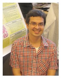 Sood Akshay合作论文数Center for Outcomes Research, Analytics and Evaluation, Vattikuti Urology Institute31
 James O Peabody合作论文数Vattikuti Urology Institute, Henry Ford Hospital27
 Daniel Pucheril合作论文数17
 Adam Stuart Kibel合作论文数Department of Urology, Brigham and Women’s Hospital;Dana-Farber Brigham Cancer Center14
 Wooju Jeong合作论文数Vattikuti Urology Institute, Henry Ford Hospital/Henry Ford Cancer Institute, Henry Ford Hospital13Schmid Marianne合作论文数10
 Anil K. Sood合作论文数Department of Gynecologic Oncology and Reproductive Medicine, Division of Surgery, The University of Texas MD Anderson Cancer Center;Graduate School of Biomedical Sciences, The University of Texas Health Science Center10
 Gandaglia Giorgio合作论文数Vita-Salute San Raffaele University;Department of Urology, San Raffaele Hospital6
 Francesco Montorsi合作论文数Department of Urology, San Raffaele Hospital;Vita-Salute San Raffaele University of Milan6
 Pucheril Dan合作论文数6
 Alberto Briganti合作论文数Faculty of Medicine, Vita-Salute San Raffaele University;Department of Urology, IRCCS San Raffaele Hospital6
 Craig Rogers合作论文数Vattikuti Urology Institute, Henry Ford Hospital6Hamilton B.D.合作论文数5
 Julian Hanske合作论文数5
 Paul L. Nguyen合作论文数Brigham and Women's Hospital;75 Francis Street;Boston, MA 02115/Dana-Farber Cancer Institute;450 Brookline Avenue;Boston, MA 022155
 Briony Varda合作论文数Department of Urology, University of Minnesota5Maxine Sun合作论文数Montreal5Nakada S.Y.合作论文数4Gerber G.S.合作论文数4Nicola Fossati合作论文数4Aaron M Potretzke合作论文数4
 Kevin Wymer合作论文数4
 Burkhard Beyer合作论文数4
 Deepansh Dalela合作论文数4
 Ali A. Dabaja合作论文数4Hans Stricker合作论文数4R.G.N. Seltzer合作论文数3Victor Kapoor合作论文数3
 Manaf Alom合作论文数Dept Urol, Mayo Clin3
 Sriram Eleswarapu合作论文数Div Androl, Univ Calif Los Angeles3
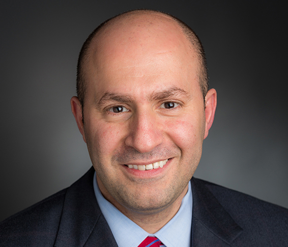 Toni Choueiri合作论文数Lank Center for Genitourinary (GU) Oncology, Dana-Farber Cancer Institute;Harvard Medical School;Brigham and Women's Hospital3Diaz Mireya合作论文数3
 Jack S. Elder合作论文数Case Western University School of Medicine;Division of Pediatric Urology, MassGeneral Hospital for Children3
 Alexander Haese合作论文数3
 Nazareno Roberto Suardi合作论文数Department of Surgical and Diagnostic Integrated Sciences (DISC), Division of Urology, University of Genoa3Markus Graefen合作论文数3Matthew H. Hayn合作论文数3
 Christian Meyer合作论文数2D. Dalela合作论文数2Christian Paul Meyer合作论文数2Tristan Juvet合作论文数2Z. Chen合作论文数2
 Jim C. Hu合作论文数Department of Urology, Weill Cornell Medical College, Cornell University;NewYork-Presbyterian Hospital2
 Gally Reznor合作论文数Boston Children's Hospital, Harvard Medical School2
 Marcelino Rivera合作论文数2




