Mutation-associated neoantigens (MANAs) are highly cancer-specific targets for immunotherapy where peptides derived from intracellular mutant proteins are presented on the cell surface via HLA molecules. T cell-engaging bispecific antibodies and CAR T cells can target MANAs to eliminate cancer cells via T cell activation. However, the low antigen density of MANAs on the cell surface can limit therapeutic efficacy. Here, we investigated whether increasing the affinity of the H2 single-chain variable fragment (scFv) targeting the p53 R175H MANA (HMTEVVRHC presented on HLA-A*02:01) improves its therapeutic effect. We identified higher-affinity H2 variants via phage biopanning and a thiocyanate elution method. Increasing bispecific antibody affinity to the low nanomolar range increased cancer cell killing and tumor control in mouse xenograft models without sacrificing antigen specificity. We next asked how increasing scFv affinity impacts CAR T cell function - a matter of debate. We appended each variant scFv to a CD28z CAR, CD3γ, or the T cell receptor. In striking contrast to the bispecific antibody results, increasing CAR affinity decreased function in each CAR format due to lower T cell activation upon interaction with target cancer cells. These results have important implications for the design of future immunotherapeutic approaches targeting low-density antigens.
Lethal toxins could become potent therapies against cancer, but their clinical utility is limited by adverse events upon systemic administration. These could be reduced if the toxins were delivered by effector cells that specifically infiltrate cancers, thereby releasing toxins locally into the tumor microenvironment. One of the challenges underlying this strategy is that cells delivering toxins would have to be resistant to them. We address this obstacle by showing that effectors derived from transformed human cell lines genetically engineered for resistance to bacterial adenosine diphos-phate ribosylating toxins (ADPRTs), including Pseudomonas aeruginosa exotoxin A (PE), can produce targeted immunotoxins that specifically kill cancer cells express-ing cognate tumor-associated antigens. Resistance to immunotoxins was achieved by knockout of genes in the diphthamide biosynthesis pathway (DPH1-4) required for the posttranslational modification of eukaryotic elongation factor 2 (EEF2) that is the target of ADPRTs or by mutation of EEF2 itself. We show that engineering resist-ance to ADPRTs, one of the most potent toxins acting on human cells, is essential to achieve robust function of armored effector cell lines. This work establishes a critical step on the path to equip effector cells with the ability to deliver powerful toxins to cancer cells and introduces a platform to investigate extension to primary autologous or allogeneic therapeutic cell types.
Efficient, cell type-selective delivery of genetic payloads remains a central challenge in the development of gene and cell therapies. Lipid nanoparticles (LNPs) offer a versatile delivery platform, but their optimization is hindered by reliance on brute-force screening methods that are laborious, resource-intensive, and focus on single targets. Here, we present FALCON (Framework for Active Learning-driven Compositional Optimization of Nanoparticles), a closed-loop pipeline that leverages iterative screening, surrogate modeling, and multi-objective optimization to accelerate LNP compositional design. In B cell-targeted validation experiments, FALCON-optimized LNPs achieved a 1.8-fold increase in splenic B cell transfection in vivo compared with reference compositions. When optimized for selectivity, FALCON LNPs displayed an 84-fold improvement in selective transfection of splenic B cells over off-target liver populations and enabled spleen-tropic behavior across factorial panels of varying ionizable and helper lipid chemistries. In vaccine studies, these LNPs induced higher IgG2c antibody titers and a more Th1-biased immune profile. FALCON was also deployed to optimize LNPs for myeloid cell-selective delivery, achieving enhanced in vivo selectivity following systemic administration both across and within spleen and liver compartments. Our results establish FALCON as a useful tool for data-driven design of LNP compositions for precision gene delivery.
While chimeric antigen receptor (CAR) T cell therapies have demonstrated therapeutic efficacy against B cell malignancies, widespread implementation of these therapies is hindered by a cumbersome, ex vivo manufacturing process. Delivery of CAR-encoding messenger RNA (mRNA) to endogenous T cells can generate these therapeutic cells in vivo and streamline this manufacturing workflow. To accomplish this, T cell-activating ligands were conjugated to a biodegradable polymeric mRNA nanoparticle to form T cell-targeted particles. By conjugating multiple activating ligands, T cell transfection and stimulation in vitro was increased, and greater T cell transfection and selectivity in vivo was achieved compared to an untargeted particle. These nanoparticles can flexibly encapsulate mRNA cargos and were used to deliver anti-CD19 CAR mRNA in vivo, enabling depletion of 95% of B cells in the peripheral blood and 50% depletion of splenic B cells in healthy mice. These findings regarding nanoparticle tropism and their potential therapeutic efficacy highlight the importance of this nonviral, polymeric platform to address key limitations associated with current CAR T practices.
Patients with radiographically detectable lesions in their brain or other symptoms compatible with brain tumors pose challenges for diagnosis. The only definitive way to diagnose such patients is through brain biopsy, an invasive and dangerous procedure. In this study, we present a new workflow termed "CSF-BAM" that simultaneously identifies B-cell or T-cell receptor sequences, aneuploidy, and mutations using amplification of both strands of the DNA from cerebrospinal fluid (CSF) samples. We applied CSF-BAM to a validation set of 209 samples from patients with brain cancers. Among the 129 samples from patients with the most common aggressive cancer types, the sensitivity of detection was 81%. None of 30 CSF-BAM assays were positive in CSF samples from patients without brain cancers (100% specificity). CSF-BAM provides an integrated approach to identify neoplasia in the central nervous system, provides information about the genetics and immune environment, and has the potential to inform patient management. SIGNIFICANCE:There is a paucity of technologies beyond surgical biopsy that can accurately diagnose central nervous system neoplasms. We developed a novel, sensitive, and highly specific assay that can detect brain cancers by comprehensively identifying somatic mutations, chromosomal copy-number changes, and adaptive immunoreceptor repertoires from samples of CSF. See related commentary by Weiss, p. 1976.
Supplementary Table S1: Cohorts for CSF-BAM components. Supplementary Table S2A: SafeBSeqS primer sequences. Supplementary Table S2B: SafeTseqS primer sequences. Supplementary Table S2C: Aneuploidy analysis primer sequences. Supplementary Table S2D: Mutation analysis primer sequences. Genomic coordinates refer to hg19. Supplementary Table S3: Summary of SafeBSeqS analysis in WBC control samples. Supplementary Table S4: Summary of CSF-BAM SafeBSeqS analysis in CSF. Supplementary Table S5: Summary of SafeTSeqS analysis in WBC control samples. Supplementary Table S6: Summary of CSF-BAM SafeTSeqS analysis in CSF. Supplementary Table S7: SafeBSeqS and SafeTSeqS analysis for CSF validation set 2. Supplementary Table S8: Summary of aneuploidy analysis in control samples. Supplementary Table S9: Summary of CSF-BAM aneuploidy analysis in CSF samples. Supplementary Table S10: Reproducibility of aneuploidy analysis. Supplementary Table S11: Comparison of aneuploidy analysis with CSF-BAM and Real-CSF. Supplementary Table S12: CSF-BAM mutation training set in non-cancer CSF samples. Supplementary Table S13: CSF-BAM mutation analysis in CSF samples. Supplementary Table S14: Demographic characteristics by sample. Supplementary Table S15: Diagnostic categories by sample. Supplementary Table S16: CSF-BAM results summary. Supplementary Table S17: CSF-BAM detection based on CSF reservoir abutment by sample. All samples with available clinical data are included. P-values were calculated by Fisher’s exact test. Supplementary Table S18A: TCR Clusters obtained via CSF-BAM. Supplementary Table S18B: TCR specificity annotation. Supplementary Table S19: CSF samples from patients with multiple sclerosis analyzed with SafeTSeqS and SafeBSeqS.
Multiple case-controlled studies have shown that analyzing fragmentation patterns in plasma cell-free DNA (cfDNA) can distinguish individuals with cancer from healthy controls. However, there have been few studies that investigate various types of cfDNA fragmentomics patterns in individuals with other diseases. We therefore developed a comprehensive statistic, called fragmentation signatures, that integrates the distributions of fragment positioning, fragment length, and fragment end-motifs in cfDNA. We found that individuals with venous thromboembolism, systemic lupus erythematosus, dermatomyositis, or scleroderma have cfDNA fragmentation signatures that closely resemble those found in individuals with advanced cancers. Furthermore, these signatures were highly correlated with increases in inflammatory markers in the blood. We demonstrate that these similarities in fragmentation signatures lead to high rates of false positives in individuals with autoimmune or vascular disease when evaluated using conventional binary classification approaches for multicancer earlier detection (MCED). To address this issue, we introduced a multiclass approach for MCED that integrates fragmentation signatures with protein biomarkers and achieves improved specificity in individuals with autoimmune or vascular disease while maintaining high sensitivity. Though these data put substantial limitations on the specificity of fragmentomics-based tests for cancer diagnostics, they also offer ways to improve the interpretability of such tests. Moreover, we expect these results will lead to a better understanding of the process-most likely inflammatory-from which abnormal fragmentation signatures are derived.
Early data have shown the potential of chimeric antigen receptor (CAR) T-cell therapies to expand the therapeutic landscape in systemic lupus erythematosus (SLE). While many CAR T-cell therapy learnings can be drawn from the experience of this modality in oncology, key questions remain regarding clinical development considerations unique to lupus. To assess and discuss these issues, the Lupus Accelerating Breakthroughs Consortium, a public–private partnership, convened a multi-partner working group to collect the diverse perspectives of academics/clinicians (including rheumatologists and oncologists), industry representatives (including SLE as well as CAR T-cell clinical development experts), regulators and people living with lupus on this potentially ground-breaking therapy. The working group considered the risk/benefit considerations for eligibility criteria in lupus, early-phase dosing and dose-limiting toxicity challenges, incorporation of comparator arms in late-phase registrational trial design, SLE-specific issues in conditioning therapy and immune monitoring and the limitations of SLE pre-clinical models for studying cell therapies. The key future ‘calls to action’ for the field include the need for well-defined severity/refractoriness-based eligibility criteria, the need for long-term monitoring infrastructure and the need for educational and logistical support for rheumatologists and patients.
Supplementary Figure S1. SafeBSeqS amplification. Supplementary Figure S2. SafeTSeqS amplification. Supplementary Figure S3. Aneuploidy reproducibility from two independent aliquots and libraries. Supplementary Figure S4. Metrics for targeted mutation panel. Supplementary Figure S5. Correlation of predicted aneuploidy neoplastic content to mutation neoplastic content. Supplementary Figure S6. A, TCR UIDs and B, BCR UIDS recovered for each CSF sample. Supplementary Figure S7. A, TCR clonality and B, BCR clonality for evaluable samples with total UIDs ≥20. Supplementary Figure S8. BCR clonality ROC and BCR IGHV4-34 gene segment usage. Supplementary Figure S9. Case reports demonstrating CSF-BAM clinical applicability.
Antibody-drug conjugates (ADCs) have been remarkably successful in treating solid and hematological malignancies. Generation of ADCs for T cell cancers is challenging because the ADCs must selectively target cancerous T cells while sparing some normal T cells necessary for immune function. T cells express one of two TRBC alleles: TRBC1 or TRBC2. Normal T cells are composed of about 40% TRBC1-expressing and 60% TRBC2-expressing cells. In contrast, T cell malignancies are characterized by the clonal expression of either TRBC1 or TRBC2. Selective targeting of TRBC1 or TRBC2 enables the killing of cancer cells but preserves about 60-40% of the normal T cells. To enable such a therapy for cancers expressing TRBC2, here we developed a high-affinity anti-TRBC2 antibody. An ADC generated with this antibody and a pyrrolobenzodiazepine dimer payload showed specific killing of TRBC2+ cancers in vitro and in mouse models. The anti-TRBC2 ADC provides a promising, off-the-shelf therapy for patients with T cell cancers.
Chimeric antigen receptor (CAR)-T cell therapies that broadly target B cells can achieve complete remission in severe systemic lupus erythematosus (SLE) but carry an increased risk of infection and cytokine-related toxicities that limit their use. Alternative strategies that combine the potency of immune effector cell therapies with more precise targeting approaches have the potential to control disease without the challenges of broad immunosuppression. B cells expressing immunoglobulin heavy variable gene 4-34 (IGHV4-34)-derived B cell receptors (BCRs) are a major source of disease-relevant autoantibodies in lupus and other autoimmune diseases. Here, we exploit a common feature of IGHV4-34 BCRs, namely the 9G4 idiotope (9G4id), to develop precision cellular immunotherapies that target autoreactive B cells. Anti-9G4 CAR-T cells and anti-9G4 chimeric T cell receptor (cTCR) T cells, integrating anti-9G4 antibody fragments into a re-engineered TCR scaffold, eliminated autoreactive Ramos B cells expressing SLE patient-derived 9G4id BCRs with equal potency, while sparing non-9G4 Ramos B cells. Using SLE patient PBMCs, autologous anti-9G4 cTCR-T cells and anti-9G4 CAR-T cells selectively depleted primary human 9G4id B cells, while maintaining total B cell numbers. Similarly, anti-9G4 T cells eliminated B cells expressing 9G4id BCRs from patients with cold agglutinin disease and Burkitt lymphoma. Compared to broad targeting with CD19 CAR-T cells, anti-9G4 T cell therapies showed lower cytokine release (~6-8-fold for interferon [IFN]-γ). In addition, anti-9G4 cTCR-T cells showed ~17-fold lower IFN-γ secretion compared to anti-9G4 CAR-T cells, despite achieving similar cytotoxicity. Together, our findings suggest that anti-9G4 precision cellular therapies provide a strategy to selectively target pathogenic B cells in SLE, while minimizing risks of infection and cytokine-related toxicities.
Lethal toxins could become potent therapies against cancer, but their clinical utility is limited by adverse events upon systemic administration. These could be reduced if the toxins were delivered by effector cells that specifically infiltrate cancers, thereby releasing toxins locally into the tumor microenvironment. One of the challenges underlying this strategy is that cells delivering toxins would have to be resistant to them. We address this obstacle by showing that effectors derived from transformed human cell lines genetically engineered for resistance to bacterial adenosine diphosphate ribosylating toxins (ADPRTs), including Pseudomonas aeruginosa exotoxin A (PE), can produce targeted immunotoxins that specifically kill cancer cells expressing cognate tumor-associated antigens. Resistance to immunotoxins was achieved by knockout of genes in the diphthamide biosynthesis pathway ( DPH1-4 ) required for the posttranslational modification of eukaryotic elongation factor 2 (EEF2) that is the target of ADPRTs, or by mutation of EEF2 itself. We show that engineering resistance to ADPRTs, one of the most potent toxins acting on human cells, is essential to achieve robust function of armored effector cell lines. This work establishes a first step on the path to equip effector cells with the ability to deliver powerful toxins to cancer cells and introduces a platform to investigate extension to primary autologous or allogeneic therapeutic cell types.
Proteinase 3 (PR3)-specific antineutrophil cytoplasmic antibody (ANCA)-associated vasculitis is one of two major ANCA-associated vasculitis variants and is pathogenically linked to granulomatosis with polyangiitis (GPA). GPA is characterised by necrotising granulomatous inflammation that preferentially affects the respiratory tract. The small vessel vasculitis features of GPA are shared with microscopic polyangiitis. Necrotising granulomatous inflammation of GPA can lead to PR3-ANCA and small vessel vasculitis via activation of neutrophils and monocytes. B cells are central to the pathogenesis of PR3-ANCA-associated vasculitis. They are targeted successfully by remission induction and maintenance therapy with rituximab. Relapses of PR3-ANCA-associated vasculitis and toxicities associated with current standard therapy contribute substantially to remaining mortality and damage-associated morbidity. More effective and less toxic treatments are sought to address this unmet need. Advances with cellular and novel antigen-specific immunotherapies hold promise for application in autoimmune disease, including PR3-ANCA-associated vasculitis. This Series paper describes the inter-related histopathological and clinical features, pathophysiology, as well as current and future targeted treatments for PR3-ANCA-associated vasculitis.
Gene therapies and cellular programming rely on effective cell transfection. Despite continuous advancements in carrier development and transfection techniques to enhance efficiency, the biophysical parameter of extracellular fluid viscosity has been largely overlooked. Here we report a substantial impact of culture media viscosity on transfection efficiency of several delivery vehicles, including lipid nanoparticles, polyplexes, adeno-associated vectors and lentiviral vectors across a range of cell types. We observed substantially increased transfection efficiencies for lipid nanoparticles and polyplexes when the media viscosity matched that of biological fluids (2.0-4.0 centipoise (cP)). This enhancement correlates with higher levels of cellular uptake and improved endosomal escape. Moreover, cells cultured in optimized viscosity conditions exhibit a different profile of uptake pathways compared with those cultured at the standard viscosity of 0.8 cP. This discovery highlights the critical role of media viscosity in the transfection process and provides an additional method to optimize gene delivery and cell programming processes, potentially reducing production costs and increasing the accessibility of gene and cell therapies.
Abstract Background: Oncogenic mutations in KRAS (mKRAS) are expressed in up to 90% of pancreatic ductal adenocarcinomas (PDAC) therefore representing a promising immunotherapeutic target. We developed a pooled mKRAS peptide vaccine targeting the 6 most common mutations in PDAC: G12V, G12A, G12C, G12R, G12D, or G13D (NCT04117087). In a phase I study, we evaluated mKRAS-specific T cell responses raised by the vaccine when given in combination with immune checkpoint inhibitors to PDAC patients following resection and adjuvant chemotherapy. Materials and Methods: Patients received the pooled mKRAS peptide vaccine (0.3mg/peptide and 0.5mg poly-ICLC weekly for 4 doses in combination with ipilimumab and nivolumab followed by boosters every 8 weeks with nivolumab. To detect vaccine-induced mKRAS-specific T cells, pre- and post-vaccine peripheral blood mononuclear cells (PBMCs) were restimulated with control or individual mKRAS peptides and IFNγ release was measured by ELISPOT. mKRAS-specific T cell activation, proliferation, memory, and exhaustion profiles were assessed by CyTOF and cytokine secretion was determined by ELISA. For a subset of patients, mKRAS-specific T cells were expanded in vitro and T cell receptor (TCR) β chain sequencing and scRNA/TCRseq were used to identify the KRAS mutation-specific TCR repertoires and define their phenotypes in circulation. Results: mKRAS-specific T cells were detected by IFNγ ELISPOT post-vaccination for all patients, although magnitude and mutation-specific responses varied between patients. CyTOF analysis identified mKRAS-specific Th1 CD4+ central memory (cm, CCR7+IFNγ+) and effector memory (em, CCR7-IFNγ +IL2+ TNFɑ+) responses as well as CD8+ effector (eff, CD137+GZMBhiKi67hi) and effector memory (CCR7-CD137+GZMB+) responses. mKRAS-specific T cells were also detectable in the extended booster phase of treatment (>1 year post initial vaccination). In vitro expanded mKRAS-specific T cells mapped to CD4+ Tcm/em and CD8+ Tem populations within single cell datasets of unstimulated PBMCs collected post-vaccine. 5-25% of the mKRAS-specific T cells significantly expanded to more than one mKRAS antigen suggesting potential cross-reactive T cell clonotypes. In addition, shared public mKRAS-specific T cell clonotypes were identified across patients. Conclusions: This study demonstrates mKRAS vaccine induction of de novo, high quality mKRAS-specific polyfunctional CD4+ and CD8+ T cells. mKRAS-specific TCR analysis identified shared public TCRs across vaccinated patients that have important implications for future “off the shelf” adoptive therapy approaches. Ongoing studies are aimed to validate antigen specificity and map HLA-restriction of mKRAS-specific T cells. Overall, this study demonstrates a positive immunogenic signal of this pooled mutant KRAS vaccine that may be amenable for interception vaccination strategies against PDAC. Citation Format: Amanda L. Huff, Alex Girgis, Saurav D. Haldar, Emily Davis-Marcisak, Thatcher Heumann, Gabriella Longway, Lalitya Andaloori, Alexei Hernandez, Maximilian F. Konig, Brian Mog, Ludmila Danilova, Luciane T. Kagohara, Julie M. Nauroth, Amy M. Thomas, Elana J. Fertig, Won Jin Ho, Elizabeth M. Jaffee, Nilofer Azad, Neeha Zaidi. An off-the-shelf vaccine activates mutant KRAS-specific T cells in patients with resected pancreatic cancer [abstract]. In: Proceedings of the American Association for Cancer Research Annual Meeting 2024; Part 1 (Regular Abstracts); 2024 Apr 5-10; San Diego, CA. Philadelphia (PA): AACR; Cancer Res 2024;84(6_Suppl):Abstract nr 1170.
Two types of engineered T cells have been successfully used to treat patients with cancer, one with an antigen recognition domain derived from antibodies [chimeric antigen receptors (CARs)] and the other derived from T cell receptors (TCRs). CARs use high-affinity antigen-binding domains and costimulatory domains to induce T cell activation but can only react against target cells with relatively high amounts of antigen. TCRs have a much lower affinity for their antigens but can react against target cells displaying only a few antigen molecules. Here, we describe a new type of receptor, called a Co-STAR (for costimulatory synthetic TCR and antigen receptor), that combines aspects of both CARs and TCRs. In Co-STARs, the antigen-recognizing components of TCRs are replaced by high-affinity antibody fragments, and costimulation is provided by two modules that drive NF-κB signaling (MyD88 and CD40). Using a TCR-mimic antibody fragment that targets a recurrent p53 neoantigen presented in a common human leukocyte antigen (HLA) allele, we demonstrate that T cells equipped with Co-STARs can kill cancer cells bearing low densities of antigen better than T cells engineered with conventional CARs and patient-derived TCRs in vitro. In mouse models, we show that Co-STARs mediate more robust T cell expansion and more durable tumor regressions than TCRs similarly modified with MyD88 and CD40 costimulation. Our data suggest that Co-STARs may have utility for other peptide-HLA antigens in cancer and other targets where antigen density may limit the efficacy of engineered T cells.
 Bettegowda Chetan合作论文数The Johns Hopkins University35
 Bert Vogelstein合作论文数Department of Molecular Biology and Genetics, Johns Hopkins School of Medicine32
 Shibin Zhou合作论文数Department of The Sidney Kimmel Comprehensive Cancer Center, School of Medicine, Johns Hopkins University26
 Nickolas Papadopoulos合作论文数Department of Oncology, School of Medicine, Johns Hopkins University26
 Suman Paul合作论文数Johns Hopkins Univ, Sidney Kimmel Comprehens Canc Ctr, Baltimore, MD USA25
 Alexander H Pearlman合作论文数25
 Kenneth W. Kinzler合作论文数Department of Oncology, School of Medicine, Johns Hopkins University;The Sidney Kimmel Comprehensive Cancer Center, Johns Hopkins University School of Medicine23Brian J Mog合作论文数Johns Hopkins Univ, Ludwig Ctr, Sidney Kimmel Comprehens Canc Ctr, Sch Med, Baltimore, MD 21287 USA22
 Felipe Andrade合作论文数Division of Rheumatology, Department of Medicine, Johns Hopkins University21
 Drew Mark Pardoll合作论文数Department of Oncology, School of Medicine, Johns Hopkins University;Bloomberg~Kimmel Institute for Cancer Immunotherapy, School of Medicine, Johns Hopkins University;Sidney Kimmel Comprehensive Cancer Center, School of Medicine, Johns Hopkins University17DiNapoli Sarah R合作论文数15Michael S Hwang合作论文数Johns Hopkins Kimmel Canc Ctr, Ludwig Ctr, Baltimore, MD 21231 USA14Jacqueline Douglass合作论文数Johns Hopkins Univ, Sch Med, Sidney Kimmel Comprehens Canc Ctr, Ludwig Ctr, Baltimore, MD 21287 USA13
 Stephanie Glavaris合作论文数School of Medicine, Johns Hopkins University12Emily Han-Chung Hsiue合作论文数Ludwig Center and Howard Hughes Medical Institute, Sidney Kimmel Comprehensive Cancer Center, Johns Hopkins University School of Medicine, Baltimore, MD;12
 Sandra Gabelli合作论文数Merck12
 Joshua T. Vogelstein合作论文数Department of Biomedical Engineering, Whiting School of Engineering, Johns Hopkins University;Department of Applied Mathematics and Statistics, Whiting School of Engineering, Johns Hopkins University10Katharine M Wright合作论文数9
 Elizabeth R. Graef合作论文数9
 Kim Alfred H J合作论文数9Philip C. Robinson合作论文数8
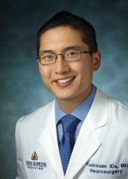 Yuanxuan Xia合作论文数Dept Neurosurg, Johns Hopkins Univ8
 Allison Koenecke合作论文数Department of Information Science, Cornell University8
 Jean W Liew合作论文数School of Medicine, Boston University8
 Manuel Francisco Ugarte-Gil合作论文数Department of Medicine, Universidad Científica del Sur8
 Jon T. Giles合作论文数Division of Rheumatology, Johns Hopkins University8Nikita Marcou合作论文数7
 Francis Berenbaum合作论文数7
 Sebastian E Sattui合作论文数Dept Rheumatol, Univ Pittsburgh7P. Aitana Azurmendi合作论文数Johns Hopkins Univ, Sch Med, Biophys & Biophys Chem, Baltimore, MD USA7
 K. Young合作论文数Div Rheumat Dis, Univ Texas Southwestern Med Ctr Dallas7
 Clifton O. Bingham合作论文数Division of Rheumatology, Johns Hopkins University;The Johns Hopkins University School of Medicine;Johns Hopkins Arthritis Center7
 Jeffrey A Sparks合作论文数Division of Rheumatology, Immunology and Allergy, Department of Medicine, Brigham and Women׳s Hospital, 45 Francis St, Boston, MA 02115.7
 Susan Athey合作论文数Golub Capital Social Impact Lab, Stanford Graduate School of Business, Stanford University7
 Michelle Petri合作论文数Johns Hopkins Lupus Center, Johns Hopkins University School of Medicine7
 Peter Korsten合作论文数Department of Nephrology and Rheumatology, Robert-Koch-Str. 40, 37075 Goettingen, Germany7
 Jinoos Yazdany合作论文数Division of Rheumatology, UCSF7Michelle S. Miller合作论文数6Jiaxin Ge合作论文数6Maria Popoli合作论文数6
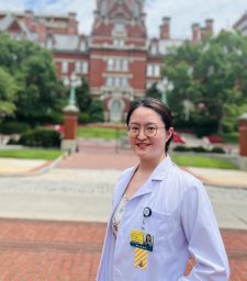 Jin Liu (津留)合作论文数Department of Pathology, Johns Hopkins University School of Medicine6Elana Shaw合作论文数Lab Clin Immunol & Microbiol, NIAID6
 Erika Lynn Darrah合作论文数Department of Medicine, School of Medicine, Johns Hopkins University6
 Laura Cappelli合作论文数Division of Rheumatology, Department of Medicine, Johns Hopkins University6
 Richard Conway合作论文数6
 Jarushka Naidoo合作论文数Royal College of Surgeons in Ireland6
 Rebecca Grainger合作论文数Wellington School of Medicine, University of Otago;Malaghan Institute of Medical Research;Malaghan Institute of Medical Research, University of Otago6Evangeline Watson合作论文数5
 Nicole Mercado Fischer合作论文数Sch Med, Johns Hopkins Univ5




