Local control remains a challenge in the treatment of recurrent glioblastomas. Our prior experience indicates that adjunctive cesium-131 brachytherapy, followed by systemic therapy, is a promising option. Here, we extend the initial findings through a multi-institutional study. Clinical information was collected for consecutive, recurrent glioblastoma (isocitrate dehydrogenase wild-type) patients treated at seven participating institutions. All patients underwent systemic treatment after surgical resection/cesium implant. Median progression-free and overall survival (mPFS and mOS) were calculated from the time of cesium tile implantation. The study cohort comprised 43 male and 17 female subjects, with a median of 1 prior glioblastoma recurrence. The mean pre-operative Karnofsky Performance Score (KPS) was 80 (± 15.7). There was one case (2
BACKGROUND:Facial nerve (CN VII) diffusion MR tractography is considered as a useful adjunct in pre-operative planning prior to vestibular schwannoma (VS) resection, especially in larger (Koos Grade III/IV) tumors. Since 2016, several systematic reviews have investigated the clinical value of CN VII tractography in VS, and all reported a "success rate" of at least 87% for predicting the pre-operative CN VII position. Yet in clinical practice, CN VII tractography has not yet been widely adopted into routine clinical practice. We suspected that underlying methodology and reporting metrics for existing tractography algorithms may be overestimating success rate. This motivated us to revisit the literature from a different perspective to unravel the caveats and nuances behind this technology. METHODS:We screened all published works on PubMed related to pre-operative CN VII tractography in VS. Twenty-two studies were reviewed in detail. RESULTS:We observed a strikingly high heterogeneity in tractography protocols in all domains of the tractography acquisition and analysis pipeline across studies. CONCLUSIONS:These findings suggest that the reliability and reproducibility of CN VII tractography in large VS has been overestimated. We believe that employing standardized reporting metrics, including sensitivity, true predictive value, and false discovery rate, would increase the transparency of benchmarking over other commonly reported metrics ("success rate" or "concordance rate"). In addition, ongoing research should aim to systematically investigate and improve each step in the acquisition and analysis pipeline for CN VII tractography in VS.
PURPOSE Laser interstitial thermal therapy is a surgical tool used to ablate brain tumors, radiation necrosis, and epileptic foci. Data from the LAANTERN prospective multicenter registry (ClinicalTrials.gov identifier: NCT02392078 ), containing patients with tumor from 25 US centers between 2015 and 2023, were analyzed to determine clinical outcomes. METHODS All patients with primary or metastatic brain tumors treated with LITT using the Monteris NeuroBlate System were included in this analysis. Demographics, intraprocedural data, adverse events, survival data, and data pertaining to functional status over time were prospectively collected and then analyzed. Both univariable and multivariable analyses were performed. RESULTS A total of 787, primary (445) and metastatic brain tumor (342), patients were included. The median age was 59.6 years and the median pediatric age 15 years (n = 18). The median length of hospital stay was 32.4 hours, and 62.6% avoided intensive care unit admission. The adverse event rate was 12.8%, with 65.5% of events considered transient. Mortality rate was 0.25%. Anticonvulsants were stopped in 30%-40% of patients, and 80% of patients stopped steroids after LITT. Quality of life remained stable up to 3 years post-LITT. Greater extent of ablation (EOA) and smaller lesion size were associated with significant improvements in survival outcomes in patients with high-grade glioma and recurrent metastases. CONCLUSION The data from this largest, prospective LITT cohort support the consideration of LITT as a cytoreductive tool for patients with primary and metastatic tumor with short hospital stays, low complication rates, and preserved functional status. The EOA in glioblastoma and lesion volume in metastatic disease emerge as factors that may guide patient selection.
Thalamic neuromodulation is clinically effective in drug-resistant epilepsy, suggesting critical contributions of the thalamus to the epileptogenic process. However, the underlying electrophysiologic mechanisms remain poorly characterized. Converging evidence implicates the thalamus in shaping large-scale functional interactions across the cortex. We hypothesized that ictal changes in thalamic activity track cortical network dynamics associated with seizure propagation. We analyzed stereo-electroencephalography recordings from 16 patients with focal epilepsy (255 seizures) with simultaneous sampling of the thalamus (anterior nucleus, N=14; pulvinar, N=11) and cortex. Cortical regions of interest included the seizure onset zone (SOZ), surrounding cortices (near-SOZ), and control regions from the contralateral hemisphere. We characterized seizure dynamics across spatial scales, from local activity within each region to network-level, inter-regional interactions. Local activity was decomposed into its periodic (oscillatory) and aperiodic components. Network interactions were characterized by directed functional connectivity computed with a multivariate method. Seizures were associated with increased broadband power (a proxy for neuronal population firing rates) and low-frequency rhythmic activity across the thalamocortical network relative to interictal baseline levels. In contrast, consistent changes in aperiodic slope (a putative marker of excitation-inhibition balance) were specific to the thalamus, which showed an early and sustained steepening (i.e., more negative slope). While local rhythms were heterogeneous across the canonical frequency bands, inter-regional interactions predominantly involved the beta band (13-30 Hz). Shortly after onset, both forward outflow from SOZ to near-SOZ and feedback inflow in the reverse direction were increased. These bidirectional effects were expressed via both a direct cortico-cortical pathway and an indirect transthalamic route, operating in parallel. These dynamics were further stratified based on seizure subtypes, leveraging the fact that there was minimal propagation of ictal activity to the near-SOZ in subclinical seizures. The ictal drop in thalamic aperiodic slope was primarily observed in clinical seizures. At the network level, whereas SOZ→near-SOZ outflow was present across seizure types, reverse feedback was particularly enhanced in clinical seizures. Multivariable regression showed that the degree of thalamic slope steepening uniquely tracked seizure-to-seizure fluctuations in the strength of near-SOZ→SOZ feedback, and further predicted seizure durations. Together these findings highlight thalamic aperiodic slope as an index of cortical network dynamics linked to seizure propagation, with potential clinical utility for further development of physiology-informed precision neuromodulation.
ObjectiveConfocal laser endomicroscopy (CLE), a handheld imaging technology, provides intraoperative real-time cellular resolution examination of tissue architecture. We evaluated the feasibility and diagnostic capability of the first clinically approved CLE system for intraoperative in vivo imaging of brain tumors.MethodsA total of 50 patients who were to undergo brain tumor surgery were prospectively enrolled. CLE images were interpreted by one CLE-experienced neuropathologist as lesional, non-lesional, or non-interpretable and compared to tissue histology acquired at the same location under neuronavigation. Diagnostic accuracy of CLE imaging was calculated using permanent sections as the standard for comparison. The neuropathologist provided real-time image interpretation using a built-in telepathology consultation platform in 27 cases.ResultsThe final pathology of the tumors in these patients included 28 gliomas, 5 meningiomas, 3 metastatic brain tumors, 5 treatment-related changes, and 10 other primary intracranial tumors. A total of 13,535 interpretable images were acquired from 304 regions of interest (ROIs). The first informative images were acquired within 10.5 s after the initiation of CLE imaging for each ROI. Mean CLE imaging time per case was 8.6 min. Using telepathology consultation extended CLE imaging per case time by 3.8 min (p=0.005). Communication between neurosurgeon and neuropathologist lasted 3.9 min per ROI. Overall sensitivity and specificity of CLE imaging were 93% and 81%, respectively. The specificity differed significantly between core and margin ROIs in glioma cases (93% vs. 50%, p=0.039). Diagnostic performance was not statistically different between new and recurrent glioma cases or between glioma and other tumor types.ConclusionsThe clinically approved CLE system allows intraoperative in vivo visualization of tissue histoarchitecture and identification of lesional tissue in real time, without the need for tissue biopsy and processing. CLE is efficient and cost-effective, with high diagnostic accuracy at the glioma core. However, CLE imaging at the tumor margin remains challenging.
Determining true recurrence versus necrosis alone after previous radiotherapy (RT) for brain metastasis based on imaging alone is challenging. Accurate diagnosis is critical to patient management, as further RT is contraindicated in the setting of radiation necrosis without tumor (TUM-). Rates of intraoperative frozen section pathology reporting tumor +/- necrosis (TUM+) or necrosis without tumor (TUM-) were examined in patients undergoing resection for presumed RBM after prior same-site RT. All cases were prospectively enrolled on a multi-institutional registry for patients undergoing resection and intraoperative cesium-131 collagen tile brachytherapy (NCT04427384)(GammaTile, GT Medical Technologies, Tempe AZ, USA). Preoperative evaluation varied by center, and patient demographics, primary site, lesion size, and prior therapies were also examined. From 10/2020 to 2/2024, 60 patients (64 lesions) underwent resection and intraoperative frozen section pathologic evaluation. Per patient, primary sites were 53% lung, 15% melanoma, 13% breast, 7% renal, and 10% other. Median age was 62, median preoperative maximum diameter was 2.9 cm, F:M ratio was 31:29, and median time from prior RT was 15.4 months. Across all histologies, TUM+ was seen in 88% (53/60) and TUM- in 12% (7/60). Rates of TUM- by primary type were highest for lung (16%), breast (13%), and melanoma (11%). The TUM- rate for lung metastasis was 16% vs 7% for non-lung origin. All TUM- patients received RT and prior chemotherapy, immunotherapy, or both. For all previously irradiated metastasis, pathologic evaluation at time of presumed radiographic recurrence demonstrated an actual 12% rate of TUM-. These findings underscore the importance of pathologic tumor confirmation before considering re-RT for presumed radiographic recurrence.
The purpose of this study was to systematically examine three different surgical approaches in treating left medial temporal lobe epilepsy (mTLE) (viz., subtemporal selective amygdalohippocampectomy [subSAH], stereotactic laser amygdalohippocampotomy [SLAH], and anterior temporal lobectomy [ATL]), to determine which procedures are most favorable in terms of visual confrontation naming and seizure relief outcome. This was a retrospective study of 33 adults with intractable mTLE who underwent left temporal lobe surgery at three different epilepsy surgery centers who also underwent pre-, and at least 6-month post-surgical neuropsychological testing. Measures included the Boston Naming Test (BNT) and the Engel Epilepsy Surgery Outcome Scale. Fisher’s exact tests revealed a statistically significant decline in naming in ATLs compared to SLAHs, but no other significant group differences. 82% of ATL and 36% of subSAH patients showed a significant naming decline whereas no SLAH patient (0%) had a significant naming decline. Significant postoperative naming improvement was seen in 36% of SLAH patients in contrast to 9% improvement in subSAH patients and 0% improvement in ATLs. Finally, there were no statistically significant differences between surgical approaches with regard to seizure freedom outcome, although there was a trend towards better seizure relief outcome among the ATL patients. Results support a possible benefit of SLAH in preserving visual confrontation naming after left TLE surgery. While result interpretation is limited by the small sample size, findings suggest outcome is likely to differ by surgical approach, and that further research on cognitive and seizure freedom outcomes is needed to inform patients and providers of potential risks and benefits with each.
INTRODUCTION: Fluorescence-guided surgery using 5-aminolevulinic acid (5-ALA) has proven effective in aiding the detection of glioma marginal zones and enhancing maximal safe resection. FDA-cleared confocal laser endomicroscopy is another imaging modality for intraoperative brain tissue discrimination on cellular level that could improve visualization at the glioma margin. METHODS: 5-ALA wide-field imaging and CLE were used in 9 patients with gliomas (6 enhancing, 3 non-enhancing). CLE imaging was enhanced by the use of a telesurgical pathology software platform to enable real-time conversation between neurosurgeons and pathologists located remotely. CLE was used for imaging tumor regions subjectively regarded as tumor margins under normal visualization with the operative microscope. After CLE imaging, 5-ALA wide-field imaging was performed in the same regions. A tissue was harvested at imaging locations, and interpretations of CLE and 5-ALA wide-field imaging were compared to permanent histological sections. RESULTS: Overall, 20 deep and superficial margin regions of interest (ROI) were imaged with CLE and 5-ALA imaging. Most of the ROIs interpreted by the neuropathologist as infiltrative glioma based on CLE imaging lacked 5-ALA-induced fluorescence. Permanent histological sections from the corresponding regions were concordant with the interpretation of CLE images in 15 of 20 (75%) ROIs and with the interpretation of 5-ALA imaging in 11 of 20 (55%) ROIs. Sensitivity/specificity of CLE and 5-ALA for interpretation of tumor margins were 79%/67%, and 50%/67%, respectively. Positive/negative predictive values for CLE and 5-ALA were 85%/57%, and 78%/37%, respectively. CONCLUSIONS: Conventional intraoperative understanding of tumor margins based on wide-field fluorescence could underestimate the invasiveness of gliomas. The ongoing study demonstrates that CLE shows higher accuracy in detecting regions with infiltrating tumor than intraoperative 5-ALA imaging.
Abstract PURPOSE/OBJECTIVE(S) Determining true recurrence versus necrosis alone after previous radiation therapy (RT) for brain metastasis based on imaging alone is difficult. Proper diagnosis is essential, as further radiation is contraindicated in the setting of radiation necrosis without tumor (TUM-). To better understand the rate of pathologic tumor positivity (TUM+) vs TUM-, we examined frozen section results from a cohort of patients with prior same-site RT undergoing resection of presumed recurrent brain metastasis (RBM). MATERIALS/METHODS Rates of intraoperative frozen section pathology disclosing tumor +/- necrosis (TUM+) or necrosis without tumor (TUM-) were examined in patients undergoing resection for presumed RBM after prior same-site RT. All cases had been prospectively enrolled on a multi-institution registry for patients undergoing resection and intraoperative cesium-131 collagen tile brachytherapy (NCT04427384)(GammaTile, GT Medical Technologies, Tempe AZ, USA). Preoperative evaluation varied by center, and patient demographics, primary site, lesion size, and prior therapies were also examined. RESULTS From 10/2020 to 2/2024 60 patients (64 lesions) underwent resection and intraoperative frozen section pathologic evaluation. Per patient, primary sites were 53% lung, 15% melanoma, 13% breast, 7% renal, and 10% other. F:M ratio was 31:29; median age 62, maximum preoperative diameter 2.9 cm, and median time from prior RT 15.4 months. Across all histologies TUM+ was seen in 88% (53/60) and TUM- in 12% (7/60). Rates of TUM- by primary type were highest for lung (16%), breast (13%), and melanoma (11%). The TUM- rate for lung metastasis was 16% vs 7% for non-lung origin. All TUM- patients received RT and prior chemotherapy, immunotherapy, or both. CONCLUSION For all previously irradiated metastasis, pathology demonstrated a 12% rate of TUM-. As all cases necessitated surgery, the adverse event grading would be ≥ Gr 4. These findings highlight the importance of pathologic confirmation before undertaking re-irradiation for presumed radiographic recurrence.
Abstract BACKGROUND Resection and intraoperative brachytherapy for operable recurrent brain metastasis allows for pathologic confirmation of recurrent disease, mass effect relief, and immediate initiation of radiotherapy (RT). In this analysis, we report patterns-of-use and treatment-related adverse events (AEs) for rBM patients treated with Cs-131 collagen tiles, an FDA-cleared intracranial brachytherapy device. METHODS Patients with rBM who underwent resection and surgically-targeted radiation therapy (GammaTile, GT Medical Technologies Inc., Tempe, AZ USA) on a prospectively enrolling phase 4 registry study (NCT04427384) were analyzed. AEs were graded per CTCAE v5.0. RESULTS Between 11/2020 and 2/2024, 56 rBM in 51 consecutive patients underwent STaRT at 19 centers, with 5 patients having 2 metastases implanted concurrently. 44 patients (86%) had prior same-site RT (median interval 14.5 mo, range 3-56). Primary tumor histologies were lung (27), melanoma (8), breast (7), renal (4), colon (2), and “other” (3). Median pre-operative maximum diameter was 3.0 cm (range 1.4-5.7); age 63 (range 28-81); 53% females; KPS median 90 (range 40-100); and median implantation time 3 minutes. 26 patients were implanted at a 1st, 15 at a 2nd, and 10 at ≥ 3rd same-site recurrence (range 1-9). At a median follow-up of 6.2 months (range <1-35.1), 6/51 patients (11.8%) experienced ≥Gr 3 AEs at a median of 12 (range 1-69) days postoperatively (POD). No radiation necrosis (RN) events were observed, and no AEs occurred in multi-implant cases or where STaRT was the initial form of RT. CONCLUSIONS In this prospective multi-institutional study, STaRT demonstrated an excellent safety profile in a cohort of larger rBM, even in the setting of multi-recurrent disease. Accrual and follow-up are on-going and will provide data on tumor control and long-term RN rates.
OBJECTIVE:Confocal laser endomicroscopy (CLE) is a US Food and Drug Administration-cleared intraoperative real-time fluorescence-based cellular resolution imaging technology that has been shown to image brain tumor histoarchitecture rapidly in vivo during neuro-oncological surgical procedures. An important goal for successful intraoperative implementation is in vivo use at the margins of infiltrating gliomas. However, CLE use at glioma margins has not been well studied. METHODS:Matching in vivo CLE images and tissue biopsies acquired at glioma margin regions of interest (ROIs) were collected from 2 institutions. All images were reviewed by 4 neuropathologists experienced in CLE. A scoring system based on the pathological features was implemented to score CLE and H&E images from each ROI on a scale from 0 to 5. Based on the H&E scores, all ROIs were divided into a low tumor probability (LTP) group (scores 0-2) and a high tumor probability (HTP) group (scores 3-5). The concordance between CLE and H&E scores regarding tumor probability was determined. The intraclass correlation coefficient (ICC) and diagnostic performance were calculated. RESULTS:Fifty-six glioma margin ROIs were included for analysis. Interrater reliability of the scoring system was excellent when used for H&E images (ICC [95% CI] 0.91 [0.86-0.94]) and moderate when used for CLE images (ICC [95% CI] 0.69 [0.40-0.83]). The ICCs (95% CIs) of the LTP group (0.68 [0.40-0.83]) and HTP group (0.68 [0.39-0.83]) did not differ significantly. The concordance between CLE and H&E scores was 61.6%. The sensitivity and specificity values of the scoring system were 79% and 37%. The positive predictive value (PPV) and negative predictive value were 65% and 53%, respectively. Concordance, sensitivity, and PPV were greater in the HTP group than in the LTP group. Specificity was higher in the newly diagnosed group than in the recurrent group. CONCLUSIONS:CLE may detect tumor infiltration at glioma margins. However, it is not currently dependable, especially in scenarios where low probability of tumor infiltration is expected. The proposed scoring system has excellent intrinsic interrater reliability, but its interrater reliability is only moderate when used with CLE images. These results suggest that this technology requires further exploration as a method for consistent actionable intraoperative guidance with high dependability across the range of tumor margin scenarios. Specific-binding and/or tumor-specific fluorophores, a CLE image atlas, and a consensus guideline for image interpretation may help with the translational utility of CLE.
ObjectivesConfocal laser endomicroscopy (CLE) is an intraoperative real-time cellular resolution imaging technology that images brain tumor histoarchitecture. Previously, we demonstrated that CLE images may be interpreted by neuropathologists to determine the presence of tumor infiltration at glioma margins. In this study, we assessed neurosurgeons’ ability to interpret CLE images from glioma margins and compared their assessments to those of neuropathologists.MethodsIn vivo CLE images acquired at the glioma margins that were previously reviewed by CLE-experienced neuropathologists were interpreted by four CLE-experienced neurosurgeons. A numerical scoring system from 0 to 5 and a dichotomous scoring system based on pathological features were used. Scores from assessments of hematoxylin and eosin (H&E)-stained sections and CLE images by neuropathologists from a previous study were used for comparison. Neurosurgeons’ scores were compared to the H&E findings. The inter-rater agreement and diagnostic performance based on neurosurgeons’ scores were calculated. The concordance between dichotomous and numerical scores was determined.ResultsIn all, 4275 images from 56 glioma margin regions of interest (ROIs) were included in the analysis. With the numerical scoring system, the inter-rater agreement for neurosurgeons interpreting CLE images was moderate for all ROIs (mean agreement, 61%), which was significantly better than the inter-rater agreement for the neuropathologists (mean agreement, 48%) (p < 0.01). The inter-rater agreement for neurosurgeons using the dichotomous scoring system was 83%. The concordance between the numerical and dichotomous scoring systems was 93%. The overall sensitivity, specificity, positive predictive value, and negative predictive value were 78%, 32%, 62%, and 50%, respectively, using the numerical scoring system and 80%, 27%, 61%, and 48%, respectively, using the dichotomous scoring system. No statistically significant differences in diagnostic performance were found between the neurosurgeons and neuropathologists.ConclusionNeurosurgeons’ performance in interpreting CLE images was comparable to that of neuropathologists. These results suggest that CLE could be used as an intraoperative guidance tool with neurosurgeons interpreting the images with or without assistance of the neuropathologists. The dichotomous scoring system is robust yet simple and may streamline rapid, simultaneous interpretation of CLE images during imaging.
The authors present the first reported case of MVNT in the thalamus in a 60-year-old man with a 20-year history of epilepsy and recent progressive neurological decline presented for neurosurgical evaluation for a non-enhancing mass predominantly in the right thalamus presumed to be a low-grade glioma. The tumor was subtotally resected using a left contralateral interhemispheric transcallosal approach. Histological and molecular assessment revealed an MVNT with MAPK pathway-activating mutation. The authors also conducted a systematic review of pathology-proven cases of MVNT to provide an up-to-date overview of the literature on the localization, presenting symptoms, and recurrence of this tumor.
Glioblastoma (GBM) is the most aggressive primary brain tumor in adults, with a universally lethal prognosis despite maximal standard therapies. Here, we present a consensus treatment protocol based on the metabolic requirements of GBM cells for the two major fermentable fuels: glucose and glutamine. Glucose is a source of carbon and ATP synthesis for tumor growth through glycolysis, while glutamine provides nitrogen, carbon, and ATP synthesis through glutaminolysis. As no tumor can grow without anabolic substrates or energy, the simultaneous targeting of glycolysis and glutaminolysis is expected to reduce the proliferation of most if not all GBM cells. Ketogenic metabolic therapy (KMT) leverages diet-drug combinations that inhibit glycolysis, glutaminolysis, and growth signaling while shifting energy metabolism to therapeutic ketosis. The glucose-ketone index (GKI) is a standardized biomarker for assessing biological compliance, ideally via real-time monitoring. KMT aims to increase substrate competition and normalize the tumor microenvironment through GKI-adjusted ketogenic diets, calorie restriction, and fasting, while also targeting glycolytic and glutaminolytic flux using specific metabolic inhibitors. Non-fermentable fuels, such as ketone bodies, fatty acids, or lactate, are comparatively less efficient in supporting the long-term bioenergetic and biosynthetic demands of cancer cell proliferation. The proposed strategy may be implemented as a synergistic metabolic priming baseline in GBM as well as other tumors driven by glycolysis and glutaminolysis, regardless of their residual mitochondrial function. Suggested best practices are provided to guide future KMT research in metabolic oncology, offering a shared, evidence-driven framework for observational and interventional studies.
OBJECTIVE:To evaluate long-term clinical outcomes among patients treated with laser interstitial thermal therapy (LITT) for predicted recurrent glioblastoma (rGBM). METHODS:Patients with rGBM treated by LITT by a single surgeon (2013-2020) were evaluated for progression-free survival (PFS), overall survival (OS), and OS after LITT. RESULTS:Forty-nine patients (33 men, 16 women; mean [SD] age at diagnosis, 58.7 [12.5] years) were evaluated. Among patients with genetic data, 6 of 34 (18%) had IDH-1 R132 mutations, and 7 of 21 (33%) had MGMT methylation. Patients underwent LITT at a mean (SD) of 23.8 (23.8) months after original diagnosis. Twenty of 49 (40%) had previously undergone stereotactic radiosurgery, 37 (75%) had undergone intensity-modulated radiation therapy, and 49 (100%) had undergone chemotherapy. Patients had undergone a mean of 1.2 (0.7) previous resections before LITT. Mean preoperative enhancing and T2 FLAIR volumes were 13.1 (12.8) cm3 and 35.0 (32.8) cm3, respectively. Intraoperative biopsies confirmed rGBM in 31 patients (63%) and radiation necrosis in 18 patients (37%). Six perioperative complications occurred: 3 (6%) cases of worsening aphasia, 1 (2%) seizure, 1 (2%) epidural hematoma, and 1 (2%) intraparenchymal hemorrhage. For the rGBM group, median PFS was 2.0 (IQR, 4.0) months, median OS was 20.0 (IQR, 29.5) months, and median OS after LITT was 6.0 (IQR, 10.5) months. For the radiation necrosis group, median PFS was 4.0 (IQR, 4.5) months, median OS was 37.0 (IQR, 58.0) months, and median OS after LITT was 8.0 (IQR, 23.5) months. CONCLUSIONS:In a diverse rGBM cohort, LITT was associated with a short duration of posttreatment PFS.
Background and objective Glioblastoma (GBM) is one of the most aggressive and lethal human cancers. Intra-tumoral genetic heterogeneity poses a significant challenge for treatment. Biopsy is invasive, which motivates the development of non-invasive, MRI-based machine learning (ML) models to quantify intra-tumoral genetic heterogeneity for each patient. This capability holds great promise for enabling better therapeutic selection to improve patient outcome. Methods We proposed a novel Weakly Supervised Ordinal Support Vector Machine (WSO-SVM) to predict regional genetic alteration status within each GBM tumor using MRI. WSO-SVM was applied to a unique dataset of 318 image-localized biopsies with spatially matched multiparametric MRI from 74 GBM patients. The model was trained to predict the regional genetic alteration of three GBM driver genes (EGFR, PDGFRA and PTEN) based on features extracted from the corresponding region of five MRI contrast images. For comparison, a variety of existing ML algorithms were also applied. Classification accuracy of each gene were compared between the different algorithms. The SHapley Additive exPlanations (SHAP) method was further applied to compute contribution scores of different contrast images. Finally, the trained WSO-SVM was used to generate prediction maps within the tumoral area of each patient to help visualize the intra-tumoral genetic heterogeneity. Results WSO-SVM achieved 0.80 accuracy, 0.79 sensitivity, and 0.81 specificity for classifying EGFR; 0.71 accuracy, 0.70 sensitivity, and 0.72 specificity for classifying PDGFRA; 0.80 accuracy, 0.78 sensitivity, and 0.83 specificity for classifying PTEN; these results significantly outperformed the existing ML algorithms. Using SHAP, we found that the relative contributions of the five contrast images differ between genes, which are consistent with findings in the literature. The prediction maps revealed extensive intra-tumoral region-to-region heterogeneity within each individual tumor in terms of the alteration status of the three genes. Conclusions This study demonstrated the feasibility of using MRI and WSO-SVM to enable non-invasive prediction of intra-tumoral regional genetic alteration for each GBM patient, which can inform future adaptive therapies for individualized oncology.
OBJECTIVE:Because gliomas have poorly defined tumor margins, the ability to achieve maximal resection is limited. To better discern these margins, fluorescence-guided surgery has been used to aid maximal safe resection. The authors describe their experience with the simultaneous use of intraoperative fluorescein sodium (FNa) confocal laser endomicroscopy (CLE) and operating microscope 5-aminolevulinic acid (5-ALA) fluorescence imaging for glioma resection to improve CLE use for better margin discrimination. METHODS:FNa CLE and 5-ALA wide-field imaging were used in 33 patients with gliomas. CLE imaging was enhanced with the use of a telesurgical pathology software platform that enables real-time conversation between the operating neurosurgeons and the pathologists located remotely. CLE was used for imaging tumor regions that were subjectively regarded as tumor margins under normal visualization with the operative microscope. After FNa CLE imaging, 5-ALA wide-field imaging was performed in the same regions. Tissue was biopsied at imaging locations, and interpretations of FNa CLE and 5-ALA wide-field imaging were compared to those of permanent histological sections. RESULTS:Eighty-eight deep- and superficial-margin regions of interest (ROIs) were imaged with FNa CLE and 5-ALA imaging. Most of the ROIs interpreted by the neuropathologist as infiltrative glioma based on FNa CLE imaging lacked 5-ALA-induced fluorescence. Permanent histological sections from the corresponding regions were concordant with the interpretation of FNa CLE images in 57 of 88 (65%) ROIs and with the interpretation of 5-ALA imaging in 43 of 88 (49%) ROIs. The sensitivity and specificity of FNa CLE for the interpretation of tumor margins were 73% and 41%, respectively, and those of 5-ALA were 38% and 82%, respectively. Positive and negative predictive values for CLE were 79% and 33%, respectively, and those for 5-ALA were 86% and 31%, respectively. CONCLUSIONS:Conventional intraoperative evaluation of tumor margins, based on MRI and wide-field fluorescence imaging, can underestimate the invasiveness of gliomas. FNa CLE showed higher accuracy in detecting regions with infiltrating tumors than intraoperative 5-ALA imaging. Future considerations should include more rigorous comparisons of FNa CLE imaging and 5-ALA-guided resections on a larger cohort of patients.
High-grade glioma continues to have dismal survival owing in part to its intra- and inter-patient heterogeneity. Standard clinical protocol collects tumor samples with the aim of providing or confirming a diagnosis and determining the status of a few key genes (e.g. IDH1, MGMT). However, this protocol is unable to capture the diversity within tumor regions, immune expression or normal cell abundances that play key roles in the development of the disease. To overcome this, during surgery we collect image-localized multi-regional biopsies to characterize this disease heterogeneity. Data collection is ongoing, and we currently have 202 samples from 58 patients with available bulk RNA-Seq. With a single-cell reference dataset from Columbia University, we used CIBERSORTx, a deconvolution method, to predict relative abundances of 7 normal, 6 glioma, and 5 immune cell subpopulations for each sample. We used Cox proportional hazard models with first-order statistics (mean, minimum, maximum) of abundances within patients to determine whether these are prognostic, then used TCGA RNA-Seq data to validate these findings. We found that one glioma proneural subtype was significantly beneficial for patient survival relative to other glioma subtypes across all statistics (and showed significance in TCGA). Proliferative and mesenchymal glioma subtypes also showed significance for one or two of the calculated statistics. Oligodendrocyte progenitor cell abundances were consistently significantly beneficial for patient survival in our cohort, while an increased abundance of abnormal (reactive) astrocytes is associated with poor prognosis. We also ran these analyses within invasive margin and core tumor regions to determine location-specific population abundances associated with patient survival. In conclusion, understanding the in vivo diversity of cellular subpopulations within high grade glioma is important for treatment stratification and patient benefit.
High grade glioma (HGG) represents a group of devastating diseases with dismal prognosis. Surgical resection of the contrast enhancing (CE) region of HGG remains the mainstay of treatment, but recurrence inevitably arises from the unresected non-contrast enhancing (NE) region, surgically inaccessible due to cancer cell invasion into healthy brain tissue. Due to its critical role in recurrence, understanding of the NE region is central to the improvement of clinical outcomes. We reveal the biological characteristics of this region through image localized multi-regional sampling. We linked microenvironmental characteristics measured by multi-parametric MRI to genomic mutations and transcriptional phenotypes using mixed effect modeling which allowed us to control for individualized patient effects. We first confirmed that T2 is a significant indicator of IDH mutation status in the NE region, being the first description of such a relationship in a HGG cohort. We found the combination of EGFR amplification and CDKN2A homozygous loss was associated with a significantly lower mean diffusivity (MD) compared to double wild type tumors in the NE region, indicating the presence of greater cellular packing and proliferation in EGFR amplification/CDKN2A loss regions. Finally, using single cell pathway based tumor classifications, we showed that nK2, a DSC-MRI metric representing cell size heterogeneity, correlated positively with neuronal signature and negatively with glycolytic/plurimetabolic signature within the NE tumor, indicating that glycolytic/plurimetobolic tumors possessed a high amount of cell size heterogeneity compared to neuronal samples. This hypothesis was supported using digital reference object (DRO) modeling which confirmed that cell size and heterogeneity drove the differential nK2 signal between neuronal and glycolytic/plurimetabolic samples. We identified immune cell infiltrate as one possible mechanism of increased cell size heterogeneity using transcriptomic signature analysis which found more immune cell signatures within glycolytic/plurimetobolic tumors compared to neuronal. Collectively this study demonstrates the central role of multi-parametric MRI as a non-invasive measure of tumor biology and a tool for understanding the clinically critical NE region which can then inform new therapies targeting this region of HGG recurrence. Citation Format: Matthew Flick, Taylor Weiskittel, Kevin Meng-Lin, Fulvio D'Angelo, Francesca Caruso, Shannon Ensign, Mylan Blomquist, Luija Wang, Christopher Sereduk, Gustavo De Leon, Ashley Nespodzany, Javier Urcuyo, Ashlynn Gonzalez, Lee Curtin, Kyle Singleton, Aliya Anil, Natenael Simmineh, Erika Lewis, Teresa Noviello, Reyna Patel, Panwen Wang, Junwen Wang, Jennifer Eschbacher, Andrea Hawkins-Daarud, Pamela Jackson, Kris Smith, Peter Nakaji, Bernard Bendok, Richard Zimmerman, Chandan Krishna, Devi Patra, Naresh Patel, Mark Lyons, Matthew Neal, Kliment Donev, Maciej Mrugala, Alyx Porter, Scott Beeman, Yuxiang Zhou, Leslie Baxter, Christopher Plaisier, Jing Li, Hu Li, Anna Lasorella, Chad Quarles, Kristin Swanson, Michele Ceccarelli, Antonio Iavarone, Nhan Tran, Leland Hu. Multi-parametric MRI maps regional heterogeneity of high grade glioma phenotypes. [abstract]. In: Proceedings of the American Association for Cancer Research Annual Meeting 2023; Part 1 (Regular and Invited Abstracts); 2023 Apr 14-19; Orlando, FL. Philadelphia (PA): AACR; Cancer Res 2023;83(7_Suppl):Abstract nr 5621.
Sampling restrictions have hindered the comprehensive study of invasive non-enhancing (NE) high-grade glioma (HGG) cell populations driving tumor progression. Here, we present an integrated multi-omic analysis of spatially matched molecular and multi-parametric magnetic resonance imaging (MRI) profiling across 313 multi-regional tumor biopsies, including 111 from the NE, across 68 HGG patients. Whole exome and RNA sequencing uncover unique genomic alterations to unresectable invasive NE tumor, including subclonal events, which inform genomic models predictive of geographic evolution. Infiltrative NE tumor is alternatively enriched with tumor cells exhibiting neuronal or glycolytic/plurimetabolic cellular states, two principal transcriptomic pathway-based glioma subtypes, which respectively demonstrate abundant private mutations or enrichment in immune cell signatures. These NE phenotypes are non-invasively identified through normalized K2 imaging signatures, which discern cell size heterogeneity on dynamic susceptibility contrast (DSC)-MRI. NE tumor populations predicted to display increased cellular proliferation by mean diffusivity (MD) MRI metrics are uniquely associated with EGFR amplification and CDKN2A homozygous deletion. The biophysical mapping of infiltrative HGG potentially enables the clinical recognition of tumor subpopulations with aggressive molecular signatures driving tumor progression, thereby informing precision medicine targeting.
 Peter Nakaji合作论文数Department of Neurosurgery, Banner – University Medical Center Phoenix59
 Eschbacher Jennifer合作论文数34
 Leland Hu合作论文数28
 Leslie C. Baxter合作论文数Barrow Neurological Institute27
 Kristin R Swanson合作论文数Department of Applied Mathematics, University of Washington24David Brachman合作论文数Oncology Services Foundation for Cancer Research and Education23
 Hawkins-Daarud Andrea合作论文数21Randall W. Porter合作论文数19Mark C. Preul合作论文数18
 Bernard R. Bendok合作论文数Comprehensive Cancer Center, Mayo Clinic;Department of Radiology, Mayo Clinic17
 Nhan Tran合作论文数Translational Genomics Research Institute17Andrew G. Shetter合作论文数17
 Jing Li合作论文数H. Milton Stewart School of Industrial and Systems Engineering, College of Engineering, Georgia Institute of Technology16
 Singleton Kyle W合作论文数Medical Imaging Informatics Group, Los Angeles, CA, USA.15
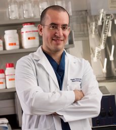 Nader Sanai合作论文数Barrow Neurological Institute, Barrow Neurological Institute15Emad Youssef合作论文数14
 Robert Fisher合作论文数Department of Neurology and Neurological Sciences, School of Medicine, Stanford University;Department of Neurosurgery, School of Medicine, Stanford University14Jackson Pamela R合作论文数Feinberg School of Medicine, Northwestern University14
 Maciej M. Mrugala合作论文数Department of Neurology, Mayo Clinic13
 Teresa Wu合作论文数School of Computing and Augmented Intelligence, Arizona State University;College of Health Solutions, Arizona State University;Fulton Schools of Engineering, Arizona State University13Stephen W. Coons合作论文数12
 Andrew S. Little合作论文数Barrow Neurological Institute12Christopher P. Sereduk合作论文数11Chandan Krishna合作论文数11Thomas Theresa A合作论文数11Pinnaduwage Dilini合作论文数11
 Robert F. Spetzler合作论文数Barrow Neurological Institute11Gustavo De Leon合作论文数10
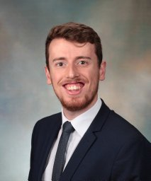 Lee Curtin合作论文数Mathematical NeuroOncology Lab, Mayo Clinic10
 Irakliy Abramov合作论文数Dept Neurosurg, Barrow Neurol Inst10
 Christopher Dardis合作论文数Department of Neurology, SUNY Downstate Medical Center10
 Patra Devi Prasad合作论文数Department of Neurosurgery, Post Graduate Institute of Medical Education and Research10John P. Karis合作论文数10
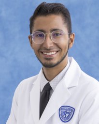 Javier Urcuyo合作论文数Mathematical NeuroOncology Lab, Mayo Clinic9
 David M. Treiman合作论文数National Institute of Neurological Disorders and Stroke9Clark-Swanson Kamala合作论文数9Adrienne C. Scheck合作论文数9Kliment Donev合作论文数8
 Leland Rogers合作论文数8
 Yuan Xu合作论文数8
 Michael T. Lawton合作论文数Department of Neurosurgery, Barrow Neurological Institute8
 Antonio De Salles合作论文数7
 Ashley Nespodzany合作论文数7
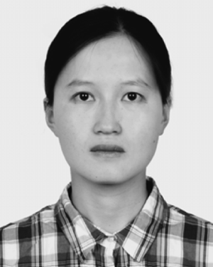 Lujia Wang合作论文数7McBride Heyoung L合作论文数7
 Clark C. Chen合作论文数Department of Neurosurgery, Warren Alpert School of Medicine, Brown University7Whiting Alexander C合作论文数7
 John R. Adler合作论文数Department of Neurosurgery, School of Medicine, Stanford University7
 François Mauguière合作论文数Centre de Recherche en Neurosciences de Lyon;Université Claude Bernard Lyon;Hôpital Neurologique de Lyon7




