Clostridium novyi has demonstrated selective efficacy against solid tumors largely due to the microenvironment contained within dense tumor cores. The core of a solid tumor is typically hypoxic, acidic, and necrotic-impeding the penetration of current therapeutics. C. novyi is attracted to the tumor microenvironment and once there, can both lyse and proliferate while simultaneously re-activating the suppressed immune system. C. novyi systemic toxicity is easily mitigated by knocking out the phage DNA plasmid encoded alpha toxin resulting in C. novyi-NT; but, after intravenous injection spores are quickly cleared by phagocytosis before accomplishing significant tumor localization. C. novyi-NT could be designed to accomplish intravenous delivery with the potential to target all solid tumors and their metastases in a single dose. This study characterizes CRISPR/Cas9 modified C. novyi-NT to insert the gene for RGD, a tumor targeting peptide, expressed within the promoter region of a spore coat protein. Expression of the RGD peptide on the outer spore coat of C. novyi-NT indicates an increased capacity for tumor localization of C. novyi upon intravenous introduction based on the natural binding of RGD with the αvβ3 integrin commonly overexpressed on the epithelial tissue surrounding a tumor, and lead to immune stimulation.
AbstractPancreatic ductal adenocarcinoma (PDAC) represents 3% of all cancer cases and 7% of all cancer deaths in the United States. Late diagnosis and inadequate response to standard chemotherapies contribute to an unfavorable prognosis and an overall 5-year survival rate of less than 10% in PDAC. Despite recent advances in tumor immunology, tumor-induced immunosuppression attenuates the immunotherapy response in PDAC. To date, studies have focused on IgG-based therapeutic strategies in PDAC. With the recent interest in IgE-based therapies in multiple solid tumors, we explored the MUC1-targeted IgE potential against pancreatic cancer. Our study demonstrates the notable expression of FceRI (receptor for IgE antibody) in tumors from PDAC patients. Our study showed that administration of MUC1 targeted-IgE (mouse/human chimeric anti-MUC1.IgE) antibody at intermittent levels in combination with checkpoint inhibitor (anti-PD-L1) and TLR3 agonist (PolyICLC) induces a robust antitumor response that is dependent on NK and CD8 T cells in pancreatic tumor-bearing mice. Subsequently, our study showed that the antigen specificity of the IgE antibody plays a vital role in executing the antitumor response as nonspecific IgE, induced by ovalbumin (OVA), failed to restrict tumor growth in pancreatic tumor-bearing mice. Utilizing the OVA-induced allergic asthma-PDAC model, we demonstrate that allergic phenotype induced by OVA cannot restrain pancreatic tumor growth in orthotopic tumor-bearing mice. Together, our data demonstrate the novel tumor protective benefits of tumor antigen-specific IgE-based therapeutics in a preclinical model of pancreatic cancer, which can open new avenues for future clinical interventions.
A highly aggressive subset of pancreatic ductal adenocarcinomas undergo trans-differentiation into the squamous lineage during disease progression. Here, we investigated whether squamous trans-differentiation of human and mouse pancreatic cancer cells can influence the phenotype of non-neoplastic cells in the tumor microenvironment. Conditioned media experiments revealed that squamous pancreatic cancer cells secrete factors that recruit neutrophils and convert pancreatic stellate cells into cancer-associated fibroblasts (CAFs) that express inflammatory cytokines at high levels. We use gain- and loss-of-function approaches to show that squamous-subtype pancreatic tumor models become enriched with neutrophils and inflammatory CAFs in a p63-dependent manner. These effects occur, at least in part, through p63-mediated activation of enhancers at pro-inflammatory cytokine loci, which includes IL1A and CXCL1 as key targets. Taken together, our findings reveal enhanced tissue inflammation as a consequence of squamous trans-differentiation in pancreatic cancer, thus highlighting an instructive role of tumor cell lineage in reprogramming the stromal microenvironment.
The tumor microenvironment (TME), or all cellular and molecular components in and around the tumor, has a well-documented impact on cancer cell proliferation, chemoresistance, and overall patient survival. However much of the research into the TME has been limited to the primary site. How the TME of metastases compares to the primary lesions remains poorly characterized, even though metastasis is responsible for 90% of all cancer deaths. Understanding these differences is critical to treating advanced cancer patients. To study this our lab examined the TME in 22 matched sets primary pancreatic tumors and liver metastasis obtained from end-stage pancreatic cancer patients via our rapid autopsy program. We used a multiplex-immunofluorescent technique that allows staining of a single slide with up to 35 antigens and characterized the presence, position and functional statues of cells that comprise the pancreatic TME. These included: tumor cells, vasculature, fibroblasts, macrophages, mast cells, basophils, eosinophils, B cells, T cells, and NK cells, along with multiple subtypes of these. Based on our preliminary data from this, we hypothesize that the organ determines the features of the TME. Analyzing primary tumors and metastases revealed that both the composition and spatial distribution of different cell types are distinct between the primary tumor and liver metastases. PAM clustering of the cellular featured grouped primary tumor with primary and metastasis with metastatis 87% of the time. As the TME has been shown to impact chemotherapeutic response, we further examined chemotherapeutic effect between sites. Comparing gemcitabine (standard of care) and untreated patients, we observed distinct site-based responses. In the primary tumors with gemcitabine treatment there was a significant increase in inflammatory cells (neutrophils and macrophages) in comparison to untreated patients, while in the liver gemcitabine induced an increase in desmoplasia (SMA). Combined this data indicates that the distinct TME’s exist within the same patient in an organ specific manner which can impact chemotherapeutic response. Understand these differences is therapeutically critical to patients whose therapeutic strategies involve targeting the TME to improve drug delivery, modulate immune suppression or otherwise modulate the microenvironment. Citation Format: Krysten E. Vance, Micheal A. Hollingsworth, Paul Grandgenett. Identifying site-specific disparities in the tumor microenvironment of primary and metastatic disease [abstract]. In: Proceedings of the American Association for Cancer Research Annual Meeting 2019; 2019 Mar 29-Apr 3; Atlanta, GA. Philadelphia (PA): AACR; Cancer Res 2019;79(13 Suppl):Abstract nr 3760.
 Michael (Tony) A. Hollingsworth合作论文数Eppley Institute for Research in Cancer and Allied Diseases, University of Nebraska Medical Center;Fred & Pamela Buffett Cancer Center, University of Nebraska Medical Center5
 Grandgenett Paul M合作论文数4Daisy Gonzalez合作论文数3Ayrianne J. Crawford合作论文数3Ragupathy Madiyalakan合作论文数3Kirsten C Eberle合作论文数3Simon Shin合作论文数3Kelly A O'Connell合作论文数3Spas Dimitrov Markov合作论文数3James A. Grunkemeyer合作论文数3Prathamesh Patil合作论文数3
 Ryan Hanson合作论文数3
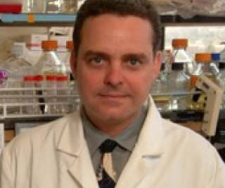 Manuel L. Penichet合作论文数Department of Microbiology, Immunology, and Molecular Genetics, College of Life Sciences, University of California3
 Kamiya Mehla合作论文数Eppley Institute for Research in Cancer and Allied Diseases, University of Nebraska Medical Center3
 Prakash Radhakrishnan合作论文数Eppley Institute for Research in Cancer/Allied Diseases,Fred & Pamela Buffett Cancer Center,University of Nebraska Medical Center3
 Shukla Surendra Kumar合作论文数Virology Group, International Centre for Genetic engineering and Biotechnology3
 Elizabeth M. Jaffee合作论文数Department of Pathology, School of Medicine, Johns Hopkins University;Institute for Clinical & Translational Research, Johns Hopkins University3
 Tracy R Daniels-Wells合作论文数3
 Jill A. Poole合作论文数Department of Internal Medicine, College of Medicine, University of Nebraska Medical Center3
 Thomas C. Caffrey合作论文数Eppley Institute for Research in Cancer and Allied Diseases, University of Nebraska Medical Center3
 Christopher F Nicodemus合作论文数3
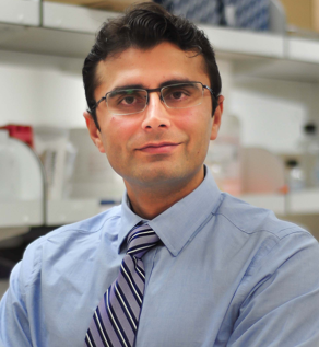 Pankaj K. Singh合作论文数Department of Oncology Science, College of Medicine, University of Oklahoma;Stephenson Cancer Center, University of Oklahoma3James M Small合作论文数1
 Jessica Pullan合作论文数Pharmaceut Sci, North Dakota State Univ1Juliane Daßler-Plenker合作论文数Cold Spring Harbor Lab1
 Tim D D Somerville合作论文数Cold Spring Harbor Laboratory, Cold Spring Harbor, NY 11724, USA.1Stella K Hur合作论文数Univ Penn, Perelman Sch Med, Dept Cell & Dev Biol, Philadelphia, PA 19104 USA1Koji Miyabayashi合作论文数1Ying Huang合作论文数1Kaitlin M Dailey合作论文数1
 Diogo Maia-Silva合作论文数Cold Spring Harbor Laboratory, Cold Spring Harbor, NY 11724, USA1
 Olaf Klingbeil合作论文数Cold Spring Harbor Lab1
 Vakoc Christopher R合作论文数Cold Spring Harbor Lab1Orr Megan C合作论文数1
 Jonathan Preall合作论文数Department of Biochemistry, Molecular Biology, and Cell Biology, Northwestern University1
 David Tuveson合作论文数Tuveson Laboratory, Cold Spring Harbor Laboratory;Cancer Center, Cold Spring Harbor Laboratory1El Demerdash Osama合作论文数1
 Kenneth W. Bayles合作论文数College of Medicine, Department of Pathology and Microbiology, University of Nebraska Medical Center1Xu Yali合作论文数1
 Biffi Giulia合作论文数Cancer Research UK Cambridge Institute, University of Cambridge1Winfree Seth合作论文数Departments of Medicine and.1
 Brooks Amanda E合作论文数1Xueyan He合作论文数1
 Mikala Egeblad合作论文数Departments of Cell Biology and Oncology, School of Medicine, Johns Hopkins University1
 Sanku Mallik合作论文数Dept Pharmaceut Sci, North Dakota State Univ1




