Phenotyping cells at transcriptomic and proteomic levels is an essential step to understanding cellular contributions to development, aging, injury, and disease. Since proteome and transcriptome level abundances modestly correlate, complementary profiling of both is needed. We report a method called simultaneous protein and RNA -omics (SPARO) to capture the cell type-specific transcriptome and proteome simultaneously in vitro using BV2 microglial and HEK293 cell lines and in vivo using astrocytic and neuronal Cre driver mice crossed with Rosa26-TurboID knock-in mice. SPARO leverages TurboID to biotinylate RNA-interacting cytosolic proteins, enabling enrichment of proteins for proteomics and protein-associated RNA for transcriptomics. We validate SPARO first using well-controlled in vitro systems to verify that the proteomes and transcriptomes obtained reflect the global proteomes and transcriptomes. The effect of neuroinflammatory activation by lipopolysaccharide is also faithfully captured. We apply SPARO to obtain native-state proteomes and transcriptomes from astrocytes and neurons, thereby validating the approach in vivo. We interrogate mRNA-protein concordance and discordance, providing insights into molecular processes that exhibit uniform or cell type-specific patterns.
BACKGROUND:Blood-based biomarkers for stroke subtyping could improve triage in emergency settings. We used cross-platform proteomics to identify plasma biomarkers differentiating major stroke diagnostic groups. METHODS:We conducted a case-control study using 2 biorepositories. Plasma was collected in the emergency department from adults with suspected stroke before therapeutic intervention. Differentially enriched proteins were identified across acute ischemic stroke, intracerebral hemorrhage, transient ischemic attack, and stroke mimics using SomaScan discovery proteomics (Grady). Differentially enriched proteins were nominated using pairwise and multigroup comparisons and adjusted for clinical covariates. Protein panels were created using least absolute shrinkage and selection operator logistic regression. Internal validation used repeated nested cross-validation (rCV) and targeted mass spectrometry (MS), while external validation used data-independent acquisition mass spectrometry in an independent cohort (Yale). RESULTS:We included 100 subjects (40 with acute ischemic stroke, 20 with intracerebral hemorrhage, 20 with transient ischemic attack, 20 with stroke mimics) in discovery and 80 subjects (20 per group) in external validation cohorts. SomaScan quantified 7307 proteins, of which 61 differentiated stroke subtypes. We identified 7 protein classifiers for acute ischemic stroke (rCV-area under the curve, 0.82 [95% CI, 0.78-0.86]), 6 for intracerebral hemorrhage (rCV-area under the curve, 0.70 [95% CI, 0.64-0.76]), 8 for transient ischemic attack (rCV-area under the curve, 0.78 [95% CI, 0.73-0.84]), and 7 for stroke mimics (rCV-area under the curve, 0.81 [95% CI, 0.77-0.86]). Targeted proteomics internally validated 11 proteins, and data-independent acquisition-mass spectrometry externally validated 32 proteins, including VTN (vitronectin), PLG (plasminogen), and S100A9 as top stroke mimics, transient ischemic attack, and intracerebral hemorrhage classifiers. CONCLUSIONS:This study highlights plasma proteomics as a valuable tool for discovering protein biomarkers of stroke diagnosis. These findings support further validation in larger, multicenter cohorts to facilitate biomarker-guided stroke diagnosis in acute care.
Proximity-based proteomics using TurboID has enabled cell-type-specific profiling without the need for cell purification, although major bottlenecks in sample lysis, biotinylated protein enrichment, digestion, and mass spectrometry (MS) parameters have limited depth of proteome coverage. Here, we systematically optimized these variables using TurboID-based labeling of BV2 microglia in vitro and brain astrocytes in vivo to define conditions that maximize proteome coverage. In microglia, the optimized protocol using 8 M urea lysis with on-bead S-Trap digestion and data-independent acquisition MS (DIA-MS) identified 4,016 proteins, double the depth of prior studies, and revealed metabolic, ribosomal, lipid-processing, autophagy, and trafficking signatures. Brain astrocyte proteomes were best recovered using SDS lysis with S-Trap digestion and DIA-MS, yielding a proteome of over 3,600 highly enriched proteins, twice the depth of prior astrocyte-TurboID studies. The expanded astrocyte proteomes captured canonical astrocyte markers as well as membrane-associated, vesicular trafficking, and presynaptic protein signatures, consistent with labeling of astrocyte-neuron interface regions, including proteins involved in receptor signaling, lipid metabolism, and plasticity at tripartite synapses, and several AD risk proteins. The increased peptide recovery following S-Trap digestion allowed the reduction of starting material to 20 µg protein for DIA-MS, and enabled multiplexed tandem mass tag (TMT-MS) proteomics using even smaller samples. When applied to synaptosomes enriched from mouse brains with neuronal TurboID labeling, our pipeline identified a synapse-specific proteome of 2,529 proteins, revealing synaptic, mitochondrial and disease-relevant signatures not detectable in prior studies. By tackling critical bottlenecks from tissue processing to MS, our optimized pipelines enable cell-type and compartment-specific proximity-labeling proteomics to obtain comprehensive biological and disease-relevant insights across various biological fields.
Understanding synapse-specific effects of neuroinflammation can provide mechanistic and therapeutically relevant insights across the spectrum of neurological diseases. We applied neuron-specific proteomic biotinylation in vivo, differential centrifugation of brain for crude synaptosome enrichment (P2 fraction) and mass spectrometry (MS) analysis of biotinylated proteins to derive native-state proteomes of Camk2a-positive neurons and their corresponding P2 synaptic compartments. Next, in an in vivo model of systemic lipopolysaccharide (LPS) dosing, we examined the effects of neuroinflammation on whole neuron and synaptic compartments using a combination of MS, network analysis, confirmatory biochemical and ultrastructural assays and integrative approaches across our mouse-derived and existing human datasets. Ultrastructural and biochemical analyses of P2 fractions verified enrichment in synaptic elements, including synaptic vesicles and mitochondria. MS of biotinylated proteins from Camk2a-specific bulk brain homogenates (whole neuron) and P2 fractions (synaptosome) showed enrichment of > 1000 proteins, consistent with neuron-specific biotinylation, also confirmed by immunofluorescence microscopy. Camk2a-specific synaptic proteome revealed molecular signatures related to mitochondrial function, synaptic transmission, protein translation. LPS-treated mice displayed body weight loss and neuroinflammation, characterized by glial activation, increased pro-inflammatory cytokine levels and upregulated expression of Alzheimer’s disease (AD)-related microglial genes. LPS-induced neuroinflammation exerted distinct effects on the synaptic proteome, including increased mitochondrial and reduced cytoskeletal-synaptic proteins, while suppressed synaptic MAPK signaling. Importantly, these changes were not observed at the whole neuron level, indicating unique vulnerability of the synapse to neuroinflammation. In line with synapse proteomic and signaling changes, LPS altered the ultrastructure of asymmetric synapses, suggesting dysregulation of excitatory neurotransmission. Co-expression network analysis of Camk2a neuronal proteins further resolved mitochondria- and synapse-specific protein modules, some of which were neuroinflammation-dependent. Neuroinflammation increased levels of a mitochondria-enriched module, and decreased levels of a pre-synaptic vesicle module, without impacting a post-synaptic membrane module. LPS-dependent mitochondrial and LPS-independent post-synaptic modules in mouse neurons mapped to post-mortem human AD brain proteomic modules which were decreased in cases with AD dementia and positively correlated to cognitive function, including pro-resilience markers for AD. Our findings using native-state proteomics of Camk2a neurons combined with synaptosome enrichment identify proteome-level mechanisms of early synaptic vulnerability to neuroinflammation relevant to AD.
Abstract Introduction Sleep disruption (SD) negatively impacts several aspects of cognitive function and increases the risk of developing dementia, including Alzheimer’s disease (AD). Synapses are particularly vulnerable to sleep loss and therefore represent a major target to prevent cognitive dysfunction. SD impairs cognition by altering synaptic molecular composition, ultrastructure, and function. Extensive evidence indicates that SD effects are especially detrimental when sleep loss is chronic. This study aims to investigate the impact of chronic SD on brain transcriptomic and cellular compartment-specific proteomic signatures to identify genes, proteins, and pathways that might confer AD risk. Methods Male wild-type mice (6.5-month-old) were randomly assigned to an undisrupted sleep control group and an SD group (N=10/group). For chronic SD, mice were placed into automated sleep fragmentation chambers that include a swipe bar, set to move every 30 sec for six hours per day, over six weeks. Next day, mice were euthanized and brains were quickly removed, bulk brain tissue was snap frozen and crude synaptosomes (P2 fraction) were prepared immediately by differential centrifugation. RNA was isolated from frozen brain tissue and then analyzed by RNAseq to identify differentially-enriched genes (DEGs). Brain homogenate and P2 fraction proteins were then analyzed by label-free quantitative mass spectrometry to identify differentially-enriched proteins (DEPs) and biological pathways by gene set variation analysis. Results Chronic SD mainly increased the gene expression of chaperones/heat shock proteins associated with cellular stress and the unfolded protein response. In contrast with a subtle transcriptional response, the effects of chronic SD on the brain proteome were much greater (518 DEPs in homogenate). Remarkably, chronic SD exerted unique proteomic effects on the synapse (556 DEPs in P2 fraction): Increased DEPs include cognitive resilience proteins, suggesting a compensatory mechanism, while reduced DEPs include mitochondrial proteins, potentially representing synaptic energy failure. The p38 MAPK pathway, implicated in the development and progression of AD, was uniquely increased in the synaptic compartment, supporting p38 inhibition as a neuroprotective strategy for improving synaptic pathology induced by chronic SD. Conclusion These results nominate synaptic-specific candidates for future mechanistic validation that might help clarify chronic SD effects linked to synapse dysfunction and AD risk. Support (if any) NIH R01AG071587
Potassium channels regulate membrane potential, calcium flux, cellular activation and effector functions of adaptive and innate immune cells. The voltage-activated Kv1.3 channel is an important regulator of T cell-mediated autoimmunity and microglia-mediated neuroinflammation. Kv1.3 channels, via protein-protein interactions, are localized with key immune proteins and pathways, enabling functional coupling between K+ efflux and immune mechanisms. To gain insights into proteins and pathways that interact with Kv1.3 channels, we applied a proximity-labeling proteomics approach to characterize protein interactors of the Kv1.3 channel in activated T-cells. Biotin ligase TurboID was fused to either N or C termini of Kv1.3, stably expressed in Jurkat T cells and biotinylated proteins in proximity to Kv1.3 were enriched and quantified by mass spectrometry. We identified over 1,800 Kv1.3 interactors including known interactors (beta-integrins, Stat1) although majority were novel. We found that the N-terminus of Kv1.3 preferentially interacts with protein synthesis and protein trafficking machinery, while the C-terminus interacts with immune signaling and cell junction proteins. T-cell Kv1.3 interactors included 335 cell surface, T-cell receptor complex, mitochondrial, calcium and cytokine-mediated signaling pathway and lymphocyte migration proteins. 178 Kv1.3 interactors in T-cells also represent genetic risk factors of T cell-mediated autoimmunity, including STIM1, which was further validated using co-immunoprecipitation. Our studies reveal novel proteins and molecular pathways that interact with Kv1.3 channels in adaptive (T-cell) and innate immune (microglia), providing a foundation for how Kv1.3 channels may regulate immune mechanisms in autoimmune and neurological diseases.
Introduction: Ischemic injury to the brain results in both acute and long-lasting effects that may trigger progressive neurodegenerative cascades mediated by glial cells such as astrocytes, the most common glial cell in the brain. However, a comprehensive understanding of proteomic changes occurring in astrocytes following ischemic injury, while retaining their native state in vivo , is lacking. Methods: We applied cell type-specific in vivo bio-orthogonal non-canonical amino acid tagging (ciBONCAT) to label newly-synthesized (nascent) proteins, specifically in astrocytes. BONCAT uses Cre-mediated recombination to express mutant Methionyl-tRNA synthetase (MetRS) that tags nascent proteins with the Methionine analog Azidonorleucine (Anl) for purification using Click-chemistry, along with GFP reporter expression. Experimental diffuse cortical ischemia was induced in Aldh1l1-Cre-ert2/MetRS-floxed mice via a 20-minute bilateral common carotid artery occlusion (BCCAO), followed by 3-week Anl water supplementation. Azide-tagged proteins from cortices were Click-labeled with biotin-alkyne, then enriched and analyzed by label-free Mass Spectrometry (LFQ-MS). Results: Immunofluorescence (IF) microscopy confirmed astrocyte-specific GFP expression in the brains of astrocyte-BONCAT mice (A). Western blot confirmed proteomic labeling in astrocyte-BONCAT mice as compared to controls (B). After enrichment of azide-tagged proteins, LFQ-MS identified an astrocyte-specific nascent proteome of over 1,200 proteins, including canonical astrocyte markers (eg. GFAP, Aqp4, Aldh1l1). BCCAO resulted in reactive astrocytosis in the cortex and hippocampus as verified by IF. LFQ-MS identified distinct proteomic changes occurring specifically in astrocytes, including 84 upregulated (eg. Fus, Adrbk1) and 100 downregulated (eg. Ndufs8, Necap1) proteins (C). Gene set enrichment analyses revealed increased translation and mitochondrial metabolic changes in astrocytes in response to BCCAO. Conclusions: We used ciBONCAT to obtain native-state astrocyte-specific nascent proteomes from the adult mouse brain. This novel approach identified unique astrocyte-specific proteomic alterations related to mitochondria, metabolism, and translation following BCCAO. These astrocytic proteomic changes are likely indicative of long-lasting progressive changes several weeks following transient global ischemic injury, representing potential targets for drug therapy to facilitate and improve stroke recovery.
Neuroinflammation plays a critical role in Alzheimer’s disease pathogenesis. Neurons are anatomically divided in subcellular compartments (axons, soma, and synapses), which may be distinctly impacted by neuroinflammation. This study aims to examine cellular compartment-specific proteomic signatures in excitatory neurons following a systemic neuroinflammatory stress. We used our innovative CIBOP (cell type-specific in vivo biotinylation of proteins) approach to selectively label Camk2a excitatory neuron proteomes in vivo . Neuron-CIBOP transgenic mice and their littermate controls were treated with lipopolysaccharide (LPS, [500 µg/kg, i.p.]) during 4 consecutive days, which induces robust microglial activation and sickness behavior. After euthanasia, brains were quickly removed, and crude synaptosomal fractions (P2 fractions) were prepared by differential centrifugation. Neuron-specific biotinylated proteins were then enriched and analyzed by label-free quantitative mass spectrometry (MS) to identify differentially-enriched proteins (DEPs) and biological pathways (by gene set variation analysis/GSVA). Neuron-derived biotinylated key cellular signaling pathways (MAPK and Akt/mTOR) were directly measured by Luminex in homogenates and P2 fractions (Fig. 1A). Electron micrographs validated the subcellular composition of the P2 fractions showing synaptosomes containing synaptic vesicles and mitochondria (Fig. 1B). MS studies confirmed that neuronal homogenates were enriched in microtubule and cytoskeleton-related proteins, while P2 fractions were enriched in mitochondria and synapse-related proteins (Fig. 1C). Interestingly, LPS induced unique compartment-specific proteomic effects, P2 fraction has 52 unique DEPs, while homogenate has 57 DEPs, and only 2 DEPs overlapped (Fig. 1D). Neuronal homogenate proteomes showed upregulation of detoxification and oxidoreductase activity, while a reduced neuron-synapse, somatodendritic compartment, and cytoskeleton organization. In P2 fraction proteomes, LPS upregulated mitochondrial envelope formation and metabolic activity, including purine containing compound metabolic process, but downregulated nucleoside triphosphate regulator activity. Increased aerobic respiration and mitochondria response overlapped among compartments (Fig. 1E). We also observed LPS-induced decrease in MAPK signaling specifically in the P2 fraction, not evident at the level of whole neurons, nor at the bulk brain tissue level (Fig. 1F). Our neuron and synaptosome-enriched proteomics approach revealed unique molecular and signaling effects of neuroinflammation that may preferentially impact the synapses of excitatory neurons.
Proteome-level investigations of distinct cell types while retaining their native states in tissue can provide key insights into disease mechanisms that may not be captured by transcriptomic studies. We describe protocols to achieve cell type-specific in vivo biotinylation of proteins (CIBOP) in mouse brain. CIBOP uses a proximity labeling approach in which biotin ligase TurboID is expressed in specific cell types, leading to broad proteomic biotinylation. Subsequently, biotinylated proteins can be enriched from bulk tissue homogenates without requiring cell type isolation, followed by mass spectrometry-based quantitative proteomics of biotinylated proteins, yielding cell type-specific proteomes. We showcase CIBOP to label neurons and astrocytes, using adenovirus-based as well as transgenic approaches. This versatile pipeline may be readily applicable to various cell types as well as to neurological and non-neurological disease model systems.
BackgroundThere is substantial interest in adding endovascular stroke therapy (EST) capabilities in community hospitals. Here, we assess the effect of transitioning to an EST-performing hospital (EPH) on acute ischemic stroke (AIS) admissions in a large hospital system including academic and community hospitals.MethodsFrom our prospectively collected multi-institutional registry, we collected data on AIS admissions at 10 hospitals in the greater Houston area from January 2014 to December 2022: one longstanding EPH (group A), three community hospitals that transitioned to EPHs in November 2017 (group B), and six community non-EPHs that remained non-EPH (group C). Primary outcomes were trends in total AIS admissions, large vessel occlusion (LVO) and non-LVO AIS, and tissue plasminogen activator (tPA) and EST use.ResultsAmong 20 317 AIS admissions, median age was 67 (IQR 57–77) years, 52.4% were male, and median National Institutes of Health Stroke Scale (NIHSS) was 4 (IQR 1–10). During the first 12 months after EPH transition, AIS admissions increased by 1.9% per month for group B, with non-LVO stroke increasing by 4.2% per month (P<0.001). A significant change occurred for group A at the transition point for all outcomes with decreasing rates in admissions for AIS, non-LVO AIS and LVO AIS, and decreasing rates of EST and tPA treatments (P<0.001).ConclusionUpgrading to EPH status was associated with a 2% per month increase in AIS admissions during the first year post-transition for the upgrading hospitals, but decreasing volumes and treatments at the established EPH. These findings quantify the impact on AIS admissions in hospital systems with increasing EST access in community hospitals.
Inhibition of voltage-gated potassium channel Kv1.3 is a therapeutic strategy to curb microglia-mediated neuroinflammation in neurodegeneration, although the cellular and signaling mechanisms of disease-modification by Kv1.3 blockers are unclear. In this study, we delineate protective mechanisms of Kv1.3 blockade in a mouse model of Alzheimer's disease (AD) pathology using comprehensive transcriptomics and proteomics profiling of brain, corresponding with neuropathological effects of two translationally relevant Kv1.3 blockers, namely small molecule PAP-1 and peptide ShK-223. Following 3 months of treatment, both molecules reduced Ab plaque burden. Single nuclear RNA seq (snRNA seq) of brain nuclei showed that PAP-1 disproportionately impacted oligodendrocytes and microglia and increased crosstalk between neurons and astrocytes with endothelial cells. In contrast, ShK-223 had pronounced effects on glutamatergic neurons and astrocytes. Both blockers increased expression of myelination genes in oligodendrocytes and synaptic genes in neurons. Neuroprotective effects of PAP-1 were further confirmed by bulk brain transcriptomics and proteomics whereby PAP-1 increased levels of synaptic, cognitive resilience and mitochondrial proteins, while decreasing glial and immune pathways including STAT1/3 phosphorylation. Using proximity labeling and co-immunoprecipitation, we found that Kv1.3 interacts with STAT1/3 in microglia. Using microglial cell lines and primary microglia, we discovered a preferential functional coupling between Kv1.3 and type 2 but not type 1 IFN signaling. Brain-level disease modification by Kv1.3 blockade was reflected in the cerebrospinal fluid (CSF) via reduced levels of neurofilament-light (NEFL) and resilience protein RPH3A, both of which are increased in human AD CSF. Together, this study demonstrates functional links between Kv1.3 channels and type 2 IFN signaling and reveals distinct cellular effects of Kv1.3 blockers in AD pathology that correspond with reduced neuropathology and neuroinflammation, augmentation of resilience and neuro-vascular pathways, along with biomarkers of therapeutic effect.
Our group has developed the innovative proximity labeling cell-type specific in vivo biotinylation of proteins (CIBOP) approach to quantify cell-specific in vivo proteomic and transcriptomic signatures that may lead to identify novel therapeutic targets for Alzheimer’s disease (AD) pathogenesis. CIBOP uses TurboID, a biotin ligase, selectively expressed in the cell type of interest using a conditional Cre/lox genetic strategy to label the cytosolic proteome. Using mass spectrometry (MS)-based proteomics, we have found that TurboID biotinylates many RNA-binding and ribosomal proteins. We extended the CIBOP approach to obtain representative cell type-specific transcriptomes and proteomes. We crossed cell-specific Cre lines (astrocytic: Aldh1l1-Cre-ert2 and neuronal: Camk2a-Cre-ert2) and Rosa26 TurboID/wt floxed mice for cell-specific proteomic labeling (astrocyte-CIBOP and neuron-CIBOP). CIBOP and control (Cre-only) mice received tamoxifen, followed by biotin-containing water. While maintaining RNA-protein interactions, cortical tissue was lysed, biotinylated proteins were enriched via streptavidin beads, and RNA and proteins were eluted. Immunofluorescent microscopy (IF), biochemical assays, MS-based proteomics, and RNA-sequencing were completed to confirm cell-specific molecular profiling. Concordance analysis of paired proteomes and transcriptomes from CIBOP mouse brains was conducted. Western blot analysis of the cortex confirmed biotinylation of the cellular proteome of CIBOP mice when compared to controls. Cell-type specificity was further validated by IF images showing that biotin-labeled proteins colocalized with corresponding astrocytic (e.g., GFAP and NDRG2) or neuronal markers (e.g., MAP2 and beta-tubulin-3). RNA gel electrophoresis displayed high levels of RNA from CIBOP brain streptavidin pulldowns and low RNA yield from control pulldowns. MS-based proteomics and RNA-sequencing analysis showed upregulation of astrocytic proteins (e.g., Hepacam, Glu, Aqp4, Plpp3) and genes (e.g., Sox9 , Aqp4 , Gfap, Apoe ) from astrocyte-CIBOP brain. In contrast, upregulation of neuronal proteins (e.g., Map2, Ncam1, Mapt) and genes (e.g., Pdyn , Tmem130 , Ptpn7 ) was observed in neuron-CIBOP brain samples, confirming specificity. Together, these results validate the CIBOP approach to capturing the cortical Aldh1l1-positive astrocytic and Camk2a-positive neuronal proteome and transcriptome. Our innovative in vivo cell type-specific and native-state dual-omics approach provides complementary transcriptomic and proteomic information that can extend to AD models to investigate disease mechanisms, discover new biomarkers, and identify therapeutic targets.
Introduction: The National Institutes of Health Stroke Scale (NIHSS) provides a clinical measure of stroke severity. Molecular biomarkers that reflect severity of neurological injury may enhance the objectivity and accuracy of stroke severity assessment. Objectives: This study aimed to discover plasma proteins indicative of stroke severity in patients with acute ischemic stroke (AIS) using aptamer-based proteomics and supervised machine learning algorithms. Methods: We used clinical and proteomics data of AIS patients aged ≥18 years lodged within a prospective plasma repository from 2010 to 2014. We collected blood from each patient at hospital admission before administering any therapeutic intervention. Our outcome was differentially expressed levels of proteins in AIS patients classified by NIHSS scores. We classified AIS patients into mild NIHSS (0-7), moderate NIHSS (8-10), severe NIHSS (11-20), and critical NIHSS (21-42) subgroups. We performed aptamer-based proteomics using the plasma 7K SomaScan assay. For comparisons between the four NIHSS subgroups, we performed feature selection by sparse partial least squares discriminant analysis (sPLS-DA) using the MixOmics R package. We determined the area under the receiver operating characteristic curves (AUC-ROC) to classify the AIS-severity subgroups. Results: We included 40 AIS patients (mean age 63.3 years, 45% males) classified into four subgroups: 10 mild NIHSS, 9 moderate NIHSS, 11 severe NIHSS, and 10 critical NIHSS (Figure 1). SomaScan quantified 7307 protein targets, including 6373 unique proteins. Using the sPLS-DA approach, we identified two components classifying critical NIHSS (component 1, 10 proteins) and mild and severe NIHSS (component 2, 35 proteins) from moderate NIHSS. The panel of 45 proteins from the two components had an AUC of 0.96 to classify mild NIHSS, 0.66 to classify moderate NIHSS, 0.96 to classify severe NIHSS, and AUC of 0.97 to classify critical NIHSS from other subgroups. The top 5 proteins for AIS risk stratification were SELENOW, ANGPTL4, FABP3, CFL2, and KDM8 (Figure 2). Conclusions: Our study revealed distinct panels of protein biomarkers capable of classifying AIS patients into NIHSS-defined severity subgroups with high accuracy, particularly for mild, severe, and critical categories. These proteins may improve stroke severity assessment, especially in conditions where a clinical exam is limited, though further validation with larger cohorts is critically needed.
Introduction: Atrial fibrillation (AFib) is a major risk factor for ischemic stroke (IS). AFib diagnosis is critical to optimize secondary prevention and reduce the recurrent stroke risk. Objectives: We undertook an exploratory cross-platform proteomics study to discover plasma biomarkers of AFib diagnosis in patients with IS or transient ischemic attack (TIA). Methods: We used clinical and proteomics data of stroke patients aged ≥18 years lodged within a prospective plasma repository from 2010 to 2014. We collected blood from each patient at hospital admission before administering any therapeutic intervention. Our outcome was differentially expressed levels of proteins in stroke patients with AFib compared to patients without AFib. We performed aptamer-based proteomics using the plasma 7K SomaScan assay. We identified the differentially expressed proteins using (i) ±1.5-fold change and unadjusted p-value <0.05 cut-offs, (ii) Boruta random forest-based machine learning algorithm, and (iii) 30% or more variation explained by AFib in variance partitioning analysis (VPA). We selected the top proteins that were identified using two of the three selection approaches and conducted multivariable adjusted analyses. We conducted internal validation on the same samples using the PeptiQuant Plus biomarker assessment kits (BAK-270) for targeted protein quantitation. Results: We included 60 patients with IS/TIA (mean age 62.9 years, 50% males) classified into 11 AFib and 49 no AFib (Figure 1). SomaScan quantified 7307 protein targets including 6373 unique proteins. We identified 171 differentially expressed proteins in stroke patients with AFib compared to no AFib. After adjusting for age, sex, diabetes, and coronary artery disease in the multivariable analysis, we identified 53 top proteins independently associated with AFib in stroke patients (adjusted p<0.05) (Figure 2). In the validation phase, we quantified 216 proteins using the BAK-270 platform, of which 185 proteins were overlapping with SomaScan. Using BAK-270, we validated increased levels of IGFBP2, B2M, and COL18A1 and decreased levels of CNDP1, AHSG, and SERPINA4 in stroke patients with AFib compared to no AFib (Figure 3). Conclusions: Our exploratory study highlights the potential of plasma proteomics as a valuable tool for discovering protein biomarkers to discriminate IS/TIA patients with AFib compared to no AFib. Further longitudinal studies with adequate sample sizes are needed to support these findings.
Cerebrospinal fluid (CSF) is an important source of protein biomarkers for diagnosis, risk stratification, and predicting treatment response in Alzheimer's disease (AD). Proximity to brain parenchyma suggests that CSF proteomic alterations may mirror brain pathological changes. Understanding the evolution of CSF proteomic changes and their alignment with concurrent brain pathology necessitates matched CSF and brain analyses, which are possible using animal models of AD pathology. CSF and brain (cortex) from 86 mice (47 wild-type (WT) and 39 5xFAD, age groups of 1.8, 3, 6, 10, 14 months, equal males and females) underwent tandem-mass-tag mass spectrometry (TMT-MS) across six TMT batches. After batch-correction of data, we identified differentially enriched proteins (DEPs, ANOVA p<0.05), comparing WT and 5xFAD across various ages. Gene Set Enrichment Analysis (GSEA) and Gene Set Variation Analysis (GSVA) were conducted to elucidate conserved and discordant pathways in CSF and brain (Figure 1A). Of 8,535 brain proteins and 3,721 CSF proteins, 3,236 proteins were present in both (Figure 1B). In CSF, most DEPs were found at 1.8-10-month ages, followed by a marked decrease at 14 months. In contrast with CSF, DEPs in brain progressively increased with age (Figures. 1C-1D). GSVA revealed that early unique CSF changes involved synaptic dysfunction and cellular stress, while later changes involved mitochondrial dysfunction and neuronal death. In brain, our analysis indicated heightened immune/glial activation and alterations in cellular structure, with late-stage changes indicative of cellular, synaptic, and vascular dysfunction. DEPs (5xFAD vs. CSF) in CSF minimally overlapped with brain DEPs (<10%) at 1.8 and 3 months, but increased to >30% at later (6-14 months) time points. Concordance between CSF and brain changes was highest at 6 and 10 months, particularly in immune response mechanisms and metabolic processes (Figures. 1E-1F). Our findings underscore distinct patterns in the CSF and brain proteomes during the progression of Aβ pathology. Although 87% of CSF proteins are shared with brain, limited DEPs overlap shows unique molecular signatures, suggesting that most CSF changes do not reflect brain-level changes. Despite this, immune, metabolic, and proteostasis-related mechanisms show temporal concordance between CSF and the brain.
Introduction: Patients with transient ischemic attack (TIA) and those with stroke mimics (MIM) are often difficult to distinguish in emergency room (ER) settings. While TIA patients are at an increased risk of stroke, MIM do not need stroke-related management. Biomarkers that distinguish TIA from MIM could guide risk-stratification and resource utilization in acute stroke care. Objectives: We undertook an exploratory proteomics study to nominate differentially enriched proteins (DEPs) as plasma biomarkers that distinguish TIA from MIM. Methods: Plasma samples from TIA and MIM were obtained from a prospective plasma repository (2010 to 2014) in which blood samples were obtained from adults ≥18 years of age who presented to the ER with acute neurological symptoms. We performed data-independent acquisition (DIA) label-free quantitative mass spectrometry (Orbitrap Astral MS) on age-matched TIA (n=20) and MIM (n=20) plasma samples, pooled into 4 TIA and 4 MIM plasma pools (5 cases per pool). Data were log 2 transformed, and random imputation for proteins with ≤50% missing values was performed (Perseus 2.0.11). We applied quantile normalization and identified DEPs using ±1.5-fold change and an unadjusted p-value <0.05, as well as the Boruta random forest-based machine learning algorithm. We conducted gene ontology analysis of the upregulated proteins in TIA (clusterProfiler package in R 4.3.2). We further validated these DEPs using data from our previous SomaScan proteomics study on the same samples. Results: We quantified 956 proteins, of which 524 proteins had ≤50% missing values and >90% had a coefficient of variation <20%. We identified 40 DEPs, with 32 increased and 8 decreased in TIA compared to MIM. Gene ontology highlighted pathways related to the extracellular matrix, intracellular lumen-related compartments, immune response, complement system, and reproductive processes in proteins increased in TIA (Figure 1). External validation identified 789 overlapping proteins between the DIA-MS and SomaScan platforms. Increased levels of IGFBP2, FTL, and RNASE1 in TIA compared to MIM were validated (Figure 2). Conclusions: Our discovery proteomics study highlights the potential of DIA-MS proteomics as a valuable tool for discovering novel protein biomarkers to distinguish TIA from MIM. These findings warrant validation in larger, longitudinal studies.
Human induced pluripotent stem cell-derived neural stem/progenitor cells are used in cell-replacement and regenerative therapeutic strategies after traumatic central nervous system injury. Traumatic injury alters the host microenvironment, which in turn affects the functionality of transplanted human neural stem/progenitor cells and potentially limits their benefits for neurorepair. However, the underlying mechanisms through which the host environment alters the fate and functionality of transplanted human neural stem/progenitor cells remain poorly understood. Here, we showed that massive deposition of blood-derived fibrinogen in a mouse model of spinal cord injury contributed to an altered lesion environment. Fibrinogen promoted human neural stem/progenitor cell differentiation into reactive astrocytes by activating the BMP receptor signaling pathway and inducing of the transcriptional regulator inhibitor of DNA binding 3. ID3-depleted human neural stem/progenitor cells, generated by CRISPR/Cas9-mediated genome editing, reduced astrocyte formation in response to astrogenic stimuli. Instead, ID3-depleted human neural stem/progenitor cells had a bipolar, immature glial progenitor cell phenotype. These modified cells secreted extracellular vesicles with a distinct miRNA profile that enhanced neurite outgrowth. We conclude that targeting inhibitor of DNA binding 3 in human neural stem/progenitor cells can beneficially modulate their functionality and cell fate in the injured central nervous system toward glial progenitor cells, potentially enhancing their capacity to promote central nervous system repair.
Introduction: Rapid stroke diagnosis is critical in the early stages to initiate stroke subtype-specific treatment soon after symptom onset. Objectives: We undertook a cross-platform proteomics study to discover plasma biomarkers of stroke diagnosis in emergency room (ER) settings. Methods: We analyzed clinical and proteomics data from stroke patients aged ≥18 years using a prospective plasma repository from 2010 to 2014. Blood samples were collected at admission to the ER before any therapeutic intervention. Our outcomes were differentially expressed protein (DEP) levels between patients with acute ischemic stroke (AIS), intracerebral hemorrhage (ICH), transient ischemic attack (TIA), and stroke mimics (MIM). We performed aptamer-based proteomics using the plasma 7K SomaScan assay. For pairwise comparisons, we identified the DEPs using ±1.5-fold change and unadjusted p-value <0.05 cut-offs, Boruta random forest feature selection, and variance partitioning analyses. We identified the top proteins and conducted multivariable logistic regression analyses. For multigroup comparisons, we performed feature selection by sparse partial least squares discriminant analysis (sPLS-DA) using the mixOmics R package. We conducted internal validation on the same samples using the PeptiQuant Plus biomarker assessment kits (BAK-270) for targeted protein quantitation (Figure 1). Results: We included 100 patients (mean age 58.6 years, 43% males) classified into four subgroups: 40 AIS, 20 ICH, 20 TIA, and 20 MIM (Figure 2). SomaScan quantified 7307 somamers targeting 6373 unique proteins. Using pairwise and multigroup comparisons, we nominated the top 58 proteins that differentiated the stroke subtypes. We identified a panel of 7 proteins as top AIS classifiers (area under the curve (AUC): 0.82, negative predictive value (NPV): 74%), 5 proteins as top ICH classifiers (AUC 0.88, NPV: 90%), 8 proteins as top MIM classifiers (AUC 0.94, NPV: 94%), and 6 proteins as top TIA classifiers (AUC 0.94, NPV: 91%) (Figure 3). In the validation phase, targeted proteomics validated VTN and PLG as top MIM classifiers against AIS, ICH, and TIA. Conclusions: Our exploratory study highlights plasma proteomics as a valuable tool for discovering protein biomarkers for stroke diagnosis. Further research is warranted to validate these findings in larger multi-center cohorts and to elucidate their clinical utility in ER settings for guiding therapeutic decision-making and improving patient outcomes.
 Rajanish Giri合作论文数58
 Srikant Rangaraju合作论文数Department of Physiology and Biophysics and the Center for Immunology, University of California35
 Taniya Bhardwaj合作论文数School of Biosciences and Bioengineering, Indian Institute of Technology Mandi22
 Neha Garg合作论文数School of Chemistry and Biochemistry, College of Sciences, Georgia Institute of Technology18
 Thomas N. Seyfried合作论文数Department of Biology, Morrissey College of Arts And Sciences, Boston College17
 Ankur Kumar合作论文数School of Biosciences and Bioengineering, Indian Institute of Technology Mandi16
 Hailian Xiao合作论文数16
 Kundlik Gadhave合作论文数School of Basic Sciences, Indian Institute of Technology Mandi14
 Duc M. Duong合作论文数Center for Neurodegenerative Diseases, Emory University13
 Claudia Espinosa合作论文数IMSS, UMAE Hospital de Especialidades11
 Levi B Wood合作论文数d'Arbeloff Laboratory for Information Systems and Technology;Department of Mechanical Engineering;Massachusetts Institute of Technology;d'Arbeloff Laboratory for Information Systems and Technology, Massachusetts Institute of Technology11Cheng Lihong合作论文数10Shivani Krishna Kapuganti合作论文数Sch Basic Sci, Indian Inst Technol Mandi9Upasna Srivastava合作论文数Pediatrics, University of California San Diego9
 Sruti Rayaprolu合作论文数9
 Vladimir Uversky合作论文数Byrd Alzheimer’s Center and Research Institute, University of South Florida;Department of Molecular Medicine, College of Medicine Molecular Medicine, University of South Florida;Institute for Biological Instrumentation9
 Bagchi Pritha合作论文数Petit Institute for Bioengineering and Bioscience, Georgia Institute of Technology8
 Ruth Nelson合作论文数7Aditya Natu合作论文数7
 Amit Kumar Verma合作论文数7Christine A Bowen合作论文数Department of Biochemistry, Emory University7
 Kumar Udit Saumya合作论文数Indian Institute of Technology Mandi7
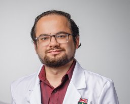 Shubham Misra合作论文数Dept Neurol, Yale Univ7
 Juliet Victoria Santiago合作论文数6
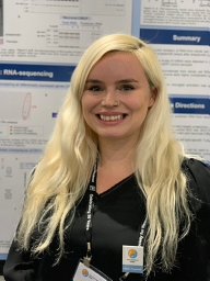 Christina C. Ramelow合作论文数6
 Aparna Bhardwaj合作论文数Sch Basic Sci, Indian Inst Technol Mandi6
 Namyashree Nayak合作论文数School of Biosciences and Bioengineering, Indian Institute of Technology Mandi6Michael Frankel合作论文数6
 Eric B Dammer合作论文数Dept Biochem, Emory Univ6
 Heike Wulff合作论文数Department of Pharmacology, University of California, Davis6Brendan R. Tobin合作论文数5
 Ashutosh Singh合作论文数5
 Nitin Sharma合作论文数5
 Annie M Goettemoeller合作论文数GDBBS Grad Program, Emory Univ5
 Sara Bitarafan合作论文数Georgia Institute of Technology5Bhuvaneshwari R Gehi合作论文数School of Basic Sciences, Indian Institute of Technology Mandi5Hein Katharina合作论文数5
 Steven A Sloan合作论文数Department of Neurobiology, Stanford University School of Medicine5
 Deepak Kumar合作论文数5
 Mathias Bähr合作论文数Department of Neurology, University Medicine Göttingen;Neurobiological Research Laboratory, University Medicine Göttingen;DZNE5
 Victor Faundez合作论文数Graduate Program in Neuroscience, Emory University5
 Sydney Sunna合作论文数4
 Sabine Herold合作论文数4
 Maureen M Sampson合作论文数Centers for Disease Control and Prevention , 4770 Buford Highway, Atlanta, Georgia 30341, United States.4Ali Tfaily合作论文数3
 Meenakshi Shegane合作论文数3Young Lin合作论文数3
 Sanjeev Kumar Singh合作论文数3Matthew JM Rowan合作论文数3




