Gene fusions are common primary drivers of pediatric leukemias and are the result of underlying structural variants (SVs). Current clinical workflows to detect such alterations rely on a multimodal approach, which often increases analysis time and overall cost of testing. In this study, we used long-read sequencing (lrSeq) as a proof-of-concept to determine whether clinically relevant (cr) SVs could be detected within a small (n = 17) pediatric leukemia cohort. We show that this methodology successfully determined all known crSVs (n = 5/5) detected through routine clinical testing. This approach also identified crSVs that resulted in the classification of a leukemia genetic subtype for four additional patients (n = 4/12), such as an ins(11;10)(q23.3;p12p12) forming a KMT2A::MLLT10 fusion, that were missed by routine clinical approaches. This study demonstrates the diagnostic potential of lrSeq as an assay for SV detection in pediatric leukemia and supports lrSeq as a valuable tool for the accurate detection of crSVs.
Gene fusions are common primary drivers of pediatric leukemias and are the result of underlying structural variant (SVs). Current clinical workflows to detect such alterations rely on a multimodal approach, which often increases analysis time and overall cost of testing. In this study, we used long-read sequencing (lrSeq) as a proof-of-concept to determine whether clinically relevant (cr) SVs could be detected within a small (n = 17) pediatric leukemia cohort. We show that this methodology successfully determined all known crSVs detected through routine clinical testing. We also identified crSVs, such as an ins(11;10)(q23.3;p12p12) forming a KMT2A::MLLT10 fusion, missed by routine clinical approaches, resulting in the classification of leukemia genetic subtypes for four additional patients. This study demonstrates the diagnostic potential of lrSeq as an assay for SV detection in pediatric leukemia and supports lrSeq as a valuable tool for the accurate detection of crSVs.
Gliomas harboring oncogenic ROS1 alterations are uncommon and primarily described in infants. Our goal was to characterize the clinicopathological features and molecular signatures of the full spectrum of ROS1 fusion-positive gliomas across all age groups. Through a retrospective multi institutional collaboration, we report a collection of unpublished ROS1 fusion gliomas along with the characterization and meta-analysis of new and published cases. A cohort of 32 new and 58 published cases was divided into the following 3 age groups: 19 infants, 40 pediatric patients, and 31 adults with gliomas. Tumors in infants and adults showed uniformly high-grade morphology; however, tumors in pediatric patients exhibited diverse histologic features. The GOPC::ROS1 fusion was prevalent (61/79, 77%) across all age groups, and 10 other partner genes were identified. Adult tumors showed recurrent genomic alterations characteristic of IDH wild-type glioblastoma, including the thorn 7/-10/CDKN2A deletion; amplification of CDK4, MDM2, and PDGFRA genes; and mutations involving TERTp, TP53, PIK3R1, PIK3CA, PTEN, and NF1 genes. Infant tumors showed few genomic alterations, whereas pediatric tumors showed moderate genomic complexity. The outcomes were significantly poorer in adult patients. Although not statistically significant, tumors in infant and pediatric patients with high-grade histology and in hemispheric locations appeared more aggressive than tumors with lower grade histology or those in nonhemispheric locations. In conclusion, this study is the largest to date to characterize the clinicopathological and molecular signatures of ROS1 fusion-positive gliomas from infant, pediatric, and adult patients. We conclude that ROS1 likely acts as a driver in infant and pediatric gliomas and as a driver or codriver in adult gliomas. Integrated comprehensive clinical testing might be helpful in identifying such patients for possible targeted therapy.& COPY; 2023 United States & Canadian Academy of Pathology. Published by Elsevier Inc. All rights reserved.
Though rare, pediatric high-grade gliomas (pHGG) are a leading cause of cancer-related mortality in children. We wanted to determine whether our currently available clinical laboratory methods could better define diagnosis for pHGG that had been archived at our institution for the past 20 years (1998 to 2017). We investigated 33 formalin-fixed paraffin-embedded pHGG using ThermoFisher Oncoscan SNP microarray with somatic mutation analysis, Sanger sequencing, and whole genome sequencing. These data were correlated with historical histopathological, chromosomal, clinical, and radiological data. Tumors were subsequently classified according to the 2021 WHO Classification of Paediatric CNS Tumours. All 33 tumors were found to have genetic aberrations that placed them within a 2021 WHO subtype and/or provided prognostic information; 6 tumors were upgraded from WHO CNS grade 3 to grade 4. New pHGG genetic features were found including two small cell glioblastomas with H3 G34 mutations not previously described; one tumor with STRN-NTRK2 fusion; and a congenital diffuse leptomeningeal glioneuronal tumor without a chromosomal 1p deletion but with KIAA1549-BRAF fusion. Overall, the combination of laboratory methods yielded key information for tumor classification. Thus, even small studies of these uncommon tumor types may yield new genetic features and possible new subtypes that warrant future investigations.
Disorders of sex development (DSD), formerly termed “intersex” conditions, arise from numerous causes. CAH secondary to 21-hydroxylase deficiency is the most common cause of DSD. Sex chromosome disorders, including sex chromosome mosaicism, are the second most common cause of DSD. We discuss a medically complex neonate with DSD presenting with ambiguous genitalia. Hormone levels suggested 21-hydroxylase deficiency. Molecular analysis revealed compound heterozygous mutations in the 21-hydroxylase gene ( CYP21A2 ), confirming the diagnosis of CAH. Chromosome analysis revealed sex chromosome mosaicism with three cell lines: 45,X[8]/45,X,tas(Y;16)(p11.32;p13.3)[8]/45,X,t(Y;8)(p11.32;p23.3)[4] had and remnants. Chromosome analyses showed mosaicism in both This is the first case of coexisting CAH and 45,X/46,XY mosaicism reported in the English literature and the third case of a constitutional chromosome Y “jumping translocation.” Our report documents the medical and genetic complexity of children such as this one with ambiguous genitalia and discusses the need for a multidisciplinary team approach.
Gliomas harboring ROS1 rearrangements comprise a rare genetic subgroup that has been poorly characterized to date. Our work aims to further characterize the histologic spectrum, molecular signature, and clinical manifestation of these rare tumors to facilitate better diagnosis and treatment approaches.
Deletions of 18q have been reported relatively frequently, with the majority of patients presenting with a deletion of the distal part including the region of 8q22q23 (distal 18q deletion). The deletion size varies between 0.5-30 Mb with unique breakpoints for each patient. While the phenotype of each individual shows great variability, there is a set of common clinical features for distal 18q deletion which include short stature, microcephaly, midface hypoplasia, hypertelorism, congenital aural atresia (CAA), foot deformities, mental retardation (MR), and hypotonia.
The most recent build of the human reference genome, GRCh38, was released in 2013. However, many laboratories performing next-generation sequencing (NGS) continue to align to GRCh37. Our aim was to assess the number of clinical diagnostic laboratories that have migrated to GRCh38 and discern factors impeding migration for those still using GRCh37. A brief, five-question survey was electronically administered to 71 clinical laboratories offering constitutional NGS-based testing and analyzed categorically. Twenty-eight responses meeting inclusion criteria were collected from 24 academic and four commercial diagnostic laboratories. Most of these (14; 50%) reported volumes of <500 NGS-based tests in 2019. Only two respondents (7%) had already migrated entirely to GRCh38; most laboratories (15; 54%) had no plans to migrate. The two prevailing reasons for not yet migrating were as follows: laboratories did not feel the benefits outweighed the time and monetary costs (14; 50%); and laboratories had insufficient staff to facilitate the migration (12; 43%). These data, although limited, suggest most clinical molecular laboratories are reluctant to migrate to GRCh38, and there appear to be multiple obstacles to overcome before GRCh38 is widely adopted.
Indolent T-lymphoblastic proliferation (iT-LBP) is a recently recognized benign pathologic condition commonly associated with Castleman disease. The lymph node of this condition shows preserved lymphoid architecture with proliferation of nonclonal T-lymphoblasts in the interfollicular area. iT-LBP is a rare condition and a diagnosis of exclusion. As a rule, it requires sophisticated techniques to exclude T-lymphoblastic leukemia/lymphoma (T-LBL/L). We present an unusual T-LBL/L in a lymph node mimicking iT-LBP. The case showed preserved nodal architecture, morphologic features of hyaline vascular Castleman disease, proliferation of T-lymphoblasts in the interfollicular area, and no clonal T-cell receptor gene rearrangement. The follow-up lymph node specimen excised 6 months later showed full-blown lymphoblastic lymphoma with an abnormal karyotype, near-early-T-precursor phenotype, partial CD19 expression, and SETD2 mutation.
Chromosomal microarray technologies, including array comparative genomic hybridization and single-nucleotide polymorphism array, are widely applied in the diagnostic evaluation for both constitutional and neoplastic disorders. In a constitutional setting, this technology is accepted as the first-tier test for the evaluation of chromosomal imbalances associated with intellectual disability, autism, and/or multiple congenital anomalies. Furthermore, chromosomal microarray analysis is recommended for patients undergoing invasive prenatal diagnosis with one or more major fetal structural abnormalities identified by ultrasonographic examination, and in the evaluation of intrauterine fetal demise or stillbirth when further cytogenetic analysis is desired. This technology also provides important genomic data in the diagnosis, prognosis, and therapy of neoplastic disorders, including both hematologic malignancies and solid tumors. To assist clinical laboratories in the validation of chromosomal microarray methodologies for constitutional and neoplastic applications, the American College of Medical Genetics and Genomics (ACMG) Laboratory Quality Assurance Committee has developed these updated technical laboratory standards, which replace the ACMG technical standards and guidelines for microarray analysis in constitutional and neoplastic disorders previously published in 2013.
Giant cell glioblastoma (GCG) is a rare histologic variant of glioblastoma that is characterized by numerous bizarre, multinucleate giant cells and abundant reticulin deposition. GCG occurs in younger patients (mean age, 44 years) and generally has a better prognosis than other variants of IDH-wildtype glioblastoma. Although prior studies have identified frequent TP53 alterations in these tumors, the genomics of GCG remain poorly characterized. Herein, we describe an inter-institutional analysis of seven patients ranging from age 9 to 68 of variable race and gender diagnosed with GCG. Across 100% (7/7) of specimens in our cohort, chromosomal microarray analysis and targeted next generation sequencing consistently identified near haploidization / massive loss of heterozygosity (LOH). Remarkably, the genomic profile of these tumors showed LOH of chromosomes 3, 4, 6, 8, 13, 14, 15, 17, 22 in all cases (7/7), and LOH of chromosomes 1, 2, 5, 9, 10, 11, 18, 19 in 6/7 cases. Notably, chromosome 7 always retained heterozygosity. We propose that this genomic profile represents a distinct subtype of GCG. These findings provide evidence of an unusual mechanism of tumorigenesis and could have the potential to reveal new tumor vulnerability.
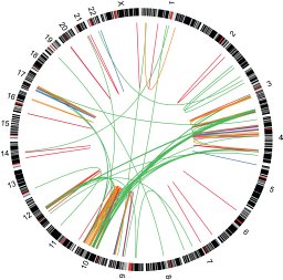
The Compendium of Cancer Genome Aberrations (CCGA, http://www.ccga.io) is a Cancer Genomics Consortium (CGC)-supported resource cataloging genomic/genetic abnormalities in neoplasia. This Wiki style interface promotes expert crowdsourcing, real-time editing and content sharing within our genomics community. The CCGA is intended to be a central hub of information with primary content plus direct links to pertinent literature and external databases for interpretation of molecular and cytogenetics cancer cases. Within the CCGA, expert authors add content to pages created based on the current World Health Organization classification designations or other entities of interest using a standardized template. Referenced information ranges from clinicopathologic descriptions of disease such as immunophenotype and clinical significance, to genomic and genetic characteristics including chromosome rearrangement/gene fusion, abnormality pattern, copy number aberrations, LOH, gene mutations, and epigenomics, with their associated affected genes and pathways. Circos and TCGA aggregate data plots summarizing genetic findings by disease type that demonstrate patterns are generated, as well as gene-specific pages for cancer-associated genes. In addition, disease overview pages for major categories such as AML allow for quick access to all AML-related content including data visualizations plus links to all gene and disease-related internal and external pages. Content has been completed for AML and B-ALL, with progress currently being made on additional myeloid disorders. Efforts to recruit volunteer experts are ongoing and an update on this resource will be provided. Continued support and input from CGC members will ensure the success of CCGA.
Background. Giant cell glioblastoma (gcGBM) is a rare histologic subtype of glioblastoma characterized by numerous bizarre multinucleate giant cells and increased reticulin deposition. Compared with conventional isocitrate dehydrogenase (IDH)-wildtype glioblastomas, gcGBMs typically occur in younger patients and are generally associated with an improved prognosis. Although prior studies of gcGBMs have shown enrichment of genetic events, such as TP53 alterations, no defining aberrations have been identified. The aim of this study was to evaluate the genomic profile of gcGBMs to facilitate more accurate diagnosis and prognostication for this entity. Methods. Through a multi-institutional collaborative effort, we characterized 10 gcGBMs by chromosome studies, single nucleotide polymorphism microarray analysis, and targeted next-generation sequencing. These tumors were subsequently compared to the genomic and epigenomic profile of glioblastomas described in The Cancer Genome Atlas (TCGA) dataset. Results. Our analysis identified a specific pattern of genome-wide massive loss of heterozygosity (LOH) driven by near haploidization in a subset of glioblastomas with giant cell histology. We compared the genomic signature of these tumors against that of all glioblastomas in the TCGA dataset (n = 367) and confirmed that our cohort of gcGBMs demonstrated a significantly different genomic profile. Integrated genomic and histologic review of the TCGA cohort identified 3 additional gcGBMs with a near haploid genomic profile. Conclusions. Massive LOH driven by haploidization represents a defining molecular hallmark of a subtype of gcGBM. This unusual mechanism of tumorigenesis provides a diagnostic genomic hallmark to evaluate in future cases, may explain reported differences in survival, and suggests new therapeutic vulnerabilities.
Giant cell glioblastoma (GCG) is a WHO-recognized rare histological variant of glioblastoma, characterized by multinucleated giant cells with marked nuclear pleomorphism and severe atypia. Clinically, GCGs occur in younger patients and are associated with a more favorable prognosis than IDH-wildtype glioblastoma, however the underlying mechanisms responsible for these clinicopathological differences remain poorly understood. Attempts to molecular characterize GCGs has been challenged by the rarity of these tumors types, misclassification and use of targeted (rather than genome-wide) genomic analyses. However, in the limited work that has been published, GCG have been shown to demonstrate a high incidence of TP53 mutations. We performed an inter-institutional analysis and identified seven patients ranging from age 9 to 68 of variable race and gender diagnosed with GCG. Across this cohort, chromosomal microarray and targeted next generation sequencing consistently identified near haploidization with massive loss of heterozygosity (LOH). LOH was observed in nearly all chromosomes, except for chromosome 7 which always retained heterozygosity. Our results demonstrate that GCGs possess a characteristic genomic profile and perhaps a unique mechanism of tumorigenesis, with diagnostic applications and the the potential to reveal new tumor vulnerabilities.
The OncoScan CNV Plus Assay (OS+) is a single-nucleotide polymorphism microarray platform that can detect 74 hotspot somatic mutations (SMs) in nine genes via molecular inversion probes. We report validation of the SM component of OS+ using a cohort of pediatric high-grade brain tumor specimens. SM calls were generated from 46 brain tumor cases, most tested orthogonally via bidirectional Sanger sequencing. The initial calling algorithm result showed that 31 tumors were positive and 15 were negative for SM, with a total of 71 OS+ SM calls [28 high-confidence (HC) and 43 low-confidence (LC)]. Sanger sequencing was performed for 54 of the 71 calls (27 HC and 27 LC), as well as for 21 randomly selected hotspots across the 15 OS+ negative cases. HC calls (except EGFR) Sanger sequencing confirmed positive, negative calls confirmed negative, but none of the LC calls were Sanger-confirmed positive. An update of the OS+ algorithm resolved the LC calls, but of the 11 HC SM EGFR calls, Sanger sequencing confirmed only one. Two PTEN SM calls by OS+ in two separate cases were also negative per Sanger sequencing. We conclude that a majority of HC OS+ SM calls were accurate, except calls identified in EGFR and PTEN. Clinically, we report SMs identified by OS+ only after Sanger sequencing verification.
AIMS:Historically, there has been no consensus on the diagnostic classification of high-grade B-cell lymphoma (HGBCL) with morphological features of Burkitt lymphoma (BL) but no MYC gene rearrangement (MYC-negative). The 2016 WHO classification of tumours of haematopoietic and lymphoid tissues has shed some light on this field with the modification of the grey-zone lymphoma with features intermediate between BL and diffuse large B-cell lymphoma, and the creation of several new entities. The aim of this study was to investigate how the revised WHO classification affects our practice in diagnosing these lymphomas in children.METHODS:We retrospectively reviewed cases of mature HGBCL diagnosed at our hospital between 2015 and 2018.RESULTS:Among 14 mature HGBCL cases with BL morphological features, 11 showed MYC rearrangement consistent with BL and 3 were MYC-negative. Two MYC-negative cases showed regions of 11q gain and loss by microarray consistent with Burkitt-like lymphoma with 11q aberration (BLL-11q). The third MYC-negative case showed diffuse and strong MUM1 expression, translocation involving 6p25 by chromosome analysis and IRF4 rearrangement by fluorescence in situ hybridisation analysis consistent with large B-cell lymphoma with IRF4 rearrangement (LBL-IRF4). All patients were treated according to applicable chemotherapeutic protocols and achieved remission.CONCLUSIONS:BLL-11q and LBL-IRF4, two newly defined entities, should be considered in paediatric MYC-negative mature HGBCL cases. Accurate diagnosis needs careful histopathological examination and proper cytogenetic testing. Since they have unique cytogenetic features, specific treatments for them may emerge in the future. Therefore, accurate diagnosis based on the 2016 WHO classification is clinically significant.
Brain tumors are the leading cause of cancer-related death in children and young adults. Molecular genetic/genomic characteristics subclassify brain tumors, which associate with different outcomes and identify pathways for therapeutic options.
Neuroblastoma (NB) in children older than 10 years is rare. We reviewed our archives for patients with NB aged 10 to 18 years and summarized their clinicopathologic/genetic records. Of 96 patients, 4 patients were identified in this age group. Four tumors were abdominal; 1 patient had 2 tumors at diagnosis, one of which was presacral. Tumor sizes ranged from 3 to 20 cm. All tumors were high risk at clinical stages 3 and 4, with metastasis to bone marrow and other areas. Four tumors were poorly differentiated with unfavorable histology and one patient with bilateral adrenal disease had an intermixed ganglioneuroblastoma on one side. Another tumor exhibited pheochromocytoma-like morphology. MYCN amplification was present in bone marrow metastasis in one case. Complex chromosomal gains and 19p deletions were common. Exome sequencing revealed ALK variants in 2 cases and previously unreported MAGI2, RUNX1, and MLL mutations. All patients received standard chemotherapy and 2 patients received ALK-targeted trial therapy. Three patients died of disease, ranging 18 to 23 months after diagnosis. One patient has active disease and is receiving trial therapy. In conclusion, NB in children older than 10 years may exhibit unusual clinicopathologic and genetic features with large tumors, bilateral adrenal disease, rare morphologic features, complex DNA microarray findings and novel mutations. Patients often have grim prognoses despite genomic profiling-guided targeted therapy.
Medulloblastoma in a Patient with Curry-Jones Syndrome with a mosaic variant, c.1234C > T (p.Leu412Phe), in SMO.
 Daynna J. Wolff合作论文数Department of Pathology and Laboratory Medicine, College of Medicine, Medical University of South Carolina16
 Farooqi Midhat S合作论文数15Melissa Gener合作论文数13
 Elena Repnikova合作论文数13Kathleen W. Rao合作论文数10Weijie Li合作论文数9
 Adrian Dubuc合作论文数Department of Pathology, Brigham and Women's Hospital and Harvard Medical School, Boston, Massachusetts, USA.9Ginn Kevin F合作论文数Division of Neuro-Oncology, St. Jude Children's Research Hospital9Herriges John C合作论文数8Donald H. Mahoney合作论文数8
 Nyla Heerema合作论文数Cytogenetics Lab, Ohio State University8
 Arthur R Brothman合作论文数7Mikhail Fady M合作论文数7Persons D L合作论文数7Hirsch Betsy合作论文数7Lisa Warren合作论文数6Byunggil Yoo合作论文数6August Keith J合作论文数Children's Mercy Hospital6
 Scott Newman合作论文数Hutchison-MRC Research Centre and Department of Pathology, University of Cambridge6Sj Lauer合作论文数5
 David Meredith合作论文数Dept Pathol, Brigham & Womens Hosp5
 Lina Shao合作论文数Department of Pathology, Medical School, University of Michigan-Ann Arbor5Camitta B M合作论文数5Jaclyn A. Biegel合作论文数5
 Merlin G. Butler合作论文数The University of Kansas Medical Center5David L. Zwick合作论文数5Nancy R. Schneider合作论文数5
 Douglas C Bittel合作论文数Children's Mercy Hospitals and Clinics, University of Missouri-Kansas City School of Medicine5Jt Mascarello合作论文数4KW RAO合作论文数4Dj Pullen合作论文数4MJ VUCHICH合作论文数4Vm Whitehead合作论文数4C Payment合作论文数4Lisa A Lansdon合作论文数aDepartment of Pathology and Laboratory Medicine, Children's Mercy - Kansas City4
 Xinyan Lu合作论文数Department of Pathology, Feinberg School of Medicine, Northwestern University4
 Scott C Smith合作论文数Kentucky Spinal Cord Injury Research Center and Department of Neurological Surgery, University of Louisville School of Medicine4
 Emily G Farrow合作论文数School of Medicine, University of Missouri-Kansas City4
 M John Hicks合作论文数Baylor College of Medicine4Neil Miller合作论文数4
 Carol J Saunders合作论文数4
 Warren Sanger合作论文数Munroe-Meyer Institute, University of Nebraska Medical Center4
 A. Thomas Look合作论文数Department of Pediatric Oncology, Dana-Farber Cancer Institute, Inc.;Department of Pediatrics, Harvard Medical School, Harvard University;Children’s Hospital Boston4
 Gordon Wayne Dewald合作论文数College of Medicine, Mayo Clinic;Division of Laboratory Genetics, Mayo Clinic4Isabelle Thiffault合作论文数4Nataliya Kibiryeva合作论文数4
 Andrew J Carroll Ill合作论文数School of Medicine, University of Alabama at Birmingham4Wilbur R. Harrison合作论文数4




