AIM:Determine the impacts of pre- and early-post-natal nutrition on selected markers of hepatic glucose and fat metabolism.METHODS:Twin-bearing ewes were fed 100% (NORM) or 50% (LOW) of protein and energy requirements during the last 6-weeks of gestation. Twin-lambs received either a high-carbohydrate high-fat (HCHF) or conventional (CONV) diet from 3 days to 6 months of age (around puberty), whereafter lambs from the four subgroups were slaughtered (16 males/3 females). Remaining lambs (19 females) were fed a moderate diet and slaughtered at 2 years of age (young adults).RESULTS:Pre-natal LOW nutrition was associated with increased hepatic triglyceride, ceramide and free fatty acid content in adulthood (not observed in lambs), which was accompanied by up-regulated early-stage insulin signalling as reflected by increased INSRβ and PI3K-p110 protein expression. The HCHF diet increased hepatic triglyceride content in lambs, associated with down-regulated expressions of energy-metabolism-related genes (GLUT1, PPARα, SREBP1c, PEPCK). These post-natal effects were not observed in adult HCHF sheep, after they had received a moderate (body-fat correcting) diet for 1.5 years. Interestingly, pre-natal LOW nutrition induced permanent alterations in hepatic phospholipids' fatty acid composition. Thus, the amount of linoleic acid (C18 : 2 ∆(9,12)) was significantly increased and composition of rumen-derived fatty acids were altered, indicating changed composition of rumenal microbiota.CONCLUSION:Hepatic insulin signalling and linoleic and microbial-derived fatty acid content in phospholipids are targets of foetal programming induced by late-gestation undernutrition. Future studies are required to explain their cause-effect associations with increased risks of developing hepatic steatosis and insulin insensitivity in adulthood.
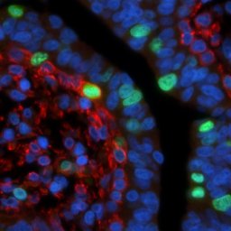
The present study aimed to 1) elucidate whether continuous milking during late gestation in dairy goats negatively affects mammary remodeling and hence milk production in the subsequent lactation, and 2) identify the regulatory factors responsible for changes in cell turnover and angiogenesis in the continuously lactating mammary gland. Nine multiparous dairy goats were used. One udder half was dried off approximately 9 wk prepartum (normal lactation; NL), and the other udder half of the same goat was milked continuously (continuous lactation; CL) until parturition or until the half-udder milk yields had dropped to below 50 g/d. Mammary biopsies were obtained from each udder half just before the NL gland was dried off (before dry period), within the first 2 wk after drying-off (early dry period, samples available only for NL glands), in the mid dry period, within the last 2 wk before parturition (late dry period), and at d 1 (the day of parturition), 3, 10, 60, and 180 of lactation. Mammary morphology was characterized in biopsies by quantitative histology, and cell turnover was determined by immunohistochemistry (terminal deoxynucleotidyl transferase dUTP nick end labeling and Ki-67). Transcription of genes encoding factors involved in mammary epithelial cell (MEC) turnover and vascular function was quantified by quantitative reverse transcription PCR. Results demonstrated that omitting the dry period was possible in goats but was not as easy as claimed before. Renewal of MEC was suppressed in CL glands, which resulted in a smaller MEC population in the subsequent lactation. At the time of parturition (and throughout lactation), the mammary glands subjected to CL had smaller alveoli, more fully differentiated MEC, and a substantially larger capillary fraction compared with NL glands. The continuously lactating gland thus resembled a normally lactating gland in an advanced stage of lactation. None of the studied genomic factors could account for these treatment differences. The described characteristics in CL glands compared with NL glands indicated a gland maintained in lactation for a longer period.
Milk production is generally lower but lactation persistency higher in primiparous (PP) than in multiparous (MP) goats. This may be related to differences in development and maintenance of mammary gland function, but the underlying mechanisms are not well understood. The present study aimed to elucidate whether differences in lactational performance between PP and MP mammary glands are related to the time course of development and maintenance, not only of the mammary epithelial cell (MEC) population, but also of the mammary vasculature that sustains synthetic activity. Mammary biopsies were obtained from both mammary glands of 3 PP and 6 MP (>or=2 parity) dairy goats at parturition (d 1), d 10, 60, and 180 of lactation. Gene transcription relating to MEC turnover and vascular function was quantified by real-time reverse transcription-PCR, mammary morphology was characterized (quantitative histology), and cell turnover was determined (terminal deoxynucleotidyl transferase dUTP nick end labeling assay and Ki-67). Primiparous glands showed higher expression for the genes involved in angiogenesis; namely, vascular endothelial growth factor receptor 2, and angiopoietin 1 and 2 and their receptor, a few days after parturition (d 10). Primiparous glands also had higher rates of MEC proliferation in early lactation. It therefore appears that initiation of lactation is associated with development and growth of the mammary gland into early lactation, which continues for a longer period in PP compared with MP glands. In addition, MEC survival was found to be higher in PP glands throughout lactation, and MEC in PP glands underwent more extensive differentiation. This could explain the reported flatter lactation curve and higher lactation persistency in PP glands. Although some of the genes included in this study were differentially expressed in PP and MP glands during the course of lactation, it was not possible to identify any specific genomic factor(s) that could account for the differences between PP and MP glands with respect to mammary development and MEC survival during lactation. It remains to be established why parity number affects MEC and vascular development and survival during lactation, and, in particular, which regulatory mechanisms are involved.
 Mette Olaf Nielsen合作论文数Department of Animal and Veterinary Sciences, Aarhus University4
 M. Engbæk合作论文数2
 Allan Vaag合作论文数Department of Biomedical Sciences, University of Copenhagen;Steno Diabetes Center Copenhagen;Lund University2
 Sina Safayi合作论文数Faculty of Life Sciences, University of Copenhagen2
 Peter Kappel Theil合作论文数Department of Animal Health;Welfare and Nutrition;University of Aarhus2
 Kristen Sejrsen合作论文数Department of Animal Science, Aarhus University2
 Jan Værum Nørgaard合作论文数Faculty of Agricultural Sciences, Aarhus University2g m crovetto合作论文数1
 Lars I Hellgren合作论文数Dept Syst Biol, Tech Univ Denmark1
 Emma Nilsson合作论文数1Kongsted A H合作论文数1Vibeke S. Elbrønd合作论文数1




