The M-shaped zygomatic arch fracture consists of anterior and posterior segments, presenting an M-shaped configuration when viewed from the skull base. However, the extent of involvement of the anterior and posterior fracture segments has not been reported. This study aims to define the range of M-shaped zygomatic arch fractures. Forty-six patients were enrolled in this study. Clinical data along with CT images were collected from the patients. Landmarks were identified on coronal CT sections using the horizontal plane at the zygomatic arch root as the reference plane. The deepest point of the fracture depression was located at the zygomaticotemporal suture in 91.30
PurposeThe migration and fusion of osteoclast precursors (OCPs) are critical steps in osteoclastogenesis and require substantial energy. Although oxidative phosphorylation (OXPHOS) is generally considered the major pathway supplying energy for these processes, it remains unclear how inhibiting OXPHOS affects the migration of OCPs.Materials and MethodsTo investigate the metabolic regulation of osteoclast migration, we used a recognized OXPHOS inhibitor rotenone to suppress the key energy-producing pathway in OCPs. We combined this intervention with bioinformatics approaches and machine learning algorithms to screen and identify genes associated with osteoclast dysfunction in patients with osteoporosis. The migratory capacity of OCPs was precisely quantified using optical tweezers.ResultsWe found that rotenone inhibited osteoclastogenesis and markedly impaired the migratory capacity of OCPs. Although the level of OXPHOS in OCPs decreased, intracellular ATP content paradoxically increased, suggesting that the impairment of migratory capacity is unlikely to be driven by an overall energy shortage. Through machine learning algorithms, we identified genes associated with abnormal osteoclast function in patients with osteoporosis and discovered their critical regulatory roles in processes such as cell migration, adhesion, and OXPHOS. Rotenone significantly suppressed the expression of these genes in OCPs, suggesting a direct mechanism for the impaired migration.ConclusionsIn summary, our study reveals a novel mechanism for rotenone-induced inhibition of osteoclastogenesis, which is achieved by impairing the migration of OCPs in a manner uncoupled from overall energy status.
Osteoarthritis (OA) is a prevalent joint disease characterizedby chronic, progressive inflammation and cartilage degeneration, for which current treatments remain limited. In this study, we propose a dual-drug delivery strategy that simultaneously suppresses inflammation and rejuvenates impaired cartilage by incorporating kartogenin (KGN) and methylprednisolone hemisuccinate (MPHS) into a single microsphere system with sequential release in the local microenvironment. To achieve coordinated dual-drug release, KGN and MPHS were loaded into the inner core and outer layer of the microspheres, respectively. Both KGN and MPHS exhibited sustained release profiles; however, MPHS showed a shorter burst-release phase than KGN due to the protective effect of the outer layer. The release of MPHS effectively suppressed interleukin-1β (IL-1β)-induced inflammation in bone marrow stromal cells (BMSCs) pellets, thereby enhancing KGN-mediated chondrogenic differentiation of BMSCs in vitro. In parallel, sustained delivery of KGN also led to the recruitment of BMSCs and subsequent chondrogenesis, ultimately leading to cartilage rejuvenation. Importantly, the sequential release of KGN and MPHS from the dual-drug microspheres synergistically enhanced in vitro chondrogenic differentiation of BMSCs, resulting in concomitant inflammation alleviation and cartilage repair. Collectively, these findings demonstrate that the KGN/MPHS-incorporated microspheres possess dual chondrogenic and anti-inflammatory functions and represent a promising therapeutic strategy for OA treatment.
Bone and cartilage defects resulting from trauma, degenerative diseases, or congenital malformations remain a major clinical challenge due to the limited intrinsic healing capacity of these tissues often leads to unsatisfactory outcomes. Piezoelectric biomaterials, which are capable of generating localized electrical signals under mechanical stimulation, have attracted considerable attention as they could mimic the electromechanical microenvironment of native tissues and modulate key cellular processes. However, conventional fabrication strategies were usually failed to meet the personalized requirements of bone and cartilage regeneration. Three-dimensional (3D) printing offers powerful tools for producing patient-specific scaffolds with complex architectures and controlled functionality. In this review, we firstly introduced the piezoelectric properties of the natural bone and cartilage tissue, and then discussed the characteristics of piezoelectric materials in regenerative medicine, with particular emphasis on the advantages and limitations of usage of 3D printing techniques in the fabrication of the piezoelectric biomaterials. Finally, we summarized the recent advances in 3D-printed piezoelectric scaffolds for bone and cartilage regeneration. Consequently, this review highlights the significant potential and practical value of 3D-printed piezoelectric scaffolds as the next generation of osteochondral implants.
Osteochondral scaffolds designed with bi-phasic and multi-phasic have typically struggled with post-implantation delamination. To address this issue, we developed a novel integrated scaffold with natural and continuous interface and heterogeneous bilayer structure. Through layer-by-layer wet electrospinning, two-dimensional (2D) bi-layer integrated membranes of silk fibroin (SF) and polycaprolactone (PCL) were fabricated. These membranes were then transformed into three-dimensional (3D) scaffolds using a CO2 gas foaming technique, followed by gelatin coating on the osteogenic layer to afford final bi-phasic porous scaffolds. In vitro studies indicated that the 3D scaffolds better-maintained cell phenotypes than conventional 2D electrospun films. Additionally, the 3D scaffolds showed superior cartilage repair and osteoinductivity potential, with increased subchondral bone volume and reduced defect area in rat osteochondral defects models at 12 weeks. Taken together, these gas-foamed scaffolds were a promising candidate for osteochondral regeneration.
In vitro mineralization has been proposed as promising approach for fabricating mineralized scaffolds and repairing bone defects. In this study, various mineralized glycidyl methacrylate-modified silk fibroin (GMA-SF) hydrogels were developed using two mineralization techniques: calcium chloride/sodium phosphate (CaCl2/Na2HPO4) solution immersion and simulated body fluid (SBF) solution immersion. Compared to the SBF immersion method, the CaCl2/Na2HPO4 method produced a greater quantity of minerals on the surface of GMA-SF hydrogels within the same mineralization duration and cycle numbers. Moreover, the mineralized phase in CaCl2/Na2HPO4 group exhibited a more uniform distribution and tighter arrangement along the hydrogel surface compared to the SBF group. Although both EDS and XPS analyses confirmed the presence of calcium (Ca) and phosphorus (P), significantly higher contents of Ca and P were observed in the GMA-SF hydrogels treated with the CaCl₂/Na₂HPO₄ method compared to the SBF method. This was also accompanied by stronger compressive strength in the CaCl2/Na2HPO4 group. Furthermore, in vitro and in vivo studies demonstrated that the mineralized GMA-SF hydrogels from both groups promoted osteogenic differentiation and bone regeneration, with the CaCl₂/Na₂HPO₄ group showing superior efficacy. These findings highlight the advantages of the CaCl₂/Na₂HPO₄ mineralization method over the SBF method for bone regeneration applications using GMA-SF hydrogels.
Periodontitis is a prevalent chronic inflammatory disease characterized by alveolar bone resorption mediated by osteoclasts. Pyruvate kinase M2 (PKM2), a key enzyme in glycolysis and pyruvate metabolism, has recently been recognized for its regulatory roles beyond metabolism, including gene expression and protein kinase activity. However, its exact role in osteoclastogenesis remains unclear. This study investigates the function of PKM2 in inflammatory osteoclastogenesis and explores its potential as a therapeutic target for periodontitis. Using murine bone marrow-derived macrophages (BMMs) stimulated with lipopolysaccharides (LPS) to mimic inflammatory conditions in vitro, we analyzed PKM2 expression and glycolytic activity during osteoclastogenesis through bioinformatics, tartrate-resistant acid phosphatase (TRAP) staining, phalloidin staining, quantitative real-time PCR (RT-qPCR), and Western blotting. Glycolysis was inhibited using 2-deoxy-D-glucose (2-DG), while TEPP-46 was used to activate PKM2. In a mouse model of periodontitis, the effects of TEPP-46 on alveolar bone loss were evaluated using micro-computed tomography, immunohistochemistry, TRAP staining, and hematoxylin-eosin (HE) staining. The results demonstrated that LPS significantly enhanced osteoclastogenesis and glycolysis, increasing PKM2 expression in osteoclasts. Inhibiting glycolysis with 2-DG suppressed osteoclast formation and osteoclast-related gene expression under inflammatory conditions. TEPP-46 treatment reduced nuclear dimeric PKM2 levels, decreased phosphorylated signal transducer and activator of transcription three (p-STAT3) expression, and inhibited osteoclastogenesis and osteoclast-related gene expression. Co-immunoprecipitation confirmed an interaction between nuclear dimeric PKM2 and p-STAT3. In vivo, TEPP-46 effectively reduced alveolar bone loss by preventing PKM2 nuclear translocation and STAT3 phosphorylation. These findings reveal that PKM2 regulates inflammatory osteoclastogenesis through modulation of glycolysis and STAT3 signaling, highlighting its potential as a therapeutic target for periodontitis.
The complex or compromised bone defects caused by osteomyelitis, malignant tumors, metastatic tumors, skeletal abnormalities, and systemic diseases are difficult to be self-repaired, leading to a non-union fracture. With the increasing demands of bone transplantation, more and more attention has been paid to artificial bone substitutes. As biopolymer-based aerogel materials, nanocellulose aerogels have been widely utilized in bone tissue engineering. More importantly, nanocellulose aerogels not only mimic the structure of the extracellular matrix but could also deliver drugs and bioactive molecules to promote tissue healing and growth. Here, we reviewed the most recent literature about nanocellulose-based aerogels, summarized the preparation, modification, composite fabrication, and applications of nanocellulose-based aerogels in bone tissue engineering, as well as giving special focus to the current limitations and future opportunities of nanocellulose aerogels for bone tissue engineering.
The healing of large bone defects remains a significant challenge in clinical practice. Accelerating both angiogenesis and osteogenesis can promote effective bone healing. In the natural healing process, angiogenesis precedes osteogenesis, providing a blood supply that supports the subsequent progression of osteogenesis. Developing a biomimetic scaffold that mimics the in vivo environment and promotes the proper sequence of vascularization followed by ossification is crucial for successful bone regeneration. In this study, a novel injectable dual-drug programmed releasing chitosan nanofibrous microsphere-based poly(D, L-lactide-co-glycolide)-b-poly(ethylene glycol)-b-poly(D,L-lactide-co-glycolide) (PLGA-PEG-PLGA) hydrogel is fabricated by incorporating vascular endothelial growth factor (VEGF) and microspheres loaded with dental pulp stem cellsderived exosomes (DPSCs-Exo). Rapid release of VEGF promotes the swift initiation of angiogenesis, while DPSCs-Exo release ensures persistent osteogenesis. Our results demonstrate that chitosan microsphere-based PLGA-PEG-PLGA hydrogel significantly promotes angiogenesis in human umbilical vascular endothelial cells and enhances the osteogenic differentiation of pre-osteoblasts. Furthermore, in vivo transplantation of this injectable chitosan microsphere-based PLGA-PEG-PLGA hydrogel into calvarial bone defects markedly promotes bone formation. Overall, our study provides a promising approach for improving bone regeneration by temporally replicating the behavior of angiogenesis and osteogenesis.
目的:探索结缔组织生长因子(CTGF)对尼克样 1 型蛋白(NELL-1)诱导的MC3T3-E1 细胞成骨分化能力的影响.方法:以MC3T3-E1 细胞为研究对象,分别使用PBS(对照)、NELL-1、CTGF和NELL-1+CTGF处理细胞,利用CCK-8 法检测各组细胞增殖情况;成骨诱导后,利用茜素红染色和定量分析、碱性磷酸酶(ALP)活性检测和ALP 染色、实时荧光定量PCR和Western blot实验检测各组细胞成骨分化情况.结果:与对照组相比,CTGF一定程度上抑制MC3T3-E1 细胞增殖;NELL-1单独处理可诱导细胞形成更多矿化结节,增强ALP活性,加深ALP染色,明显提高Runt相关转录因子 2(RUNX2)、ALP、成骨细胞特异性转录因子Osterix(OSX)等成骨形成基因的转录和蛋白表达水平(P<0.01).与NELL-1单独处理组相比,NELL-1+CTGF联合处理组中细胞内矿化结节形成减少,ALP活性降低,ALP染色变浅,RUNX2、ALP、OSX等基因的转录和蛋白表达水平显著降低(P<0.05).结论:CTGF抑制NELL-1诱导MC3T3-E1 细胞的成骨分化能力.
Objective: This study was performed to explore bone remodelling in children with intracapsular condylar fractures after the condylar fracture fragments were fixed using long screws and to offer possible explanations about the underlying mechanism. Patient and Methods: Records of children (less than 12 y old) who sustained intracapsular condylar fractures and fixed with long screws from May 2012 to January 2015 were retrieved. Age, gender, dates of injury, admission, and discharge, mechanism of trauma, location and pattern of fracture, other mandibular fractures, treatment methods, and time of review were recorded and analyzed. Image dates of pretreatments and posttreatments, including the date of review, were also recorded. Results: A total of 8 patients completed their follow-up, and all patients (n=5) who were followed up after more than 3 months showed serious resorption of the condylar head. The condylar head resorbed until the height (or articular surface) dropped and aligned with the surface of the screw. The shortest time of absorption, as shown by the computed tomography scan was 106 days, and the longest time was 171 days (average time of 141.8 d). Conclusions: Intracapsular condyle fractures in children should be managed conservatively as much as possible. However, if the height of the fracture fragments drops remarkably, open reduction and rigid internal fixation become possible choices.
This study aimed to explore and impart understanding of bone remodelling in children with intracapsular fractures treated conservatively. Records of children (less than 12 years), who sustained intracapsular fractures and treated conservatively, were retrieved consecutively for the period of March 2011 to February 2016. Data about age, gender, date of injury, dates of admission and discharge, mechanism of trauma, location and pattern of fracture, other mandibular fractures, treatment methods and time of review were recorded and analysed. Image dates of pre- and post-treatments, including date of review, were also recorded. A total of 22 patients complete their follow-up and show bone remodelling process. During their follow-up, all the displaced condylar fragments fused with the ramus stump at the displaced position. Regardless of the type of conservative procedure, both treatments cannot promote the spontaneous fracture reduction in patients with intracapsular condylar fractures. During follow-up, the absorption of the lateral process of the condyle after the closed treatment becomes close to the 'horizontal absorption', until the height (or articular surface) of the lateral condylar process dropped and aligned to the articular surface of the medial process. In children with intracapsular condylar fractures, the fracture fragment of the condyle determines the ramus height of the mandible. Closed treatment cannot restore the fracture fragment. If the height of the fracture fragments dropped remarkably, then open reduction and rigid internal fixation become more suitable.
PURPOSE:Screw osteosynthesis is advocated for the treatment of sagittal fracture of mandibular condyle (SFMC). This study aimed to explore the applicability of resorbable-screw osteosynthesis in the treatment of SFMC. METHODS:A retrospective cohort study was performed in patients with SFMC treated with resorbable-screw osteosynthesis (group A) from June 2011 through June 2021. The patients who had undergone titanium-screw osteosynthesis served as the control group (group B). The primary outcome variable was fracture healing, defined as follows: 1) normal mouth opening and restoration of pretrauma occlusion; 2) without complications or discomfort of temporomandibular joint symptoms; and 3) fracture union without abnormal reactions or bone resorption in computed tomography images. The secondary outcome variable was condylar morphological changes including radiographic imaging appearance of the condyle, mandibular ramus height (MRH), anteroposterior diameter (APD), and mediolateral diameter (MLD) of the condyle, which were assessed by comparing the computed tomography images 1 week after surgery with those of 3 months after surgery. The collected data of the outcome variables of the 2 groups were analyzed correspondingly using Student's paired t test and Student's t test. RESULTS:There were 24 patients in group A and 71 patients in group B. All the patients displayed an evident improvement in mouth opening and restored pretrauma occlusion. Few patients had complications (group A, 8.33%; group B, 9.86%) and discomfort of temporomandibular joint symptoms (group A, 16.67%; group B, 15.49%). Fracture union without abnormal reactions or bone resorption was observed during the follow-up. The radiographic evaluation revealed no significant difference in the MRH, the maximum APD, and MLD of the condyles between 1 week and 3 months after surgery in both groups. There were no significant intergroup differences in the changes in the MRH, APD, and MLD of the condyles. CONCLUSIONS:Resorbable-screw osteosynthesis is a viable option for the treatment of SFMC.
The poor mechanical performance of the aerogels hampered their potential applications in bone tissue engineering. To reinforce their mechanical properties, an electrospinning nanofibers-incorporated aerogels were fabricated in this study. Here, silk fibroin solution is blended with chitin in various proportions, and then electrospun to produce the hybrid nanofibers. The silk fibroin/chitin hybrid nanofibers were incorporated into the SF-based composite aerogels to enhance their mechanical properties. nHA was deposited onto the surface of the nanofiber-incorporated aerogels via in situ biomineralization, leading to the nanofibers/nHA-reinforced composite aerogels. The as-prepared composite aerogels exhibit favorable mineralization abilities through immersing in simulated body fluid. SEM observation suggested that the mineralization degree of the SF-based aerogels was positively related with the content of electrospinning nanofibers in the composite aerogels. The results obtained from the compress stress-strain curves revealed that the incorporation of nanofibers and in vitro biomineralization of nHA both enhanced the mechanical performance of the composite aerogels. The osteogenic ability of the prepared aerogels was also improved after incorporation of NFSF-0.75%CT and nucleation of nHA in SBF. Therefore, the composite aerogels prepared by the incorporation of NFSF-0.75%CT and immersing into SBF exhibited an enhanced mechanical properties and improved osteogenic ability.
OBJECTIVE:This study aimed to explore bone remodeling after condylar fracture fragments have been removed from patients with intracapsular condylar fractures. it also evaluated whether condyle fracture removal can be used alternatively when the authors treat patients with comminuted or small pieces of fracture or in extremely difficult operations.METHODS:Records of patients who sustained intracapsular condylar fractures and treated by removal of fragments for the period of February 2013 to September 2019 were retrieved. Data about age, gender, date of injury, dates of admission and discharge, mechanism of trauma, location and pattern of fracture, other mandibular fracture, treatment methods and time of review were recorded and analyzed. Image dates of pre- and post-treatment (including the time of review) were also recorded.RESULTS:The data of a total of 103 patients ranging from 5 to 84 years old were retrieved during this study. A total of 135 sides of condylar fragments were removed. Almost all of the patients with comminuted condyle head fracture or type A fracture presented apparent shortening of the ramus height, and none of them showed osteogenesis (or new bone formation) during their follow-up. Present study only observed osteogenesis in few patients who sustained type B/C intracapsular condylar fractures during their follow-up. The younger the patient was, the longer the follow-up time was, and the higher the possibility of new bone formation was. No correlation was found between the amount of osteogenesis and follow-up time, the amount of osteogenesis was generally small, and no patient could form a new condyle head similar to the normal (or original) condyle head. Condylar hypertrophy only occurred in children. Four patients developed temporomandibular joint ankylosis.CONCLUSIONS:Removal of fracture fragments is an alternative treatment option for patients who sustained comminuted or small pieces of fracture or in extremely difficult operations.
BACKGROUND:Isolated depressed zygomatic arch fractures are often treated with closed reduction. Reduction is usually performed through the Gillies approach or Keen approach. comminuted zygomatic arch fractures generally require open reduction and fixation to achieve good results. This article describes how to use a pair of surgical templates to assist in the fixation of comminuted zygomatic arch fractures with absorbable plates.METHODS:A pair of computer-designed surgical templates were applied to restore the main part of zygomatic arch. Placing a surgical template on the medial side of the zygomatic arch can provide a supporting force and improves the stability of the reduced bone fragments. The lateral template of zygomatic arch limits the excessive uplift of bone fragments.RESULTS AND DISCUSSION:The operation was performed according to the predetermined procedure. Postoperative computed tomography showed satisfactory reduction effect. In conclusion, with the aid of surgical templates, the reduction and fixation of comminuted zygomatic arch fractures can be more easily performed using absorbable plates.
Objective:This study aims to reveal the reconstruction process in pediatric patients with extracapsular condylar fractures after conservative treatment. We clarify that the "upright" position (or "recontouring" or favorable prognosis) of condyles is not a result of the anatomical reduction of the deviated condylar processes but originates from the remodeling of the skeleton. We also explore the related mechanism.Methods:The sample consisted of 27 pediatric patients aged less than 12 years who presented with extracapsular condylar fractures and were treated conservatively within an 8-year period (June 2011-April 2019). Data on the age, gender, date of injury, mechanism of trauma, location and pattern of mandibular condylar fracture and associated injuries and treatment methods of the patients were obtained. The process of bone remodeling in condyles was also recorded and analyzed.Results:The 27 children in this study sustained 33 extracapsular condylar fractures over the 8-year period of record retrieval. Amongst these fractures, 8 (24.2%) and 25 (75.8%) were condylar neck and condylar base fractures, respectively. Deviation and green-stick fractures were the predominant types and accounted for over 3 quarters of the condylar neck and base fractures (28, 84.8%), followed by dislocation fracture (3, 9.1%), displacement fracture (1, 3.0%), and non-displaced fracture (1, 3.0%). The period of follow-up ranged from 2 days to 257 days (average, 58.78 days). Only 1 patient with bilateral extracapsular condylar fractures showed vertically reconstructed condyles, which indicates an upright position of the condylar processes. One patient showed less angulation after treatment than before treatment, 1 patient revealed greater angulation after treatment than before treatment and all other patients (20 patients) showed the same angulation pre- and post-treatment. Both patients with only extracapsular condylar fractures showed no obvious deviations in dentition and facial asymmetry after their injury and treatment. The shortest and longest times observed for bone remodeling were 33 and 256 days, respectively. Children whose condylar head remained completely or at least partly inside the glenoid fossa showed satisfactory remodeling results during follow-up. Computed tomography scan during follow-up generally showed bone regeneration in the lateral condyle articular surface and the medial portion of the ascending ramus and bone resorption in the displaced direction (ie, the medial condyle head became sharp). Condylar heads displaced completely outside of the glenoid fossa showed serious shortening of the ascending ramus, and no obvious bone remodeling was observed. Only 1 patient with bilateral extracapsular condylar fractures showed a normal contour (ie, a vertically reconstructed condyle reflecting the upright position of the condylar processes) after 8 months.Conclusion:Stress stimulation originating from the glenoid fossa and ascending ramus of the mandible is a prerequisite for good condylar reconstruction. Conservative treatment could be carried out if the condylar head remains completely or at least partly inside the glenoid fossa. When the condylar head is dislocated completely outside the glenoid fossa, the glenoid-condylar relationship ceases to exist, joint function is lost and the height of the ascending ramus is significantly reduced. In this case, open reduction may be suitable.
BACKGROUND:Adenosine is a purine nucleoside involved in regulating bone homeostasis through binding to A1, A2A, A2B, and A3 adenosine receptors (A1R, A2AR, A2BR, and A3R, respectively). However, the underlying mechanisms by which adenosine and receptor subtypes regulate osteoclast differentiation remain uncertain. This study aims to assess the role of exogenous adenosine and receptor subtypes in receptor activator of NF-κB ligand (RANKL)-induced osteoclast formation and explore the underlying molecular mechanisms.METHODS AND RESULTS:The nanofibrous mats incorporated with adenosine exhibited robust ability to facilitate rat critical-size calvarial defect healing with decreased number of osteoclasts. Moreover, exogenous adenosine substantially enhanced the expression of A2AR and suppressed tartrate-resistant acid phosphatase-positive osteoclast formation and expression of osteoclast-related genes Ctsk, NFATc1, MMP9, and ACP5. This enhancement and suppression could be reversed by adding an A2AR antagonist, ZM241385, in RAW264.7 cells. Finally, RNA sequencing showed that the expression of Fos-related antigen 2 (Fra2) was distinctly downregulated through stimulation of adenosine in RAW264.7 cells treated with RANKL. This downregulation was reversed by ZM241385 according to real-time PCR, Western blot, and immunofluorescence analyses.CONCLUSIONS:These findings demonstrated that exogenous adenosine binding to A2AR attenuated osteoclast differentiation via the inhibition of activating protein-1 (AP-1, including Fra2 subunit) pathway both in vitro and in vivo.
Objective: To explore a digital solution for long screw fixation of condylar sagittal fracture, and to achieve accurate positioning of the long screw. Methods: The CT data of the patient with condylar sagittal fracture was imported into Materialise Mimics, and the fractures were reduced by virtual surgery. The surgical guide for long screw fixation was designed in Materialise 3-matic, and then 3D printed for intraoperative assistance. Results: With the help of the 3D printed surgical guide, the long screw used to fix condylar sagittal fracture was accurately positioned, which was completely consistent with the preoperative design. Conclusion: The digitally designed 3D printed surgical guide is an effective way to achieve accurate positioning of the long screw fixation of condylar sagittal fracture.
埋伏牙拔除术作为口腔颌面外科专业临床训练项目之一,是实习医生必须掌握的一项复杂手术技能.由于埋伏牙一般与邻牙牙根、神经等重要解剖结构关系密切,使用传统手术拔除的方法对术者临床操作要求较高,需要准确判断埋伏牙的位置.而实习医生由于临床经验不足,常发生定位不精确而导致手术时间过长、创伤过大甚至发生邻牙损伤等并发症.因此,在埋伏牙拔除术的教学培训过程中,术前进行充足的模拟训练十分必要.本研究联合运用数字化外科导板和3D打印技术制备仿真教学模型,并以此模型辅助实习医生精确了解埋伏牙的形态、位置和方向,进而决定开窗位置、去骨范围,并最终顺利拔除埋伏牙.其中,数字化外科导板能有效提高实习医生在训练拔除埋伏牙手术过程中的准确性,降低邻牙及附近神经血管损伤的概率.而3D打印个性化模型能直观地模拟埋伏牙的形态及其在牙槽骨中的位置,便于实习医生精准定位埋伏牙,迅速积累临床经验并掌握拔除技巧.该教学模型有助于埋伏牙虚拟拔除手术的训练,其设计符合埋伏牙的临床特征,能有效提高实习医生的临床动手能力,适合推广到医学院校用于临床教学.
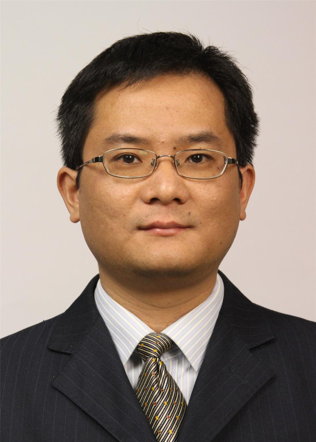 Zhi Li (李智)合作论文数School of Stomatology, Wuhan University85
 YaoJun Dong (东耀峻)合作论文数50
 Rongtao Yang (杨荣涛)合作论文数39
 Gu Cheng (程谷)合作论文数34Haihua Zhou (周海华)合作论文数Hospital of Stomatology, Wuhan University34
 Xuewen Yang (杨学文)合作论文数Orthopedic surgery of orthognathic and cleft lip and palate, School of Stomatology, Wuhan University33
 Jihong Zhao (赵吉宏)合作论文数Department of Oral and Maxillofacial Surgery, Hospital of Stomatology, Wuhan University29JinRong Li (李金荣)合作论文数26Lv Kun合作论文数25
 Zhengjun Shang (尚政军)合作论文数Hospital of Stomatology, Wuhan University18
 Yifang Zhao (赵怡芳)合作论文数Hospital of Stomatology, Wuhan University18
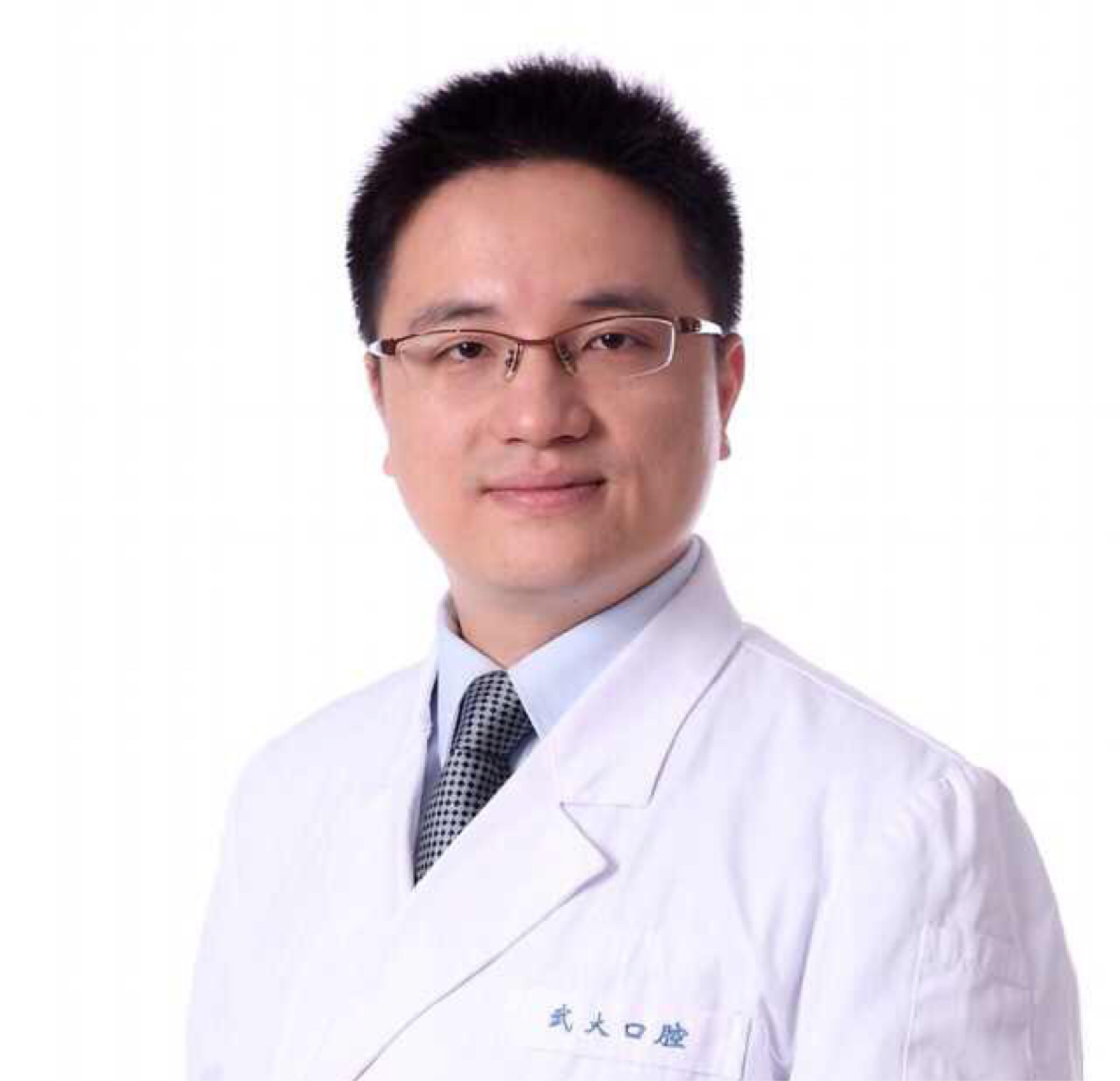 Qilong Wan (万启龙)合作论文数School of Stomatology, Wuhan University18
 Qi Liu (刘琦)合作论文数School of Stomatology, Wuhan University15
 Xin Xing合作论文数Dept Pathol, Johns Hopkins Univ14
 Hongbing Deng (邓红兵)合作论文数School of Resource and Environmental Sciences, Wuhan University13Yuet Cheng合作论文数11
 Wenfeng Zhang (张文峰)合作论文数School of Stomatology, Wuhan University11Zhong-xing Wu合作论文数10
 Chengcheng Yin合作论文数10TuQiang Hu (胡图强)合作论文数10ShiBin Yu (余世斌)合作论文数9XiaoMing Jin (金晓明)合作论文数9Ongodia David合作论文数9
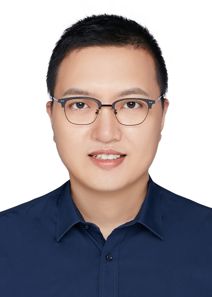 Qingbin Zhang (张庆斌)合作论文数School of Physics, Huazhong University of Science and Technology9YaXin Li (李雅馨)合作论文数8Xinming Chen (陈新明)合作论文数Hospital of Stomatology, Wuhan University8
 Xin Cheng (程鑫)合作论文数School of Information Engineering, Wuhan University of Technology7
 Dianqi Li合作论文数7Yifeng Ni合作论文数6XinCai Zhou (周鑫才)合作论文数6
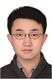 Wei Huang (黄伟)合作论文数上海交通大学6
 Chuanbin Guo (郭传瑸)合作论文数Department of Oral and Maxillofacial Surgery, School and Hospital of Stomatology, Peking University6
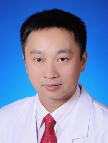 Bo Cheng (程波)合作论文数Zhongnan Hospital of Wuhan University6Wei Zhang合作论文数5TieXia Hu (胡铁霞)合作论文数5GuoWen Sun (孙国文)合作论文数5
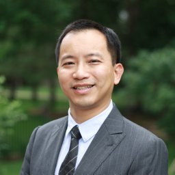 Wang Qun合作论文数Dept Chem & Biol Engn, Iowa State Univ5
 Xue Zhou (周雪)合作论文数School of Public Health, Tongji Medical College, Huazhong University of Science and Technology5
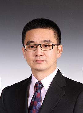 Xin Peng (彭歆)合作论文数Hospital of Stomatology, Peking University5Fusheng Dong (董福生)合作论文数河北医科大学口腔医学院5Xiao-Jing Liu合作论文数4Wen-Bo Zhang合作论文数4Yan Pu Liu合作论文数4
 Guangyan Yu (俞光岩)合作论文数School of Stomatology, Peking University4Chan-Yuan Yang合作论文数4
 Yuhua Yan (闫玉华)合作论文数4Li Lu合作论文数4Shuang Han合作论文数4
 Xin Yu合作论文数4




