The larynx sits at the crossroads between gastrointestinal and respiratory tracts. Besides its intrinsic importance in breathing, swallowing and voice production, the larynx is also exposed to unique immunological challenges. Given the propensity of chronic inflammatory conditions such as chronic laryngitis, which affects up to 20% of Western populations, it is surprising that our understanding of the immunology of this organ remains relatively limited. Recent work on the immunological architecture of the laryngeal mucosa, and its changes that result from external challenges and inflammatory conditions, provided valuable insight into the fascinating immunology of this organ. The lessons learnt from these investigations may go beyond devising improved therapy for chronic laryngeal inflammation. Establishing whether and how the laryngeal mucosa may be involved in the modulation of wider mucosal responses may provide novel routes to the treatment of inflammatory diseases of the respiratory and alimentary tracts such as asthma and inflammatory bowel disease.
BACKGROUND:Matrix metalloproteinases (MMPs) have roles in inflammation and other processes relevant to the architectural disturbances seen in the gastric mucosa in response to Helicobacter pylori infection. Upregulation of MMPs has been reported in H pylori infection, but there are no detailed reports regarding altered production of their inhibitors, the tissue inhibitors of metalloproteinases (TIMPs). AIMS:To investigate changes in the abundance of TIMPs in human gastric corpus mucosa and murine stomach in Helicobacter infection, and to study cellular sources in man. METHODS:Gastric corpus biopsy samples were assessed for abundance of mRNA or protein for TIMP-1 to -4 by real-time quantitative PCR or western blotting, respectively. Antral and corpus biopsies were processed for histology, H pylori status and inflammatory scoring. Cellular sources of TIMP-1, -3 and -4 were examined by indirect immunohistochemistry. Circulating gastrin was measured by radioimmunoassay. Also, abundance of TIMP-1, -3 and -4 mRNA in the stomach of Helicobacter felis infected mice post-infection was compared with that of uninfected control animals. RESULTS:Compared with uninfected patients, mRNA and protein for TIMP-1, -3 and -4 were significantly more abundant in the gastric corpus of H pylori infected subjects. Gastric TIMP expression did not differ significantly between hyper- and normogastrinaemic subjects within the H pylori negative and positive groups. There was no difference in mRNA abundance for MMP-3 or -8. Immunohistochemistry showed TIMP proteins localised to gastric epithelial, stromal cells and inflammatory cells. Murine H felis infection was associated with upregulation of TIMP-1 and -3 mRNA. CONCLUSIONS:Helicobacter infection is associated with upregulation of specific TIMPs (TIMP-1 and -3) in glandular epithelium and stroma. It is suggested that increased expression of specific protease inhibitors in the corpus mucosa may exert important effects on extracellular matrix remodelling and influence the outcome of H pylori infection.
Objectives. We recently demonstrated an increase in the MHC‐associated molecule, β‐2‐microglobulin in laryngeal epithelium from patients with laryngopharyngeal reflux (LPR). This could not be accounted for by an increase in MHC Class I, particularly in the superficial layers where Class I is weakly expressed. We hypothesised that this reflected an increase in CD1d, a non‐classical MHC molecule. Methods. Posterior laryngeal biopsies were obtained from 10 controls and 11 LPR patients. Multicolour immunofluorescence microscopy was used to measure expression of CD1d, CD161 (NK/NKT marker), CD4/8 T‐cells, macrophage, neutrophil and eosinophil antigens. Two‐way analysis of variance and unpaired T‐tests were performed on transformed data. Results. Expression of CD1d increased from basal to superficial layers, and was marginally raised in LPR ( P = 0.067), whilst CD161 increased significantly ( P = 0.02). While CD161(+) cells were distributed throughout the epithelium, CD8(+) T‐cells were localised to the deep, MHC class I‐expressing layers. EMBP was significantly reduced in LPR ( P = 0.01). Conclusions. CD1d and MHC class I exhibit reciprocal expression patterns, which are reflected by the distribution of cells expressing their respective ligands (NKT cells and CD8(+) T‐cells). An increase in CD1d in LPR may be responsible for the increase in β‐2‐microglobulin observed, and is associated with a significant increase in NKT‐cells. The CD1d‐NKT cell axis may be a crucial part of maintaining tolerance in the laryngeal mucosa and changes in this system may be responsible for the persistence of inflammation in LPR.
Objectives. Proteomics could provide important insights into pathogenesis and potential new targets for diagnosis, prognosis, prediction and therapy for head and neck squamous cell carcinoma (HNSCC). However, study of solid tissue is problematic.1 Cystic degeneration that occurs in advanced nodal metastasis can be difficult to differentiate from benign disease, especially in the absence of primary tumour.2 As with other malignant effusions, HNSCC cystic fluid may prove a rich source of novel tumour associated proteins: possible targets for immunotherapy, diagnosis, prediction or prognosis. Design. Feasibility study of 2D‐gel electrophoresis (2DGE) and matrix‐assisted laser desorption‐ionisation mass spectrometry (MALDI‐MS) to analyse cyst fluid within squamous metastasis. Method. Cystic fluid was aspirated from a cervical metastasis in a patient with advanced squamous cancer. Proteins were separated by 2DGE, stained with Coomassie brilliant blue and identified by MALDI‐MS. Results. Initial analysis showed an abundance of haemoglobin, albumin and immunoglobulin, as expected. The gel also revealed unexpected proteins, particularly periredoxin‐2. There were also some unidentified spots, possibly containing novel proteins. In order to increase the potential for identifying lower abundance proteins, albumin and immunoglobulin were depleted by affinity chromatography and further 2DGE undertaken. Conclusions. 2DGE is applicable to the study of fluid from cystic cervical metastases. Unexpected proteins identified in this pilot study require confirmation in a larger series. Depletion of abundant proteins will allow unidentified spots to be characterised. These approaches are likely to open a new window to proteins ‘at the coal‐face’ of head and neck cancer. References 1 Wu W., Tang X., Hu W., et al. (2002) Clin. Exp. Metastasis19, 319‐‐3262 Raghavan U. & Bradley P.J. (2003) Curr. Opin. Otolaryngol. Head Neck Surg. 11(2), 124‐‐128
Conference Abstract| February 01 2004 Helicobacter Pylori Infection Suppresses the Expression of Gastric Ghrelin A. Bassi; A. Bassi 1 Aintree Centre for Gastroenterology, University of Liverpool, Liverpool, United Kingdom Search for other works by this author on: This Site PubMed Google Scholar H. Kamarova; H. Kamarova 2 Dept of Medicine, University Hospital Aintree, University of Liverpool, Liverpool, United Kingdom Search for other works by this author on: This Site PubMed Google Scholar L. Pazmany; L. Pazmany 2 Dept of Medicine, University Hospital Aintree, University of Liverpool, Liverpool, United Kingdom Search for other works by this author on: This Site PubMed Google Scholar K. Bodger K. Bodger 3 Dept of Medicine, University of Liverpool, Liverpool, United Kingdom Search for other works by this author on: This Site PubMed Google Scholar Clin Sci (Lond) (2004) 106 (s50): 1P. https://doi.org/10.1042/cs106001Pa Views Icon Views Article contents Figures & tables Video Audio Supplementary Data Peer Review Share Icon Share Twitter LinkedIn Cite Icon Cite Get Permissions Citation A. Bassi, H. Kamarova, L. Pazmany, K. Bodger; Helicobacter Pylori Infection Suppresses the Expression of Gastric Ghrelin. Clin Sci (Lond) 1 February 2004; 106 (s50): 1P. doi: https://doi.org/10.1042/cs106001Pa Download citation file: Ris (Zotero) Reference Manager EasyBib Bookends Mendeley Papers EndNote RefWorks BibTex toolbar search Search Dropdown Menu toolbar search search input Search input auto suggest filter your search All ContentAll JournalsClinical Science Search Advanced Search This content is only available as a PDF. © 2004 The Biochemical Society and the Medical Research Society2004 Article PDF first page preview Close Modal You do not currently have access to this content.
Human NK cells adhere to and lyse porcine endothelial cells (pEC) and therefore may contribute to the cell-mediated rejection of vascularized pig-to-human xenografts. Since MHC class I molecules inhibit the cytotoxic activity of NK cells, the expression of HLA genes in pEC has been proposed as a potential solution to overcome NK cell-mediated xenogeneic cytotoxicity. HLA-G, a minimally polymorphic HLA class I molecule that can inhibit a wide range of NK cells, is an especially attractive candidate for this purpose. In this study we tested whether the expression of HLA-G on pEC inhibits the molecular mechanisms that lead to adhesion of human NK cells to pEC and subsequent xenogeneic NK cytotoxicity. To this end two immortalized pEC lines (2A2 and PED) were stably transfected with HLA-G1. Rolling adhesion of activated human NK cells to pEC monolayers and xenogeneic cytotoxicity against pEC mediated by polyclonal human NK lines as well as NK clones were inhibited by the expression of HLA-G. The adhesion was partially reversed by masking HLA-G on pEC with anti-HLA mAbs or by masking the HLA-G-specific inhibitory receptor ILT-2 on NK cells with the mAb HP-F1. The inhibition of NK cytotoxicity by HLA-G was only partially mediated by ILT-2, indicating a role for other unknown NK receptors. In conclusion, transgenic expression of HLA-G may be useful to prevent human NK cell responses to porcine xenografts, but is probably not sufficient on its own. Moreover, the blocking of rolling adhesion by HLA-G provides evidence for a novel biological function of HLA molecules.
Natural killer (NK) cells have been extensively studied in their traditional roles in host defense against tumor or virally infected cells. Uterine NK cells are of 2 distinct subsets: endometrial NK (eNK) cells, found in the uterus during the menstrual cycle, and decidual NK (dNK) cells, found in the decidua during pregnancy. This review will explore the immunosurveillance and cytotoxicity profiles of NK cells, the inert nature of eNK cells, and the role of dNK cells as builders at the maternal-fetal interface that create a pregnancy-favorable environment by inducing angiogenesis, trophoblast invasion, and vascular remodeling.
 Robert John Moots合作论文数Liverpool University Hospital;National Centre for Behçet's Syndrome, Liverpool12
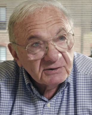 Jack Strominger合作论文数Department of Stem Cell and Regenerative Biology, Faculty of Arts and Sciences, Harvard University10
 Ofer Mandelboim合作论文数9
 Daniel M Davis合作论文数Division of Immunology, Immunity to Infection and Respiratory Medicine, School of Biological Sciences, University of Manchester/Manchester Institute for Collaborative Research on Ageing8Valés-Gómez Mar合作论文数Department of Immunology and Oncology, Centro Nacional de Biotecnología/Consejo Superior de Investigaciones Científicas8Hugh T. Reyburn合作论文数8
 Andrew McMichael合作论文数Nuffield Department of Medicine, University of Oxford4
 Martin Birchall合作论文数Ear Institute, University College London;Faculty of Brain Sciences, University College London;The Royal College of Surgeons of England4Eric G. Sheu合作论文数Department of Surgery, Brigham and Women's Hospital, Boston, MA4
 Jörg D Seebach合作论文数Hopitaux Universitaires de Geneve;Division of Immunology and Allergology, University of Geneva3A. L. Khan合作论文数3
 Keith Bodger合作论文数3C Pridgeon合作论文数2Rn Thompson合作论文数2SE Christmas合作论文数2Rs Langley合作论文数2HM Massey合作论文数2
 Paolo Rovero合作论文数Chemistry Department, A. Menarini Pharmaceuticals2
 Doriana Fruci合作论文数Istituto di Biologia Cellulare, CNR2L. E. N. Rees合作论文数2
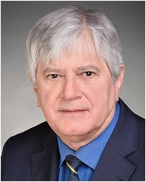 András Varró合作论文数Department of Pharmacology and Pharmacotherapy, University of Szeged2Giovanni Greco合作论文数Dipartimento di Chimica;Farmaceutica e Tossicologica;Studi di Napoli;Dipartimento di Chimica|Farmaceutica e Tossicologica, Studi di Napoli2Eliana Vigneti合作论文数2
 Danuta Gutowska-Owsiak合作论文数2K Badger合作论文数1MKJ Schneider合作论文数1A Bassi合作论文数1Avs Hill合作论文数1Richard H Butler合作论文数1Rh Butler合作论文数1Julian K Sutton合作论文数1Andy Cross合作论文数1GL WANECK合作论文数1
 Nikki Johnston合作论文数1Stephen E Christmas合作论文数1CEM ALLSOPP合作论文数1Roberto Tosi合作论文数1D Brewster合作论文数1E. Williams合作论文数1A Hughes合作论文数1R Dimaline合作论文数1
 Christian Leguern合作论文数1R. N. Thompson合作论文数1NR KITTERINGHAM合作论文数1M Bunce合作论文数1S Ahmed合作论文数1Anne Phillips合作论文数1J K Dawson合作论文数1




