The scope, organization, and biological significance of innate immune functions across structural cell types remain poorly defined. To address these fundamental knowledge gaps, we analyze experimental data of transcriptomes generated by our group and others. Our findings demonstrated the following: 1) the 46 identified innate immune structural cell types exhibit a dual functional identity: under physiological conditions, they maintain tissue architecture and organ-specific homeostasis, whereas under immune stress, they transition into environment-supporting innate immune cells that actively participate in pathogen/damage-associated molecular patterns (PAMP/DAMP) sensing and immune effector responses; 2) The majority of environment-supporting innate immune structural cell types are anatomically positioned to be directly exposed to PAMPs and DAMPs; 3) Endothelial cells as prototypic environment-supporting innate immune structural cells redefine the architecture of innate immunity; 4) Environment-supporting innate immune structural cells have been historically underrecognized for their immune functions; and 5) Neuron-associated structural cell types exhibiting innate immune features suggests a potential role for neural cells in tissue-specific inflammatory regulation. This work establishes a new concept that environment-supporting innate immune structural cells are indispensable components of innate immunity that bridge tissue physiology and immune defense. By redefining immunity as a cooperative network of migratory immune cells and resident structural sentinels with endothelial cells and vascular smooth muscle cells serving as prototypic examples, this paradigm provides new mechanistic insight into tissue-specific inflammation, immune responses and immune privilege, and chronic inflammatory disease pathogenesis. Importantly, it opens new therapeutic avenues aimed at selectively targeting structural cell-intrinsic immune programs in cardiovascular disease, infection, inflammation, autoimmunity, transplantation, and cancer.
Clinical maturation of arteriovenous fistulas (AVF) is largely determined by the adaptive response of the vein to extreme hemodynamic conditions. Histological and bulk transcriptomic analyses of human veins indicate that this is a profound mechanosensitive and inflammatory reparative process, with subtle molecular differences separating maturation and failure. To look for these subtle changes, we analyzed the microanatomy of veins and AVFs that matured and failed using single-cell RNA sequencing (70,281 cells from 20 individuals) and immunofluorescence (IF, n=48 cross-sections). Comparisons of vein and AVF pairs from the same individuals confirmed an increase in area and cell count per layer after AVF creation. However, in all vascular layers, cell density decreased after anastomosis, reflecting an increase in extracellular matrix (ECM) deposition but a slow rate of proliferation. A reduction in density was specifically observed in MYH11+ smooth muscle cells (SMCs) in the intima and media, and CD31+ endothelial cells (ECs) in the lumen. In contrast, ECM-producing myofibroblasts, PDGFRA+ fibroblasts, and CD68+ macrophages maintained their preoperative density after anastomosis. Single-cell expression analyses further highlighted the role of inflammation and ECM remodeling and defined the cell-to-cell interactions responsible for wall repair in AVFs. These were characterized by the expansion of inflammatory and glycosaminoglycan-producing fibroblasts after anastomosis, as well as the activation of wound healing programs in macrophages, myofibroblasts, and ECs. Of interest, these analyses uncovered an enhanced cross-talk among all these cells in AVFs that failed but not in those that matured. Both single cell sequencing and IF stainings confirmed a higher density of infiltrated macrophages, more ACKR3+ ECs, and upregulation of inflammatory factors in myofibroblasts and fibroblasts in association with AVF failure. This study highlights the importance of the inflammatory-ECM remodeling axis in AVF remodeling and identifies the key cellular players for therapeutic targeting to prevent maturation failure.
Achieving a mature arteriovenous fistula (AVF) for hemodialysis remains a significant challenge, even for the most experienced vascular surgeons. Despite decades of research, ≈40% of new AVFs require salvage interventions or can never be used for dialysis. All clinical trials aimed at improving early AVF maturation have failed. This underscores the limitations of the existing biological model, which continues to frame stenosis through a reductionist lens centered on intimal hyperplasia, despite emerging evidence that this explanation is insufficient. This review seeks to redefine the biological framework of early AVF failure by adopting a human-centered perspective. In contrast to the prevailing paradigm, primarily built upon experimental data, our model is centered on human observational research and clinical trials. We then incorporate experimental data to provide mechanistic insights and contextualize or contrast the differences between human and animal biology. We unravel the biology of AVF maturation through 2 tightly connected phases: the acute biomechanical response, encompassing immediate structural and hemodynamic changes after AVF creation, and the subsequent vascular healing and remodeling processes that determine the long-term adaptation of the vein to supraphysiological circulation. By integrating these phases into a cohesive framework, this review advances a more comprehensive model of early AVF maturation failure, highlights therapeutic opportunities, and underscores that meaningful innovation in AVF biology remains both necessary and achievable.
Background: Arteriovenous fistula (AVF) is the preferred hemodialysis access for individuals with end-stage renal disease (ESRD); however, approximately 40% of AVFs fail to mature for hemodialysis without intervention. Despite this significant failure rate, our understanding of the cellular and molecular mechanisms underlying post-anastomosis vein remodeling remains limited. In this study, we present a detailed cellular atlas capturing the temporal changes in the mouse jugular vein following AVF anastomosis at the single-cell level. Methods: Nephrectomy (Nx) was performed on male and female AGE C57BL/6 mice. AVF was created 21 days post-Nx by anastomosing the right jugular vein to the adjacent carotid artery. Animals were euthanized at 3, 10, and 21 days post-AVF for bulk RNA-seq analysis, and at 7 and 30 days for scRNA-seq analysis. The contralateral jugular vein served as a control. Results: Bulk RNA-seq analysis revealed that, acutely (day 3), AVF formation significantly increased the expression of genes related to leukocyte activation (e.g., Cd44 and Itgam ), alongside a decrease in genes associated with vascular smooth muscle contraction (e.g., Myh11 , Cnn1 , and Tagln ). By days 10 and 21, there was a notable upregulation of genes involved in extracellular matrix organization (e.g., Lox , Fn1 , and Eln ). scRNA-seq analysis confirmed an increase in myeloid cells, particularly profibrotic macrophages (identified by high expression of Spp1 and Lgas3 ) and neutrophils, coupled with a reduction in endothelial cells, T-cells, and Schwann cells. Remarkably, in the steady state, 82% of fibroblasts in the vein remained inactive, with only 4.7% displaying an activated state (characterized by high expression of Fap , Postn , and Acta2 ). However, by day 7 post-AVF, activated fibroblasts constituted ~50% of the fibroblast population. Transcription factor enrichment analysis identified Smad3 as the top enriched factor in fibroblast clusters post-AVF. Further intercellular communication analysis demonstrated that, at 7 and 30 days post-AVF, profibrotic macrophages were the primary source of TGFβ ligands (canonical activators of Smad3 ) acting on activated fibroblasts. Conclusion: AVF formation induces significant changes in the vein cellular composition, particularly by increasing profibrotic macrophages, which in turn activate fibroblasts through the TGFβ/Smad3 signaling pathway.
IntroductionR-loops, RNA-DNA hybrid structures with a displaced single-stranded DNA loop, are key regulators of transcriptional control, chromatin architecture, and genome stability and have emerging roles in inflammatory signaling. However, the relationship between R-loop abundance and strongly modulated inflammatory effector genes in metabolic inflammation and influenza virus infection remains underexplored.MethodsWe performed a locus-centric integrative analysis combining robust differentially expressed genes (DEGs) from multiple inflammatory and infection-related murine and human transcriptomic disease models with experimentally validated multi-cell R-loop annotations from the reference atlas RLoopBase. Our correlation framework evaluated the directional relationship between R-loop abundance and inflammatory gene expression rather than assuming disease-sample-matched R-loop measurements. We further analyzed R-loop regulatory proteins, NRF2-associated R-loop regulators, and overlaps between R-loop regulators and CRISPRi-identified mitochondrial and cellular reactive oxygen species (ROS) regulators.ResultsIn angiotensin II-infused apolipoprotein E-deficient (ApoE−/−) mice, a model of abdominal aortic aneurysm (AAA), genomic regions encoding the top significantly upregulated genes exhibited significantly fewer R-loops than those encoding downregulated genes at days 14 and 28. Similarly, in atherosclerotic ApoE−/− mice fed a high-fat diet for 32 and 78 weeks, upregulated genes were associated with fewer R-loops than downregulated genes. Reduced R-loop abundance was also observed in genomic regions encoding the top significantly upregulated genes in liver tissues from patients with non-alcoholic steatohepatitis (NASH), as well as in monosodium urate (MSU)-stimulated lymphatic endothelial cells (LECs) and influenza virus-infected human umbilical vein endothelial cells (HUVECs). R-loop regulatory proteins upregulated during metabolic inflammation were enriched in immune and inflammatory pathways. NRF2 was identified as a regulator of 27 R-loop regulatory proteins, including 10 positively and 17 negatively regulated proteins. Furthermore, 54 R-loop regulatory proteins overlapped with CRISPRi-identified mitochondrial and cellular ROS regulators, suggesting potential reciprocal regulation between R-loop homeostasis and ROS signaling. Disease-associated changes in pro-ROS and anti-ROS R-loop regulatory proteins further linked R-loop regulation to inflammatory and oxidative stress pathways.DiscussionThese findings identify reduced R-loop abundance at genomic regions encoding strongly upregulated inflammatory genes as a shared feature across multiple models of metabolic inflammation and influenza virus infection. The results further suggest that immune-associated R-loop regulatory proteins and the NRF2–ROS axis may contribute to R-loop remodeling during inflammatory disease. This integrative framework provides new insight into the potential role of R-loops and ROS-sensitive R-loop regulators in inflammatory and metabolic diseases and identifies candidate pathways for future mechanistic investigation and therapeutic targeting.
Background Alzheimer's disease (AD) is a neurodegenerative disorder characterized by memory impairment. Neuroinflammatory processes, mediated by glial and immune cells, contribute to neuronal damage. Emerging evidence implicates innate immune mechanisms, including trained immunity and cell trans-differentiation, in AD pathogenesis, though their roles remain unclear. Objective To investigate transcriptomic changes in the 3xTg-AD mouse model, focusing on trained immunity and cell trans-differentiation in disease mechanisms. Methods RNA-sequencing was performed on brain tissue (cortex plus hippocampus) from 11-month-old female 3xTg-AD and wild-type mice (n = 3/group). Differentially expressed genes (fold change > 1.5, p < 0.05) were identified and followed by bioinformatics and knowledge-based transcriptomic profiling. Public AD datasets were also analyzed. Results 3xTg-AD mice exhibited 316 upregulated and 412 downregulated genes. Downregulated genes included those for blood-brain barrier protein, while upregulated genes related to cerebrospinal fluid. Increased expression of proinflammatory markers, as well as genes related to cell differentiation, proliferation, activation, and adhesion. Upregulation of genes associated with cell migration and trans-differentiation suggests a potential role for inflammation and cellular plasticity. Additionally, genes involved in inflammasome pathways, immunometabolism, and trained immunity were upregulated. Mechanistically, these genes were modulated by knockdown of trained immunity promoter SET-7, overexpression of trained immunity inhibitor IL-37, and knockout of inflammasome genes IL-1 receptor, caspase-1, and pattern recognition receptor CD36. Conclusions The finding underscore the potential role of trained immunity and cell trans-differentiation in AD, revealing a mechanistic framework in which danger-associated molecular patterns drive innate immune responses, inflammasome activation, and cell plasticity contribute to AD, offering therapeutic targets for neuroinflammation and cellular reprograming.
The autogenous arteriovenous fistula (AVF) remains the preferred vascular access for hemodialysis, despite its high rate of maturation failure, primarily due to occlusive stenosis driven by fibrotic remodeling and neointimal hyperplasia. Currently, no therapies effectively improve AVF outcomes, largely due to limited understanding of the cellular and molecular mechanisms underlying venous remodeling. To address this knowledge gap, we conducted single-nucleus (sn) RNA/ATAC-seq analysis using a translational aortocaval fistula model in mice. This analysis showed significant heterogeneity among venous cells in the AVF outflow tract and identified unique venous smooth muscle cells (vSMC) reprogramming from a quiescent to a previously uncharacterized, proliferative and matrix organizing phenotype that retains a contractile state. The presence of these cells was further validated using SMC lineage tracing and multiplex RNA in situ hybridization. Additionally, snATAC-seq demonstrated a significant increase in the motif activity of ATF3 in fibrotic vSMC within AVF compared to controls. Notably, ATF3 is positively regulated by MRTFA, a key co-transcription factor that activates multiple gene programs. MRTFA expression was markedly upregulated in vSMC following AVF creation. Both SMC-specific knockout and pharmacological inhibition of MRTFA/B in mice significantly reduced AVF wall thickness, neointimal formation, and fibrosis while enhancing AVF flow. Consistently, MRTFA and its target genes (COL1A1, COL8A1, PDGFRB) were upregulated in neointimal vSMC in human AVF. Single-cell RNA-seq analysis of human AVF mirrored the contractile-to-fibrotic vSMC reprogramming observed in mouse AVF. Furthermore, CosMx single-cell spatial omics identified distinct localization patterns of multiple vSMC clusters, with fibrotic and stressed vSMC clusters predominantly situated in the neointima, while contractile vSMC clusters were primarily localized in the medial layer of human AVF. Collectively, these studies, for the first time, integrate multiple advanced single-cell omics approaches to define the occurrence and regulation of vSMC reprogramming during AVF remodeling and its contribution to AVF pathology. They also suggest that targeting vSMC-MRTF could provide a novel therapeutic strategy to prevent adverse AVF remodeling, reduce failure rates, and improve AVF patency.
Introduction The biological mechanisms underlying arteriovenous fistula (AVF) maturation in patients receiving hemodialysis remain poorly understood despite decades of research. Methods To address this gap, we first investigated the cellular changes in the venous wall after fistula creation in histological biopsies of longitudinal veins and AVF samples (23 patients). Using single-cell RNA sequencing of 70,281 cells from independent pre-access veins, early resections, mature, and failed AVFs (20 patients), we then created a complementary transcriptomic atlas of the human vein before and after anastomosis. Results AVFs had increased wall area and cell number but reduced cell density in histological sections, suggesting that postoperative wall thickening occurs predominantly through extracellular matrix (ECM) deposition. The early remodeling of the AVF was characterized by a loss of smooth muscle cells, increased monocyte infiltration, and the reprograming of myofibroblasts and fibroblasts toward reparative phenotypes. In contrast, later stages of remodeling were dominated by ECM-producing myofibroblasts and fibroblasts, occurring in the context of low cell proliferation. Failed AVFs displayed persistent inflammation and exaggerated healing responses as defining features. Specifically, these AVFs contained abundant proinflammatory and adhesive macrophages with upregulation of the Myddosome signaling complex, proinflammatory vasa vasorum endothelial cells, and hyperactivated fibroblasts and myofibroblasts. Macrophage-derived osteopontin emerged as a key paracrine signal driving vascular cell activation in failed AVFs. Additional signals derived from fibroblasts, myofibroblasts, and endothelial cells, including chemokines, semaphorins, fibroblast growth factors, angiopoietin-like proteins, periostin, and transforming growth factor-β, were also enriched within the inflammatory microenvironment sustaining AVF failure. Conclusions Our findings uncover previously unrecognized cellular and molecular patterns in human veins following AVF creation, providing novel insights and potential therapeutic targets to improve AVF maturation outcomes.
IntroductionEndothelial-to-mesenchymal transition (EndoMT), cell death, and fibrosis are increasingly recognized as contributing factors to Alzheimer’s disease (AD) pathology, but the underlying transcriptomic mechanisms remain poorly defined. This study aims to elucidate transcriptomic changes associated with EndoMT, diverse cell death pathways, and fibrosis in AD using the 3xTg-AD mouse model.MethodsUsing RNA-seq data and knowledge-based transcriptomic analysis on brain tissues from the 3xTg-AD mouse model of AD. This included pathway-level analysis of gene expression changes across multiple brain cell types. Mechanistic insights were further validated using single-cell RNA sequencing (scRNA-Seq) dataset from human AD brain.ResultsOur analysis showed that in the 3xTg-AD model: (i) multiple brain cell type genes are altered, promoting EndoMT through upregulation of RGCC and VCAN; (ii) genes related to various types of cell death, including apoptosis, ferroptosis, necrosis, anoikis, mitochondrial outer membrane permeability programmed cell death, mitochondrial permeability transition-driven necrosis, NETotic, and mitotic cell death, are upregulated in the several brain cell types; (iii) fibrosis-related genes are upregulated across multiple brain cell types. Further mechanistic analysis revealed: (1) mitochondrial stress through upregulation of mitochondrial genes in the brain cells; (2) upregulation of cellular, oxidative, and endoplasmic reticulum (ER) stress genes; (3) nuclear stress via upregulation of nuclear genes, transcription factors (TFs), and differentiation TFs FOSB and MEOX1; (4) metabolic reprogramming/stress through the upregulation of genes related to lipid and lipoprotein metabolism, fatty acid oxidation (FAO), glucose metabolism, and oxidative phosphorylation (OXPHOS); (5) catabolic stress via upregulation of catabolic genes. Single-cell RNA-Seq data indicated that many of these were also increased in AD patients’ brain cells. These changes were reversed by knockdown of the ER stress kinase PERK (EIF2AK3) and deficiencies in FOSB and MEOX1.DiscussionThis study uncovers previously unrecognized molecular signatures of organelle stress and bioenergetic reprogramming that drive EndoMT, cell death, and fibrosis in AD. The reversal of these changes via PERK, FOSB, and MEOX1 inhibition highlights potential therapeutic targets for mitigating neurodegenerative processes in AD.
The mouse carotid–jugular arteriovenous fistula (AVF) is a widely adopted surgical model to study venous remodeling after AVF creation. Despite its increasing use, the extent to which this model recapitulates the cellular and molecular remodeling processes observed in humans remains uncertain, which is essential for validating its translational relevance. Using bulk and single-cell RNA sequencing, we have depicted the transcriptional and cellular evolution of the mouse jugular vein after AVF anastomosis. Global transcriptomic profiling revealed that venous remodeling begins with a robust inflammatory response, followed by a prominent extracellular matrix (ECM) remodeling phase that peaks at postoperative day 10. Single-cell analyses confirmed the role of macrophage (3-fold) and neutrophil infiltration (12-fold) in sustaining the onset of venous remodeling. These monocytes/macrophages exhibited marked upregulation of pro-inflammatory and pro-fibrotic genes, including Il1b, Spp1, Fn1, Thbs1, and Tgfb1. Evidence of the differentiation of fibroblasts into myofibroblasts positive for Postn, Col8a1, and Thbs1 emerged by postoperative day 5. The temporal dynamics of differentially expressed genes in these myofibroblasts closely mirrored the ECM gene expression patterns identified by bulk RNA-seq, indicating that they are the principal source of ECM deposition in the AVF. Cell-to-cell communication analyses highlighted macrophages and fibroblasts as the main populations driving postoperative remodeling. Comparative analysis with single-cell data from human pre-access veins and AVFs demonstrated that the mouse model reproduces the core inflammatory–fibrotic axis of fibroblast activation observed in humans, supporting its utility for mechanistic studies of postoperative ECM remodeling.
Hyperlipidemia and chronic kidney disease (CKD) are well-established risk factors for cardiovascular disease and act synergistically to promote vascular inflammation and disease progression. However, the mechanisms underlying this synergetic effect remain largely unknown. Using a mouse model combining hyperlipidemia (via high-fat diet feeding, HFD) with 5/6 nephrectomy-induced CKD, we made the following significant findings: 1) HFD + CKD upregulated 1179 genes in mouse aortas and induced prominent reactive oxygen species (ROS), far more than either HFD or CKD alone. 2) HFD + CKD upregulated 86 CRISPRi-identified mitochondrial ROS regulators, 36 CRISPRi-identified cellular ROS regulators, and 19 GSEA-collected ROS regulators. These changes were associated with the upregulations of 48 cytokines, 7 highest toxicity uremic toxin receptors—including CD1D, FCGRT, AHR, IL6RA AGER, NR1H3 and NPY5R—in aortas. 3) These uremic toxin receptors emerged as novel promoters of inflammation and trained immunity. Deficiencies in CD1D, AHR, AGER, and the trained immunity promoter SET7 each downregulated up to 5.5 % of the genes upregulated by HFD + CKD. Conversely, activation of NR1H3 using an agonist upregulated up to 12.2 % of these genes. 4) The expression of 46 cytokine genes was strongly associated with NR1H3 upregulation. 5) The NR1H3 agonist also induced the expression of 28 ROS regulators, including YBX2, a novel anti-ROS transcription factor and RNA-binding protein, suggesting a potential negative feedback mechanism. YBX2 deficiency increased the cellular ROS level, while YBX2 overexpression suppressed 27 proinflammatory genes induced by HFD + CKD. Our findings provide novel insights into the role of the NR1H3-YBX2 axis in regulating inflammation accelerated by hyperlipidemia and CKD.
Introduction: Hypertension (HTN) is highly prevalent among patients with chronic kidney disease (CKD) and end-stage kidney disease (ESKD), uncontrolled HTN as well as high blood pressure variability (BPV) can significantly impact cardiovascular health. This study aims to understand the impact of BPV in patients with ESKD following arteriovenous fistula (AVF) creation for hemodialysis, to better understand its association with age, sex, BMI, and cardiovascular risk. Methods: A retrospective analysis was conducted on 83 patients who underwent AVF creation between February 2019 and October 2020 at University of Miami Hospital. During 20 months, blood pressure measurements were collected from medical records at four different times during AVF access creation process, and BPV was quantified using machine learning clustering techniques. Results: The study identified three distinct clusters among the 83 patients with ESKD who underwent AVF creation. Cluster 1 ( n = 28) was characterized by high blood pressure variability, with a mean systolic blood pressure standard deviation of 16.2 mmHg. This cluster exhibited a significantly higher cardiovascular risk rate of 42.9% compared to Cluster 2 ( n = 30) and Cluster 3 ( n = 25), which had lower variability with mean systolic blood pressure standard deviations of 10.3 and 8.7 mmHg, respectively, and cardiovascular risk rates of 16.7% and 12.0%. Age, sex, and BMI did not differ significantly across the clusters. Conclusion: This study underscores the association between blood pressure variability and the increased risk of cardiovascular events, including myocardial infraction, stroke or heart failure in patients with ESKD undergoing AVF creation. Our findings highlight the critical need for vigilant monitoring of blood pressure fluctuations in this population and demonstrate how these variations are influenced by individual demographic factors. Further research is needed to develop targeted interventions to mitigate this risk.
Arteries and veins develop different types of occlusive diseases and respond differently to injury. The biological reasons for this discrepancy are not well understood, which is a limiting factor for the development of vein-targeted therapies. This study contrasts human peripheral arteries and veins at the single-cell level, with a focus on cell populations with remodeling potential. Upper arm arteries (brachial) and veins (basilic/cephalic) from 30 organ donors were compared using a combination of bulk and single-cell RNA sequencing, proteomics, flow cytometry, and histology. The cellular atlases of six arteries and veins demonstrated a 7.8× higher proportion of contractile smooth muscle cells (SMCs) in arteries and a trend toward more modulated SMCs. In contrast, veins showed a higher abundance of endothelial cells, pericytes, and macrophages, as well as an increasing trend in fibroblasts. Activated fibroblasts had similar proportions in both types of vessels but with significant differences in gene expression. Modulated SMCs and activated fibroblasts were characterized by the upregulation of MYH10, FN1, COL8A1, and ITGA10. Activated fibroblasts also expressed F2R, POSTN, and COMP and were confirmed by F2R/CD90 flow cytometry. Activated fibroblasts from veins were the top producers of collagens among all fibroblast populations from both types of vessels. Venous fibroblasts were also highly angiogenic, proinflammatory, and hyper-responders to reactive oxygen species. Differences in wall structure further explain the significant contribution of fibroblast populations to remodeling in veins. Fibroblasts are almost exclusively located outside the external elastic lamina in arteries, while widely distributed throughout the venous wall. In line with the above, ECM-targeted proteomics confirmed a higher abundance of fibrillar collagens in veins vs. more basement ECM components in arteries. The distinct cellular compositions and transcriptional programs of reparative populations in arteries and veins may explain differences in acute and chronic wall remodeling between vessels. This information may be relevant for the development of antistenotic therapies.
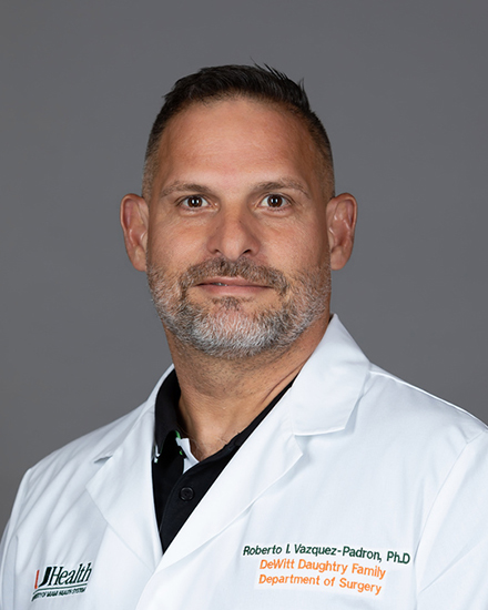 Roberto I. Vazquez-Padron合作论文数DeWitt Daughtry Family Department of Surgery, University of Miami Miller School of Medicine66
 Marwan Riad Tabbara合作论文数53
 Loay Salman合作论文数38
 Juan Duque合作论文数Leonard M. Miller School of Medicine, University of Miami34
 Miguel Rojas合作论文数30
 Roberto Vazquez-Padron合作论文数28
 Xiao-Feng Yang合作论文数Lemole Center for Integrated Lymphatics and Vascular Research, Lewis Katz School of Medicine, Temple University;Center for Metabolic Disease Research, Temple University;Sol Sherry Thrombosis Research Center, Temple University21Zachary M Zigmond合作论文数19
 Omaida C. Velazquez合作论文数Division of Vascular Endovascular Surgery, DeWitt Daughtry Family Department of Surgery, Leonard M. Miller School of Medicine, University of Miami;University of Pennsylvani16Eric L. Greidinger合作论文数Miami Department of Veterans Affairs Medical Center, University of Miami Miller School of Medicine16Simone Pereira-Simon合作论文数16Keman Xu合作论文数12Saaoud Fatma合作论文数Department of Cell Biology and Anatomy, University of South Carolina School of Medicine12
 Ying Shao合作论文数Shanghai Medical College, Fudan University12
 Diana Hernandez合作论文数11Rj Herrera合作论文数11Nieves Santos Falcon合作论文数10
 Hong Wang合作论文数Lewis Katz School of Medicine, Temple University;Center for Metabolic Disease Research, Temple University10Robert W. Hoffman合作论文数10Yuntao Wei合作论文数10Yunjuan Zang合作论文数10Guillermo Selman合作论文数9
 Yifan Lu (陆一凡)合作论文数School of Mechatronics Engineering, Harbin Institute of Technology9Roberta M. Lassancesoares合作论文数9Filipe F. Stoyell Conti合作论文数7Alghidak Salama合作论文数7Adriana Dejman合作论文数7
 Sheng Wu (吳盛)合作论文数7
 Luis A Escobar合作论文数Univ Miami7Xiaohua Jiang合作论文数Department of Medicine;Baylor College of Medicine;Department of Medicine, Baylor College of Medicine7
 Irina Fernandez合作论文数Department of Biomedical Engineering, The University of Texas at Austin7Akshara Sree Challa合作论文数6
 Xiaochun Long合作论文数6
 Lei Song合作论文数China Pharmaceutical University, China Pharmaceutical University6Kimberly Jaimes合作论文数6
 Karen Manzur Pineda合作论文数5
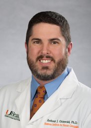 Anthony J. Griswold合作论文数Department of Biology, Indiana University5Yan-Ting Shiu合作论文数5
 Ali Mir Arif Mir Asif合作论文数5Fotios M. Andreopoulos合作论文数Molecular and Cellular Pharmacology;Vascular Biology Institute;School of Medicine;School of Medicine, Vascular Biology Institute5Angela Paez合作论文数4Suzanne Laboyrie合作论文数4Brandon Applewhite合作论文数4
 Mohd Ahmar Rauf合作论文数Miller School of Medicine, University of Miami4
 Xiaolei Liu合作论文数Lemole Center for Integrated Lymphatics and Vascular Research, Lewis Katz School of Medicine, Temple University;Guillermo Oliver Lab, Feinberg School of Medicine, Northwestern University;St. Jude Children's Research Hospital4
 Beata Kosmider合作论文数4
 Joris Rotmans合作论文数Department of Internal Medicine, Leiden University Medical Centre;Subdepartment of Nephrology, Leiden University Medical Center4Christina Manning合作论文数3
 Natasha Fernandez合作论文数3




