Training of the perineal muscles was attempted in our centers on the basis of our previous findings indicating that when the appropriate source of stimulation is used, subjects exhibit peaks of activity suggesting a rehabilitation potential. Muscular training was considered because pressure stimulation of the glans penis triggers contractions of perineal muscles which propel the blood throughout the penis to ensure optimal tumescence. The technique was combined with biofeedback and home exercises and was tested on 10 patients initially presenting erectile difficulty but maintaining perineal innervation. The results showed significant improvements in average and maximal tumescence and maintenance of these improvements. The results are discussed in terms of their clinical application to a cognitive-behavioral approach to sexual dysfunction in spinal cord-injured men.
OBJECTIVETo study the extent to which atrophy of muscle and progressive weakening of the long bones after spinal cord injury (SCI) can be reversed by functional electrical stimulation (FES) and resistance training.DESIGNA within-subject, contralateral limb, and matching design.SETTINGResearch laboratories in university settings.PARTICIPANTSFourteen patients with SCI (C5 to T5) and 14 control subjects volunteered for this study.INTERVENTIONSThe left quadriceps were stimulated to contract against an isokinetic load (resisted) while the right quadriceps contracted against gravity (unresisted) for 1 hour a day, 5 days a week, for 24 weeks.MAIN OUTCOME MEASURESBone mineral density (BMD) of the distal femur, proximal tibia, and mid-tibia obtained by dual energy x-ray absorptiometry, and torque (strength).RESULTSInitially, the BMD of SCI subjects was lower than that of controls. After training, the distal femur and proximal tibia had recovered nearly 30% of the bone lost, compared with the controls. There was no difference in the mid-tibia or between the sides at any level. There was a large strength gain, with the rate of increase being substantially greater on the resisted side.CONCLUSIONOsteopenia of the distal femur and proximal tibia and the loss of strength of the quadriceps can be partly reversed by regular FES-assisted training.
Colonic transit time (CTT) was measured with abdominal radiographs using Chaussade's technique in 30 spinal cord injured patients (ASIA A and B) following ingestion of 20 radiomarkers per day for three days. A significant increase in total CTT (p = 0.0001) and segmental CTT of the right colon (p = 0.0004) and of the left colon (p = 0.0001) was shown. While using on the average only 2.3 films of the abdomen per patient, we obtained results comparable with other radiologic techniques which use radiomarkers to measure CTT. The clinical relevance of these results is not clear and their correlation with intestinal symptoms remains to be investigated.
Background and Purpose. The effect of therapeutic massage on the H-reflex amplitude in persons without neurological impairment has been established. To investigate its effects in a sample of persons with a spinal cord injury (SCI), two independent but interrelated studies were undertaken. Study 1 investigated whether the recorded response (H-reflex amplitude) to massage with the subjects in the supine testing position was similar to that recorded in previous studies in which the subjects were tested in the prone position. This study was undertaken because the prone testing position was considered inappropriate for persons with SCI. In study 2, the therapeutic effect of massage (petrissage) on H-reflex amplitude in persons with SCI was examined. Subjects. Seven persons without neurological impairment volunteered to participate in study 1, and 10 individuals with a traumatic SCI volunteered to participate in study 2. Methods. The two studies shared many methodological features and involved the recording of 10 H-reflex and M-response peak-to-peak amplitudes from the triceps surae muscle during each of five sequential, 3-minute time periods. Massage treatment (MASS) was given during third time period, and the premassage time periods (C1, C2) and postmassage time periods (C3, C4) served as control conditions. Study 2, in addition to recording the peak-to-peak amplitudes of the recorded responses, also included the recording of the H-reflex latencies. Results. The results of study 1 showed that massage applied with the subjects in the supine position decreased the H-reflex amplitude during the massage. A 56% decrease in the H-reflex amplitude was recorded. Study 2 demonstrated a 27% mean group decrease in the H-reflex peak-to-peak amplitude during the massage for all subjects, with variations in individual responses ranging from an increase in the H-reflex amplitude of 20% to a decrease of 84%. An analysis of variance revealed that the H-reflex means of the five conditions were significantly different. Newman-Keuls post hoc analyses revealed that the mean of the MASS condition (2.01 mV) was significantly different from the means of C1, C2, and C4 (2.79, 2.81, 2.58 mV). The mean of C3 (2.42 mV) was not found to be statistically different from the means of the other conditions. These changes were noted against a stable M-response. Conclusion and Discussion. The results recorded in study 1 are comparable to those obtained with the subjects in the prone position. Based on these results, the supine position was adopted as the testing position for study 2. Study 2 further showed a decrease in H-reflex amplitude concomitant with massage in persons with SCI, but no long-term effects were noted. [Goldberg J, Seaborne DE, Sullivan SJ, Leduc BE. The effect of therapeutic massage on H-reflex amplitude in persons with a spinal cord injury. Phys Ther. 1994;74:728-737.]
(1990) Comparison of three methods of electrical stimulation for converting skeletal muscle to a fatigue resistant power source suitable for cardiac assistance. Abstract: Twelve dogs were sorted into 3 equal groups, and the in-situ right latissimus dorsi muscle of each dog was stimulated via its motor nerve for a period of 6 weeks. The resulting isotonic contractions were used to pump fluid in an implanted, 2-chambered, compressible pouch system. Three methods of electrical stimulation were used: (a) continuous 2 sec-1 single pulses that caused muscle twitching, (b) a 250 msec train of pulses (36 sec-1) that caused tetanic muscle contractions and was repeated every 2 sec for 15 min followed by a 15 min period of rest, and (c) alternating 15 min periods of the above 2 stimulation methods to cause alternating twitch and tetanic contractions. The 2 sec-1 twitch stimulation and the combined twitch/tetanic stimulation methods resulted in a 100% conversion to fatigue-resistant fibers within 6 weeks. Standardized muscle function tests were performed weekly. With the twitch stimulation (Method 1), the time to fatigue increased from 9 to 116 min (p less than 0.001), but fluid pumping ability of the muscle decreased substantially from 0.25 to 0.14 liters min-1 (p less than 0.05). With the intermittent tetanic stimulation (Method 2), the fatigue resistance increased only slightly from 7 to 11 minutes (p = NS), and pumping ability was unchanged. With the combined (twitch-tetanic) stimulation (Method 3), the time to fatigue increased from 9 to 107 min (p less than 0.001), and the pumping ability did not significantly change from 0.20 to 0.22 liters min-1 (p = NS). These results suggest that a combined electrical stimulation method which produces both twitches and tetanic contractions can achieve rapid fiber conversion and increased fatigue resistance without loss of muscle strength Bajuk S., Jelnikar T., and Ortar M. (1996) Rehabilitation of patient with brachial plexus lesion and break in axillary artery. Case study. Abstract: This paper describes the physiotherapy and occupational therapy used in treating a 74-year-old woman with a left brachial plexus lesion, a break in the axillary artery, dislocation of the acromioclavicular joint, a broken scapula and clavicula, serial left rib fractures, and lacerations on the upper and lower arm. After testing the patient, the following goals were set: reduce pain, soften scar tissue, and improve joint motion, muscle strength, and functionality of the hand. A 12-month outpatient program was used. …
 Marc Bélanger合作论文数3T C Chalmers合作论文数1G. HEGER合作论文数1M L Harris合作论文数1D Kyroussis合作论文数1
 Rupert Koller合作论文数1G WHEELER合作论文数1Ch Hamnegard合作论文数1Kh Kiessling合作论文数1DE Seaborne合作论文数1L. A. Geddes合作论文数1M Pope合作论文数1A DELITTO合作论文数1U Losert合作论文数1B. Bake合作论文数1
 Monroe Hinds合作论文数1Tessa Gordon合作论文数1
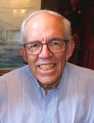 Richard B. Stein合作论文数Department of Physiology, Faculty of Medicine & Dentistry, University of Alberta1
 John Moxham合作论文数King's College London;King's College Hospital1Goldberg J合作论文数1
 Malcolm Granat合作论文数School of Health and Society, University of Salford1
 Mark Delargy合作论文数1Michael I. Polkey合作论文数1William B. Stason合作论文数1
 Winfried Mayr合作论文数Center for Biomedical Engineering and Physics Austria, Medical University Vienna1Kathleen F Charvier合作论文数1Manfred Bijak合作论文数1M Glanz合作论文数1
 Werner Girsch合作论文数1Yuanming Luo合作论文数1Robert Edward Day合作论文数Medical Engineering & Physics, Royal Perth Hospital1
 S. John Sullivan合作论文数Centre for Physiotherapy Research, University of Otago1Frédérique Courtois合作论文数Institut de réadaptation de Montréal1Tom Haggmark合作论文数1Robert a Shively合作论文数1
 Joseph Abiodun Balogun合作论文数School of Physical Therapy, Texas Woman's University1
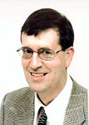 Brian James Andrews合作论文数Oxford1
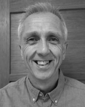 Gerrard F. Rafferty合作论文数MRC & Asthma UK Centre in Allergic Mechanisms of Asthma, King’s College London1
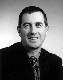 Garry D Wheeler合作论文数1Taka Ashikaga合作论文数1
 Stephen F. Badylak合作论文数Department of Surgery, School of Medicine, University of Pittsburgh;Center for Pre-Clinical Tissue Engineering, McGowan Institute for Regenerative Medicine, University of Pittsburgh;ACell, Inc.1D. K. Marzouk合作论文数1Herwig Thoma合作论文数1S Klawansky合作论文数1
 Hermann Lanmueller合作论文数Department of Biomedical Engineering & Physics, University of Vienna1Catherine Berkey合作论文数1




