Chimeric antigen receptor (CAR) redirected T cells are potent therapeutic options against hematological malignancies. The current dominant manufacturing approach for CAR T cells depends on retroviral transduction. With the advent of gene editing, insertion of a CD19-CAR into the T cell receptor (TCR) alpha constant (TRAC) locus using adeno-associated viruses for gene transfer was demonstrated, and these CD19-CAR T cells showed improved functionality over their retrovirally transduced counterparts. However, clinical-grade production of viruses is complex and associated with extensive costs. Here, we optimized a virus-free genome-editing method for efficient CAR insertion into the TRAC locus of primary human T cells via nuclease-assisted homology-directed repair (HDR) using CRISPR-Cas and double-stranded template DNA (dsDNA). We evaluated DNA-sensor inhibition and HDR enhancement as two pharmacological interventions to improve cell viability and relative CAR knockin rates, respectively. While the toxicity of transfected dsDNA was not fully prevented, the combination of both interventions significantly increased CAR knockin rates and CAR T cell yield. Resulting TRAC-replaced CD19-CAR T cells showed antigen-specific cytotoxicity and cytokine production in vitro and slowed leukemia progression in a xenograft mouse model. Amplicon sequencing did not reveal significant indel formation at potential off-target sites with or without exposure to DNA-repair-modulating small molecules. With TRAC-integrated CAR+ T cell frequencies exceeding 50%, this study opens new perspectives to exploit pharmacological interventions to improve non-viral gene editing in T cells.
The intestine mediates a delicate balance between tolerogenic and inflammatory immune responses. The continuous pathogen encounter might also augment immune cell responses contributing to complications observed upon intestinal transplantation (ITx). We thus hypothesized that ITx patients show persistent signs of immune cell activation affecting both the adaptive and innate immune cell compartment. Information on the impact of intestinal grafts on immune cell composition, however, especially in the long-term is sparse. We here assessed activated and differentiated adaptive and innate immune subsets according to time, previous experience of cellular or antibody-mediated rejections or type of transplant after ITx applying multi-parametric flow cytometry, gene expression, serum cytokine and chemokine profiling. ITx patients showed an increase in CD16 expressing monocytes and myeloid dendritic cells (DCs) compared to healthy controls. This was even detectable in patients who were transplanted more than 10 years ago. Also, conventional CD4+ and CD8+ T cells showed persistent signs of activation counterbalanced by increased activated CCR4+ regulatory T cells. Patients with previous cellular rejections had even higher proportions of CD16+ monocytes and DCs, whereas transplanting higher donor mass with multi-visceral grafts was associated with increased T cell activation. The persistent inflammation and innate immune cell activation might contribute to unsatisfactory results after ITx.
AbstractBackgroundMost current cell‐based regenerative therapies are based on the indirect induction of the affected tissues repair. Xenogeneic cell‐based treatment with expanded human placenta stromal cells, predominantly from fetal origin (PLX‐RAD cells), were shown to mitigate significantly acute radiation syndrome (ARS) following high dose irradiation in mice, with expedited regain of weight loss and haematopoietic function. The current mechanistic study explores the indirect effect of the secretome of PLX‐RAD cells in the rescue of the irradiated mice.MethodsThe mitigation of the ARS was investigated following two intramuscularly (IM) injected 2 × 10⁶ PLX‐RAD cells, 1 and 5 days following 7.7 Gy irradiation. The mice survival rate and their blood or bone marrow (BM) cell counts were followed up and correlated with multiplex immunoassay of a panel of related human proteins of PLX‐RAD derived secretome, as well as endogenous secretion of related mouse proteins. PLX‐RAD secretome was also tested in vitro for its effect on the induction of the migration of BM progenitors.ResultsA 7.7 Gy whole body mice irradiation resulted in ~25% survival by 21 days. Treatment with two IM injections of 2 × 10⁶ PLX‐RAD cells on days 1 and 5 after irradiation mitigated highly significantly the subsequent lethal ARS, with survival rate increase to nearly 100% and fast regain of the initial weight loss (P < 0,0001). This was associated with a significant faster haematopoiesis recovery from day 9 onwards (P < 0.01). Nine out of the 65 human proteins tested were highly significantly elevated in the mouse circulation, peaking on days 6–9 after irradiation, relative to negligible levels in non‐irradiated PLX‐RAD injected mice (P < 0.01). The highly elevated proteins included human G‐CSF, GRO, MCP‐1, IL‐6 and lL‐8, reaching >500 pg/mL, while MCP‐3, ENA, Eotaxin and fractalkine levels ranged between ~60–160pg/mL. The detected radiation‐induced PLX‐RAD secretome correlated well with the timing of the fast haematopoiesis regeneration. The radiation‐induced PLX‐RAD secretome seemed to reinforce the delayed high levels secretion of related mouse endogenous cytokines, including GCSF, KC, MCP‐1 and IL‐6. Additional supportive in vitro studies also confirmed the ability of cultured PLX‐RAD secretome to induce accelerated migration of BM progenitors.ConclusionsA well‐regulated and orchestrated secretion of major pro‐regenerative BM supporting secretome in high dose irradiated mice, treated with xenogeneic IM injected PLX‐RAD cells, can explain the observed mitigation of ARS. This seemed to coincide with faster haematopoiesis regeneration, regain of severe weight loss and the increased survival rate. The ARS‐related stress signals activating the IM injected PLX‐RAD cells for the remote secretion of the relevant human proteins deserve further investigation.
Bone marrow (BM) failure occurs in individuals who fail to produce sufficient red blood cells, white blood cells, or platelets. This may be a result of damage to hematopoietic stem cells by a congenital defect or by exposure to a noxious substance or factor.
We previously showed that the T cell activation inhibitor, mitochondrial (Tcaim) is highly expressed in grafts of tolerance-developing transplant recipients and that the encoded protein is localized within mitochondria. In this study, we show that CD11c(+) dendritic cells (DCs), as main producers of TCAIM, downregulate Tcaim expression after LPS stimulation or in vivo alloantigen challenge. LPS-stimulated TCAIM-overexpressing bone marrow-derived DC (BMDCs) have a reduced capacity to induce proliferation of and cytokine expression by cocultured allogeneic T cells; this is not due to diminished upregulation of MHC or costimulatory molecules. Transcriptional profiling also revealed normal LPS-mediated upregulation of the majority of genes involved in TLR signaling. However, TCAIM BMDCs did not induce Il2 mRNA expression upon LPS stimulation in comparison with Control-BMDCs. In addition, TCAIM overexpression abolished LPS-mediated Ca2+ influx and mitochondrial reactive oxygen species formation. Addition of IL-2 to BMDC-T cell cocultures restored the priming capacity of TCAIM BMDCs for cocultured allogeneic CD8(+) T cells. Furthermore, BMDCs of IL-2-deficient mice showed similarly abolished LPS-induced T cell priming as TCAIM-overexpressing wild type BMDCs. Thus, TCAIM interferes with TLR4 signaling in BMDCs and subsequently impairs their T cell priming capacity, which supports its role for tolerance induction.
Background and Purpose— Temporary immunosuppression has been identified as a major risk factor for the development of pneumonia after acute central nervous system injury. Although overactivation of the sympathetic nervous system was previously shown to mediate suppression of systemic cellular immune responses after stroke, the role of the parasympathetic cholinergic anti-inflammatory pathway in the antibacterial defense in lung remains largely elusive. Methods— The middle cerebral artery occlusion model in mice was used to examine the influence of the parasympathetic nervous system on poststroke immunosuppression. We used heart rate variability measurement by telemetry, vagotomy, α7 nicotinic acetylcholine receptor–deficient mice, and parasympathomimetics (nicotine, PNU282987) to measure and modulate parasympathetic activity. Results— Here, we demonstrate a rapidly increased parasympathetic activity in mice after experimental stroke. Inhibition of cholinergic signaling by either vagotomy or by using α7 nicotinic acetylcholine receptor–deficient mice reversed pulmonary immune hyporesponsiveness and prevented pneumonia after stroke. In vivo and ex vivo studies on the role of α7 nicotinic acetylcholine receptor on different lung cells using bone marrow chimeric mice and isolated primary cells indicated that not only macrophages but also alveolar epithelial cells are a major cellular target of cholinergic anti-inflammatory signaling in the lung. Conclusions— Thus, cholinergic pathways play a pivotal role in the development of pulmonary infections after acute central nervous system injury.
Human cytomegalovirus infections have a major negative effect on morbidity and mortality of immunosuppressed allograft recipients and indirectly on graft function and survival. The adoptive antiviral T-cell therapy is a novel therapeutic tool to restore immune competence after solid organ transplantation. Till now, the antiviral T-cell products mainly focused on cytotoxic CD8(+) T cells, whereas CD4(+) T cells played a minor role. Here, we demonstrate the importance of CD4(+) T cells within T-cell lines specific for human cytomegalovirus besides its essential support for the quality of CD8(+) T-cell memory. Virus-specific CD4(+) T cells elicit profound functionality after rechallenge (multicytokine secretors, CD137, CD154, and CD107a expression and killing of infected target cells). The CD4(+) T cells show predominantly a Th1 phenotype with cytolytic properties that is mainly perforin-dependent. The data demonstrate the significance of CD4(+) T cells within T-cell products to achieve a successful adoption with enhanced efficacy.
A subset of T cells inhibits bone regeneration in humans.
Pulmonary infection is a highly relevant complication and leading cause of death in patients with acute CNS injury. The high incidence of pneumonia in these patients is likely to be the result of temporary stroke induced immunodepression. We here analysed changes in cellularity and function of lung immune and epithelial cells by FACS and ex vivo stimulation at different time points after experimental stroke in mice. In addition, we monitored pulmonary host responses in vivo during the course of pneumococcal pneumonia by analyzing cellular changes, cytokine/ chemokine levels and barrier function in lungs of stroke mice and sham controls. Analysis of cellularity at different time points after stroke showed a loss of interstitial macrophages within the first day after stroke as well as decreased numbers of dendritic cells and lymphocytes in lung. Functional analysis of immune cells indicates an impaired cytokine production in lung cells after ex vivo TLR stimulation. In a model of induced pneumonia we analysed the in vivo host response to Streptococcus pneumonia after stroke. Our data suggest that pulmonary phagocytes in stroke mice are not able to clear the bacteria. It seems that this results in a prolonged hyper-inflammatory response with increased cytokine levels in lungs followed by a disruption of the epithelial cell barrier function. The correlation between impaired activation of pulmonary antimicrobial immune responses and development of infectious complications is still incompletely understood. A detailed knowledge of leukocyte recruitment and defects in anti-bacterial defences may lead to new therapeutic strategies such as local immunostimulatory treatment to prevent infections and improve outcome.
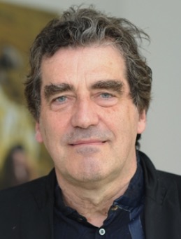 Hans-Dieter Volk合作论文数Institute of Medical Immunology, Charité – Universitätsmedizin Berlin;TC Balance Biopharmaceutics8
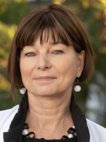 Petra Reinke合作论文数Berlin Center for Advanced Therapies, Charité – Universitätsmedizin Berlin;Tcell Europe5
 Christian Meisel合作论文数Charite, Inst Med Immunol, D-13353 Berlin, Germany3
 Michael Schmueck-Henneresse合作论文数Institute for Medical Immunology, Charité University Medicine Berlin;Department of Nephrology and Internal Intensive Care, Charité University Medicine Berlin;Berlin-Brandenburg Center for Regenerative Therapies, Charité University Medicine Berlin;michael.schmueck-henneresse@charite.de.;Charité University Medicine Berlin, Charité University Medicine Berlin3
 Dimitrios L. Wagner合作论文数2
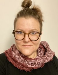 Katarzyna Winek合作论文数Charite, Ctr Stroke Res, D-10098 Berlin, Germany2Engel Odilo合作论文数2Dames Claudia合作论文数2Vogt Katrin合作论文数2
 Lena Pinzur合作论文数2
 Racheli Ofir合作论文数2
 Leila Amini合作论文数2Aberman Zami合作论文数2Andreas Meisel合作论文数2Christine Appelt合作论文数2Stephan Schlickeiser合作论文数2
 Birgit Sawitzki合作论文数Charité — Universitätsmedizin Berlin Institute of Medical Immunology2
 Raphael Gorodetsky合作论文数Immunology Laboratory, Hadassah University Hospital2Andreas Wilhelm合作论文数1Kim-Long Truong合作论文数1Michal Blumenfeld合作论文数1Evgenia Volinsky合作论文数1Ben Hammoud合作论文数1
 Simone Zazie Vogel合作论文数1Tatiana Zittel合作论文数1
 Tony Hartwig合作论文数1Andreas Pascher合作论文数1Joerg C Schefold合作论文数1Su-Jin Park合作论文数1Alina Pruene合作论文数1Nadja Stobutzki合作论文数1mareike thielke合作论文数1Jonas Kath合作论文数1Lisa-Marie Burkhardt合作论文数1
 Jacqueline Wendering合作论文数1
 Stefania Martini合作论文数1
 Kerstin Juelke合作论文数1
 Katarina Stanko合作论文数Charite Univ Med Berlin, D-13353 Berlin, Germany1Weijie Du合作论文数<institution>Cell Biology, Deutsches Rheuma-Forschungszentrum Berlin (DRFZ), an Institute of the Leibniz Association</institution>, Berlin, <country>Germany</country>1Bernice Thommandru合作论文数Integrated DNA Technol, Coralville, IA USA1
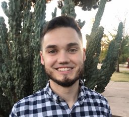 Gavin Kurgan合作论文数Integrated DNA Technologies, Inc.1
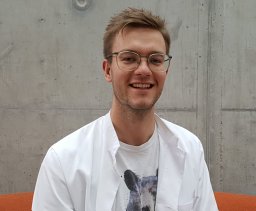 Lennard Ostendorf合作论文数Charite, Neuropathol, D-10117 Berlin, Germany1
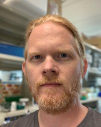 Rolf Turk合作论文数Integrated DNA Technol Inc, Coralville, IA 52241 USA1Olga Blau合作论文数1Axel Pruss合作论文数1
 Josef Priller合作论文数Department of Psychiatry and Neurosciences, Charité Campus Mitte, Universitätsmedizin Berlin;Centre for Clinical Brain Sciences, Edinburgh Medical School, The University of Edinburgh1
 Susanne Herold合作论文数Department of Medicine V, Internal Medicine, Infectious Diseases and Infection Control, University Hospital Giessen and Marburg1
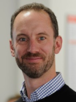 Bill Taylor合作论文数Department of Health Sciences and Technology, Eidgenössische Technische Hochschule Zürich1
 Uta Elisabeth Höpken合作论文数Max-Delbruck-Centrum fur Molekulare Medizin (MDC) Berlin-Buch1




