Non-alcoholic steatohepatitis (NASH) is rapidly surpassing viral hepatitis as the primary cause of hepatocellular carcinoma (HCC). However, understanding of NASH-progressed HCC remains poor, which might impede HCC diagnosis and therapy. In this study, we aim to identify shared transcriptional changes between NASH and HCC, of which we focused on E3 ligase TRIM45. We found TRIM45 exacerbates HCC cells proliferation and metastasis in vitro and in vivo. Further transcriptome analysis revealed TRIM45 predominantly affects fatty acid metabolism and oleic acid restored impaired proliferation and metastasis of TRIM45-deficient HCC cells. IP-tandem mass spectrum and FABP5 depriving experiment indicated that TRIM45 enhance fatty acid synthesis depending on FABP5 presence. Interestingly, we found TRIM45 directly added K33-type and K63-type poly-ubiquitin chains to FABP5 NLS domain, which ultimately promoted FABP5 nuclear translocation. Nuclear FABP5 interacted with PPARγ to facilitate downstream lipid synthesis gene expression. We observed TRIM45 accelerated NASH-to-HCC transition and exacerbated both NASH and NASH-HCC with the enhanced fatty acid production in vivo. Moreover, high concentration of fatty acid increased TRIM45 expression. The established mechanism was substantiated by gene expression correlation in TCGA-LIHC. Collectively, our research revealed a common lipid reprograming process in NASH and HCC and identified the cyclical amplification of the TRIM45-FABP5-PPARγ-fatty acid axis. This signaling pathway offers potential therapeutic targets for therapeutic intervention in NASH and NASH-progressed HCC.
INTRODUCTION AND OBJECTIVES:Cavin1 is a cell membrane caveolin, with controversial function in different tumors. Meanwhile, the role of Cavin1 in hepatocellular carcinoma (HCC) progression remains unclear. In this study, we attempted to elucidate the significance of Cavin1 in HCC occurrence and progression.MATERIALS AND METHODS:Cavin1 content was examined in HCC tissues and paired adjacent normal liver tissues by qRT-PCR and IHC among 81 HCC patients. The Cavin1-mediated regulation of HCC proliferation and metastasis was assessed through in vitro and in vivo experiments. Finally, using GSEA, we found out Cavin1 could be a potential regulator of the Wnt pathway. The alterations of the Wnt pathway-related proteins were identified by Western Blot analysis.RESULTS:Cavin1 was lower expressed in HCC, which implied poor survival outcomes in HCC patients. Phenotypic experiments revealed that Cavin1 strongly suppressed HCC proliferation and migration in vitro and in vivo. Besides, altered epithelial-mesenchymal transition (EMT)-related protein expressions were detected. Based on our GSEA analysis, Cavin1 activated the Wnt pathway, and Western Blot analysis revealed diminished β-catenin, c-Myc, and MMP9 contents upon Cavin1 overexpression.CONCLUSIONS:Cavin1 suppresses HCC progression by modulating HCC proliferation and migration via inhibiting the Wnt/β-catenin axis activation.
Background and Aims: Hyperlipidemia has been extensively recognized as a high-risk factor for NASH; however, clinical susceptibility to NASH is highly heterogeneous. The key controller(s) of NASH susceptibility in patients with hyperlipidemia has not yet been elucidated. Here, we aimed to reveal the key regulators of NASH in patients with hyperlipidemia and to explore its role and underlying mechanisms. Approach and Results: To identify the predominant suppressors of NASH in the setting of hyperlipidemia, we collected liver biopsy samples from patients with hyperlipidemia, with or without NASH, and performed RNA-sequencing analysis. Notably, decreased Lineage specific Interacting Motif domain only 7 (LMO7) expression robustly correlated with the occurrence and severity of NASH. Although overexpression of LMO7 effectively blocked hepatic lipid accumulation and inflammation, LMO7 deficiency in hepatocytes greatly exacerbated diet–induced NASH progression. Mechanistically, lysine 48 (K48)-linked ubiquitin-mediated proteasomal degradation of tripartite motif-containing 47 (TRIM47) and subsequent inactivation of the c-Jun N-terminal kinase (JNK)/p38 mitogen-activated protein kinase (MAPK) cascade are required for the protective function of LMO7 in NASH. Conclusions: These findings provide proof-of-concept evidence supporting LMO7 as a robust suppressor of NASH in the context of hyperlipidemia, indicating that targeting the LMO7-TRIM47 axis is a promising therapeutic strategy for NASH.
Abstract Our previous work had suggested that LncRNA SNHG6 interacted with COPS5 and their expression was positively correlated in hepatocellular carcinoma(HCC). In this study we found that SNHG6 and COPS5 was upregulated in HCC patients, and patients with high expression of SNHG6 and COPS5 had worse overall survival(OS). After RNA pulldown experiments and mass spectrometry analysis of SNHG6, and CO-IP experiments and mass spectrometry analysis of COPS5, we finally determined that EEF1D interacted both SNHG6 and COPS5. EEF1D, which had high expression in HCC tissues and cell lines, was related to poor prognosis. Meanwhile, SNHG6, EEF1D and COPS5 could affect the cell cycle and promote proliferation of HCC. Subcutaneous tumors grown from SNHG6-knockdown cells were inhibited compared to control cells. Knocking down SNHG6 or EEF1D leads to faster degradation of COPS5 protein, and activate COPS5/P27 pathway. The first domain leucine zipper of EEF1D was the binding site to COPS5. Eventually, SNHG6 binds to EEF1D and stabilize COPS5 and activate the P27 pathway, affecting the cell cycle to accelerate cell proliferation. SNHG6 may serve as a new target for the diagnosis and treatment of HCC.
Pancreatic adenocarcinoma (PAAD) carries the lowest survival rate of all major organ cancers, which is of dismal prognosis and high mortality rate. Thus, the present study attempted to identify a few novel prognostic biomarkers and establish an immune-related prognostic signature which could predict the prognosis of PAAD. Four prognostic immune-related genes (IRGs) including S100A6, S100A10, S100A16, and SDC1 were screened by differentially expressed gene (DEG) identification and weighted gene coexpression network analysis (WGCNA). Subsequent analysis proved the high expression of these IRGs in PAAD tissues, suggested by TCGA-PAAD data, merged microarray-acquired dataset (MMD), GEPIA, and Oncomine webtool. By using MMD and TCGA-PAAD data, S100A6 (MMD: AUC = 0.897; TCGA: AUC = 0.843), S100A10 (MMD: AUC = 0.880; TCGA: AUC = 0.780), S100A16 (MMD: AUC = 0.878; TCGA: AUC = 0.838), and SDC1 (MMD: AUC = 0.885; TCGA: AUC = 0.812) exhibited excellent diagnostic efficiency for PAAD. By conducting connectivity map (CMap) analysis, we concluded that three molecule drugs (sulpiride, famotidine, and nalidixic acid) might have worked in the treatment of PAAD. Then, an immune-related prognostic index was constructed, which was validated as an independent prognostic factor for PAAD patients (P=0.004). We further constructed a nomogram by using this immune-related signature and age, the prognostic value of which was validated by using concordance index (C-index = 0.780) and area under curve (AUC = 0.909). Moreover, the immune-related prognostic signature was associated with response to anti-PD-1/L1 immunotherapy. To sum up, four IRGs were screened out and verified to be novel immune-related prognostic biomarkers in PAAD. Besides, sulpiride, famotidine, and nalidixic acid might be potential choices in the treatment of PAAD. An immune-related signature was established to show great potential for prognosis prediction for PAAD, independently, which might guide more effective immunotherapy strategies. A nomogram is further established by using this immune-related prognostic index, which might contribute to more effective prognosis prediction in PAAD patients.
Laparoscopic right posterior hepatectomy is considered difficult on the basis of the surgery difficulty scoring system. In this study, we evaluated the safety and effectiveness of the technical application of indocyanine green (ICG) fluorescence imaging-guided laparoscopic right posterior hepatectomy. Twenty-six patients who underwent ICG fluorescence imaging-guided laparoscopic right posterior hepatectomy at Hepatobiliary and Pancreatic Surgery Department of Zhongnan Hospital, Wuhan University, from June 2018 to December 2019, were included. The influence of patient position, trocar placement, hepatic inflow occlusion, central venous pressure (CVP), and the ICG fluorescence imaging-guided method were analyzed. In 17 patients, the left lateral position was maintained when the main tumor was in the S7, and in the remaining nine patients, the supine position was maintained with the right side of the body raised when the main tumor was in the S6. Ten patients who underwent preoperative injection of ICG were successfully developed for nonanatomical hepatectomy. Sixteen patients received intraoperative ICG injection for anatomical hepatectomy (2 cases had positive imaging findings, 14 cases had negative imaging findings, and 2 cases had failed imaging findings). All patients underwent the Pringle maneuver during the procedure. Four patients were preset with subhepatic vena cava blocking and one patient with suprahepatic inferior vena cava blocking. CVP was controlled at 3.00 ± 0.63 (mean ± SD) cmH2O. The operative time was 216.14 ± 52.05 min, and the bleeding volume was 128.57 ± 75.55 ml. Four patients had Clavien–Dindo level I complications, and one had level III complications. Postoperative hospitalization duration was 6.19 ± 1.40 days. There were 14 patients with hepatocellular carcinoma, 9 with metastatic liver malignancies, 2 with hepatic hemangioma, 1 with focal nodular hyperplasia of the liver, and 10 with hepatitis B liver cirrhosis. ICG fluorescence imaging guidance could be helpful for the safe implementation of laparoscopic right posterior hepatectomy.
Despite of advances in treatment options, hepatocellular carcinoma (HCC) remains nearly incurable and has been recognized as the third leading cause of cancer-related deaths worldwide. As a deubiquitinating enzyme, the antitumor effect of ubiquitin-specific peptidase 53 (USP53) has been demonstrated on few malignancies. In this study, we investigated the potential antitumor role of USP53 in HCC. The results showed that USP53 was downregulated in HCC tissues as well as in HCC cell lines using both in silico data as well as patient samples. Furthermore, the ectopic expression of USP53 inhibited the proliferation, migration and invasion, and induced the apoptosis of HCC cells. Co-immunoprecipitation (CO-IP) assay and mass spectrometry (MS) combined with the gene set enrichment analysis (GSEA) identified cytochrome c (CYCS) as an interacting partner of USP53. USP53 overexpression increased the stability of CYCS in HCC cells following cycloheximide treatment. Finally, the overexpression of CYCS compensated for the decreased apoptotic rates in cells with USP53 knocked down, suggesting that USP53 induced the apoptosis in HCC cells through the deubiquitination of CYCS. To summarize, we identified USP53 as a tumor suppressor as well as a therapeutic target in HCC, providing novel insights into its pivotal role in cell apoptosis.
目的 探讨吲哚菁绿(indocyaninegreen,ICG)荧光显影技术在腹腔镜胆囊手术中的临床应用价值.方法 前瞻性选取术前诊断为胆囊结石合并急性胆囊炎 、慢性萎缩性胆囊炎或残余胆囊的56例病人,其中48例于气腹建立后经外周静脉注射ICG,按剂量不同分为0.5 mg组、1 mg组、5 mg组和10 mg组四组(每组各12例);另外8例于术中根据目标胆道容积经胆道注射浓度为0.025 mg/ml ICG,观察术中目标胆管荧光显影的时间 、荧光强度及对手术的指导作用.结果 48例经外周静脉注射ICG的病人,均成功实施腹腔镜胆囊切除术,除0.5 mg组2例病人显影失败外,余均显影成功.0.5 mg组、1 mg组、5 mg组、10 mg组显影时间分别为(53.60±13.73)min、(42.83±7.80)min、(24.92±6.95)min、(23.17±8.11)min.显影时间显示:0.5 mg组长于1 mg组(P=0.01),1 mg组长于5 mg组(P<0.001),差异均具有统计学意义;5 mg组与10 mg组比较差异无统计学意义(P=0.647).0.5 mg组、1 mg组、5 mg组、10 mg组荧光强度分别为(13.03±4.46)AU、(28.38±7.31)AU、(30.44±6.91)AU、(33.16±6.34)AU.荧光强度显示:0.5 mg组弱于1 mg组,差异具有统计学意义(P<0.001),1 mg组与5 mg组、5 mg组与10 mg比较差异均无统计学意义(均P>0.05).8例术中ICG胆道直接注射者,除1例腹腔镜下肝穿刺经肝内胆管注射病人目标胆管显影失败,其余全部显影成功.结论 准确把握ICG用药剂量、途径和时机,有利于提高腹腔镜胆囊手术的安全性,减少胆道并发症的发生.
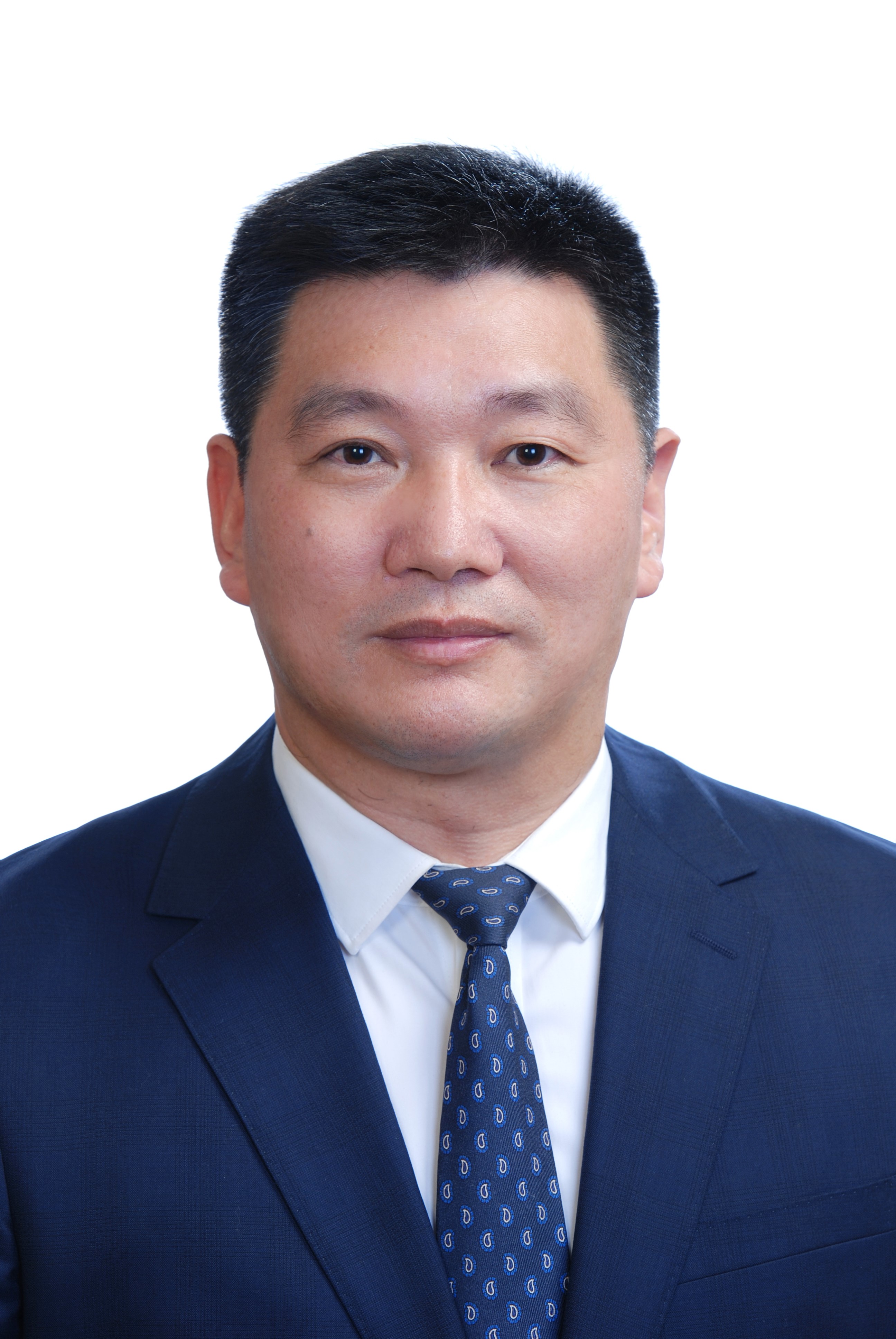 Yufeng Yuan (袁玉峰)合作论文数Taikang Center for Life and Medical Sciences of Wuhan University;Zhongnan Hospital of Wuhan University9
 Weijie Ma合作论文数6
 Chen Xi合作论文数5Fusheng Liu合作论文数4Yonghua Guo合作论文数4Peng Xia合作论文数4Jinghua Li (李景华)合作论文数4
 Zhonglin Zhang (张中林)合作论文数Zhongnan Hospital of Wuhan University4Yang-Wen-Qing Zhang合作论文数3Yingyi Liu合作论文数3Chengjie Mei合作论文数2Xingyuan Hao (郝星源)合作论文数2Tiangen Wu合作论文数2Xiaoling Wu合作论文数2Meng Gao合作论文数2Ganggang Wang合作论文数2Yiran Chen合作论文数2HaiTao Wang (王海涛)合作论文数2
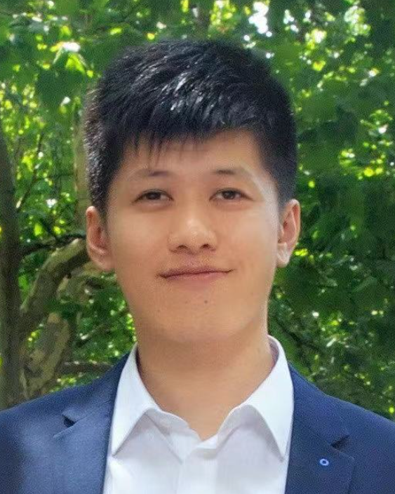 Ye Yao (姚烨)合作论文数Department of Biostatistics, School of Public Health, Fudan University;Research Institute of Intelligent Complex Systems, Fudan University2
 Wang Kunlei合作论文数2
 Zhisu Liu (刘志苏)合作论文数Zhongnan Hospital of Wuhan University2
 Yueming He (何跃明)合作论文数The Second Clinical College of Wuhan University2Xutao Zhang合作论文数1Caixia Dai合作论文数1Jia Zhang合作论文数1Kequan Xu合作论文数1Zhiwen He合作论文数1Bin Liu合作论文数1Xiao Zhang合作论文数1Qian Zhu合作论文数1Jun XIE (谢君)合作论文数1
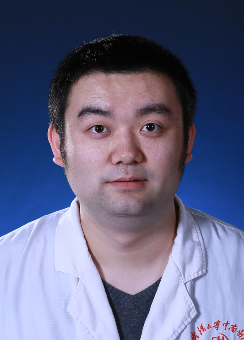 Zhen Li (李臻)合作论文数Zhongnan Hospital of Wuhan University1
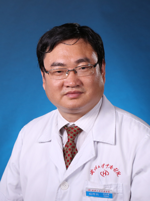 Bicheng Wang (汪必成)合作论文数The Second Clinical College of Wuhan University1
 Jing Wei (魏晶)合作论文数School of Life Science and Technology, Xi'an Jiaotong University1
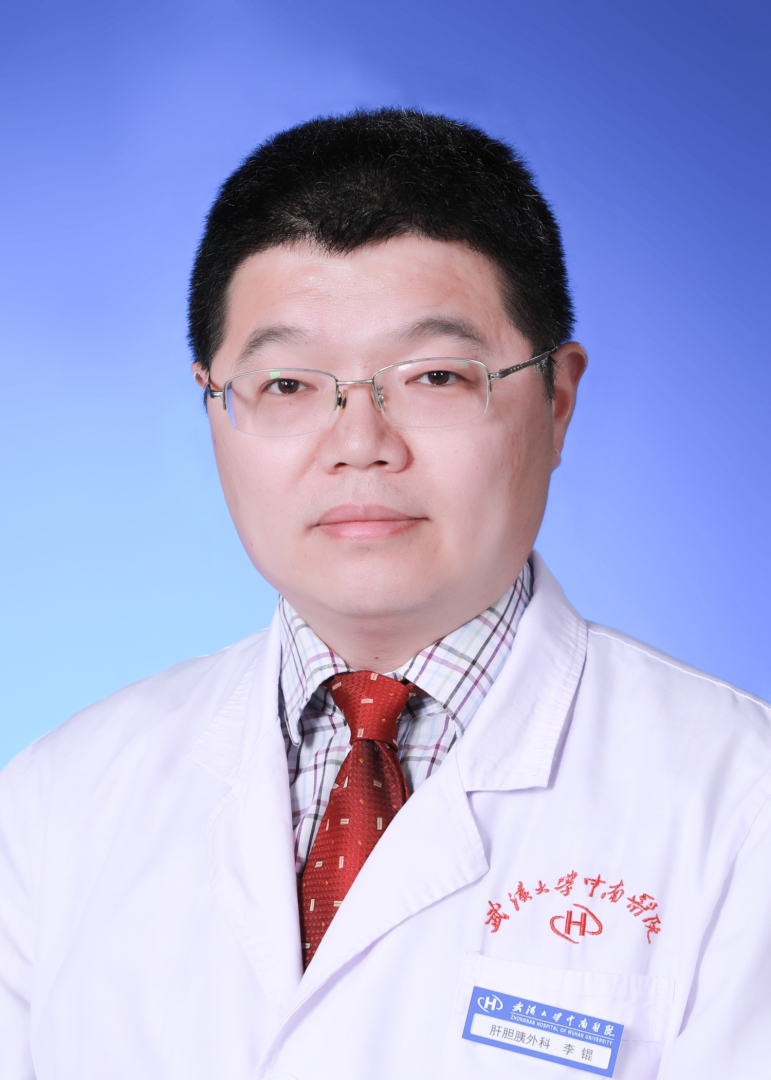 Kun Li (李锟)合作论文数Zhongnan Hospital of Wuhan University1
 Haitao Wang (王海涛)合作论文数College of Smart Materials and Future Energy, Fudan University1




