Inflammatory bowel disease (IBD) is a chronic relapsing disorder driven by complex interactions between genetic susceptibility, immune dysregulation, and environmental factors. Ferroptosis has been identified as a key regulator in the progression of IBD. While much research focuses on endogenous signaling pathways, extrinsic mechanisms—particularly the modulation of IBD through the gut microbiota-induced ferroptosis remain underexplored. Dysregulated ferroptosis, influenced by gut microbiota, exacerbates microbial imbalance, creating a vicious cycle. Notably, the gut microbiota plays a critical role in IBD progression through multidimensional mechanisms, including regulation of metabolites, maintenance of immune homeostasis, and protection of the intestinal barrier. This review examines the microbiota–ferroptosis axis in IBD pathogenesis, aiming to provide insights into potential therapeutic strategies. In particular, we discuss emerging treatments targeting ferroptosis inhibition, iron homeostasis regulation, and microbiota interventions, which hold promise for improving clinical outcomes and promoting pathological recovery in IBD patients.
Abstract Although the role of neutrophils in modulating antitumor T-cell responses has been extensively studied, their direct effects on tumor cells remain less well understood. In this study, we investigated whether neutrophils have the capacity to directly kill tumor cells independently of T cells. We found that anti-CD40–based therapy, when combined with IL10 receptor blockade, initiates a Batf3-dependent pathway in which IL12 and IFNγ secretion results in oncolytic neutrophil activity. Using a combination of microscopy, single-cell, and functional assays, we observed that killing of tumor cells by neutrophils is dependent on physical contact and degranulation. This degranulation-mediated killing is associated with an atypical dynamic invasive neutrophil phenotype. In line with our preclinical findings, our phase I trial of anti-CD40 shows that circulating IL12, IFNγ, and IL10 increase in response to anti-CD40, whereas our phase Ib/2 PRINCE study shows that lower circulating IL10 is associated with favorable overall survival (OS) specifically among anti-CD40–treated patients. Finally, we found that neutrophil expansion with granulocyte colony-stimulating factor is associated with improved OS, specifically in patients treated with anti-CD40, suggesting that this pathway may be amenable to therapeutic intervention in patients with advanced cancer.
BackgroundHuman papillomavirus 33 (HPV33) is among the five most prevalent HPV genotypes in China and is commonly involved in co-infections. However, the synergistic effects of specific genotype combinations on cervical carcinogenesis remain incompletely understood. This study aimed to characterize HPV33 co-infection patterns and their associated genetic variations in relation to cervical lesion progression.MethodsWe enrolled 1,770 HPV33-positive patients from the Obstetrics and Gynecology Hospital of Fudan University between 2018 and 2023, including 852 with HPV33 single infections and 918 with multiple infections. Logistic regression was used to assess associations between co-infection characteristics and cervical histopathological results. In a subset of 90 cases, the full-length L1 gene of HPV33 was sequenced and phylogenetically analyzed to evaluate genomic variation by infection status.ResultsThe number of HPV33 co-infecting genotypes was positively correlated with cervical lesion severity (p < 0.05). HPV52, HPV16, and HPV58 were the most frequent co-infecting genotypes and were associated with an increased risk of cervical intraepithelial lesion progression: HPV16 (OR 1.97, 95% CI: 1.17–3.34), HPV52 (OR 2.07, 95% CI: 1.26–3.39), HPV58 (OR 2.83, 95% CI: 1.42–5.64). However, among HPV33 multiple infections (≥3 genotypes), only those that involved HPV16 were directly and significantly associated with an increased risk of cervical lesions, with ORs increasing from 1.72 (95% CI: 1.03–2.85) for LSIL to 3.16 (95% CI: 1.75–5.72) for HSIL or worse. Phylogenetic analysis classified most HPV33 sequences into sublineage A1, with no significant difference in L1 gene mutations between single and multiple infections.ConclusionHPV33 co-infection patterns, particularly those involving HPV16, are consistently associated with an elevated risk of high-grade cervical lesions in this Chinese cohort. These findings underscore the differential risks associated with distinct HPV33 co-infection patterns and support genotype-specific risk stratification in cervical cancer screening programs.
Although the role of neutrophils in modulating antitumor T-cell responses has been extensively studied, their direct effects on tumor cells remain less well understood. In this study, we investigated whether neutrophils have the capacity to directly kill tumor cells independently of T cells. We found that anti-CD40-based therapy, when combined with IL10 receptor blockade, initiates a Batf3-dependent pathway in which IL12 and IFNγ secretion results in oncolytic neutrophil activity. Using a combination of microscopy, single-cell, and functional assays, we observed that killing of tumor cells by neutrophils is dependent on physical contact and degranulation. This degranulation-mediated killing is associated with an atypical dynamic invasive neutrophil phenotype. In line with our preclinical findings, our phase I trial of anti-CD40 shows that circulating IL12, IFNγ, and IL10 increase in response to anti-CD40, whereas our phase Ib/2 PRINCE study shows that lower circulating IL10 is associated with favorable overall survival (OS) specifically among anti-CD40-treated patients. Finally, we found that neutrophil expansion with granulocyte colony-stimulating factor is associated with improved OS, specifically in patients treated with anti-CD40, suggesting that this pathway may be amenable to therapeutic intervention in patients with advanced cancer.
Volumetric visualization of tumor cell and interacting neutrophil shown in Figure 7A. Tumor cell is shown in green, interacting neutrophil in purple, and overlapping volume in red.
Volumetric visualization of interacting neutrophil (inner box) and tumor cell shown in Supplemental Video 2.
Contact depicted in circle C of Figure 7A between tumor cell and interacting neutrophil.
Time-lapse SDCM, at 20-minute intervals, of treated neutrophils engaged in DIP prestained with lipophilic membrane dye (magenta) in coculture with B16-YFP (green).
Time-lapse SDCM, at 20-minute intervals, of treated neutrophils engaged in DIP prestained with lipophilic membrane dye (magenta) in coculture with B16-YFP (green).
Background:Chordomas are rare bone malignancies originating from remnants of the notochord, primarily occurring at the base of the skull or sacrum. While typically slow growing with low-grade histology, chordomas can be locally aggressive and highly recurrent. Materials and methods:We retrospectively identified 29 patients with sacral chordomas and utilized archival tissue to uncover novel insights into chordoma molecular heterogeneity, using a combination of DNA methylation profiling, spatial transcriptomics and multiplexed immunofluorescence. Results:Unsupervised clustering of methylation probes revealed two distinct epigenetic clusters, Cluster1 and Cluster2. Cluster2 was enriched for recurrent tumors and exhibited significant hypomethylation across most differentially methylated probes in intergenic and open sea regions. Multivariate linear regression analysis indicated that exon boundary and intergenic regions primarily drove these methylation changes. Digital Spatial Profiler (DSP) analysis identified differential gene expression among 9489 genes between tumor and stromal regions across 120 regions of interest, revealing 824 significantly differentially expressed genes. Tumor regions showed reduction of expression of major histocompatibility complex (MHC) class II genes and up-regulation of MHC class I genes compared with stroma. Multiplexed immunofluorescence revealed stromal enrichment of CD3+ and CD14+ cell populations, which was also associated with distinct survival groups. Stromal regions exhibited significant populations of immune-activated T cells and expression of immune checkpoint factors T-cell immunoglobulin and mucin-domain containing protein 3 and programmed cell death protein 1 (PD-1) in T-cell subsets. Stromal myeloid cells showed increased CD47 and PD-1 expression. Conclusions:Our study identified distinct epigenetic profiles in sacral chordomas, which were associated with recurrence, and revealed expression of checkpoint markers TIM3, CD47 and PD-1, warranting further investigation through functional validation.
Complete differentially expressed gene list between CMI WT and CMI KO in neutrophils (Related to Figure 5I)
Distribution of neutrophil scores by study phase and anti-CD40 treatment among patients in the PRINCE Study (Related to Figure 3D)
 Jedd Wolchok合作论文数Graduate School of Medical Sciences, Weill Cornell Medicine;Meyer Cancer Center, Weill Cornell Medicine47
 Taha Merghoub合作论文数Graduate School of Medical Sciences, Weill Cornell Medicine;Meyer Cancer Center, Weill Cornell Medicine45
 Sadna Budhu合作论文数32Hollmann Travis J合作论文数Dept Pathol, Mem Sloan Kettering Canc Ctr29Danny N. Khalil合作论文数22Daniel Hirschhorn合作论文数20
 Ronan Chaligne合作论文数20Liu Cailian合作论文数20Schulze Isabell合作论文数Department of Hematology and Oncology, University of Halle;Department of Hematology and Oncology, University of Muenster;University of Halle, University of Muenster19
 Cabanski Christopher R合作论文数Dept Stat & Operat Res, Univ N Carolina18
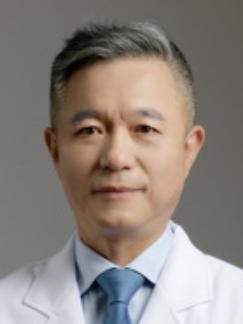 Long Sui (隋龙)合作论文数Obstetrics & Gynecology Hospital of Fudan University18
 Robert H. Vonderheide合作论文数Abramson Cancer Center, Perelman School of Medicine, University of Pennsylvania;Department of Medicine, Perelman School of Medicine, University of Pennsylvania18Deena M. Maurer合作论文数16Fadi Samaan合作论文数16
 Nathan Suek合作论文数Columbia Ctr Translat Immunol, Columbia Univ16
 Eileen O'Reilly合作论文数David M Rubenstein Ctr Pancreat Canc, MSK16
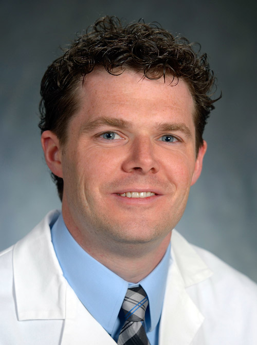 Mark H. O'Hara合作论文数Division of Hematology and Oncology, Department of Medicine, Perelman School of Medicine, University of Pennsylvania16
 Wungki Park合作论文数Yonsei University, Yonsei University16Monette Sebastien合作论文数16Maltez Vivien I合作论文数16
 Ghassan Kanazi合作论文数Memorial Sloan Kettering Cancer Center/Weill Cornell Medical College16Tibor Keler合作论文数16
 Margaret K. Callahan合作论文数Neag Comprehensive Cancer Center, University of Connecticut;Carole and Ray Neag Comprehensive Cancer Center, University of Connecticut15
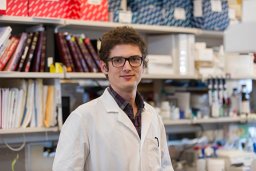 Billel Gasmi合作论文数Ludwig Collaborative Laboratory, Memorial Sloan Kettering Cancer Center15
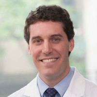 Michael A. Postow合作论文数Memorial Sloan Kettering Cancer Center14Qi Zhou合作论文数13
 Katherine Panageas合作论文数Memorial Sloan Kettering Cancer Center13David A Schaer合作论文数12Roberta Zappasodi合作论文数Joan and Sanford I. Weill Department of Medicine, Weill Cornell Medicine, Cornell University12Zhong Hong合作论文数12Romin Yevgeniy合作论文数11
 Congjian Xu (徐丛剑)合作论文数School of Medicine, Westlake University11YingXin Gong (宫颖新)合作论文数10Hongwei Zhang合作论文数10Yan Wang合作论文数9
 Matthew Hellmann合作论文数Memorial Sloan Kettering Cancer Center9
 Vamsidhar Velcheti合作论文数8Wenjie Qu合作论文数7Nathaniel Aleynick合作论文数7
 Lavery Jessica合作论文数College of Physicians and Surgeons, Columbia University7
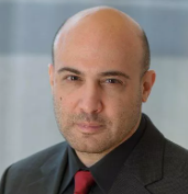 Nicholas D. Socci合作论文数Sloan Kettering Institute, Memorial Sloan Kettering Cancer Center7
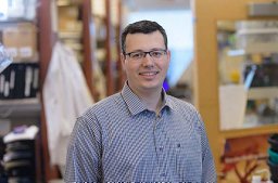 Yasin Senbabaoglu合作论文数University of Michigan7
 Gabrielle Rizzuto合作论文数Memorial Sloan Kettering Cancer Center7Tianyi Bi合作论文数6Rami Vanguri合作论文数Computat Oncol, Mem Sloan Kettering Canc Ctr6
 Jacob Ricca合作论文数6
 Olivier de henau合作论文数6
 Cynthia Sirard合作论文数6
 Raphael Bueno合作论文数Brigham and Women's Hospital Heart and Vascular Center;Brigham and Women's Lung Center;Harvard Medical School6




