Controlling the self-assembly of mesoporous materials beyond equilibrium remains a fundamental challenge. Conventional templating systems form ordered structures through energy-minimizing coassembly but lack the kinetic and spatial freedom required to produce asymmetric or topologically complex architectures. Here, we report a bioinspired coacervate-directed silicification strategy that enables diffusion-limited and spatially asymmetric condensation within soft templates, yielding ordered hexagonal mesoporous nanotoroids. In this system, poly(acrylic acid) (PAA) electrostatically associates with the cationic surfactant cetyltrimethylammonium bromide (CTAB), driving liquid-liquid phase separation and forming disc-like coacervate assemblies. Reaction-diffusion imbalance across the inner and outer interfaces within these templates induces asymmetric silicification, driving an interior collapse and toroidal self-transformation. By tuning PAA concentration, the diffusion-condensation kinetics can be precisely modulated, allowing programmable control over collapse dynamics and final topology, with enlarged central cavities (14-71 nm) and reduced rim thickness (15-35 nm). These nanotoroids exhibit uniform sub-100 nm size, high surface area (846 m2 g-1), and abundant mesopores (∼2.0 nm). The nanotoroids (rim thickness ∼15 nm, height ∼40 nm) display markedly prolonged blood circulation, enhanced tumor accumulation (+173%), improved vascular extravasation, and deeper intratumoral penetration, while reducing hepatic and splenic uptake by 19% and 14%, respectively, compared with spherical analogues. These combined advantages translate into potent antitumor efficacy in both subcutaneous and spinal metastasis models. This work establishes a new paradigm for sol-gel topology control by bridging reaction-diffusion dynamics with bioinspired silicification based on the chemistry of LLPS (liquid-liquid phase separation), thereby unlocking the untapped biomedical potential of toroidal topologies that were rarely accessible.
Peripheral nerve injury (PNI) imposes a substantial burden due to its high disability rate. Although drug-delivering nerve guidance conduits (NGCs) are widely used for PNI repair, their efficacy remains limited, largely because of inefficient, poorly controllable drug release and the hostile early microenvironment driven by oxidative stress and iron metabolism imbalance. Ferroptosis is a key manifestation of this post-injury iron dysregulation. Here, we identify Schwann cell ferroptosis as an early, temporally confined pathological process after PNI and accordingly develop an electro-responsive conductive silk fibroin/polypyrrole (SF/PPy) NGC loaded with menaquinone-4 (MK-4) for temporally targeted local anti-ferroptotic therapy. Electrical stimulation (ES) markedly enhanced MK-4 release, increasing the 14-day cumulative release from 11.20 +/- 0.43% to 23.5 +/- 0.84%. In vitro, under ES, MK-4 suppressed Schwann cell ferroptosis mainly via activation of ferroptosis suppressor protein 1 (FSP1). In vivo, SF/PPy/MK-4 conduits combined with ES modulated the early post-injury microenvironment, inhibited Schwann cell ferroptosis, and significantly improved sensory and motor functional recovery. Overall, this study establishes Schwann cell ferroptosis as an early therapeutic target in PNI and presents a conductive NGC platform integrating ES responsiveness with temporally targeted microenvironmental modulation to promote peripheral nerve regeneration.
Peripheral nerve injury (PNI) poses significant challenges due to the complex structure and regenerative microenvironment of peripheral nerves, which limit self-repair capabilities. Artificial nerve conduits have been widely used for nerve repair. Here, a conductive-piezoelectric integrated microstructured conduit is designed, using poly(lactic glycolic acid) (PLGA) and poly(vinylidene fluoride) (PVDF) via electrostatic spinning to obtain an implantable, biodegradable piezoelectric nanofibrous membrane. This membrane is further enhanced with a reduced graphene oxide/methacrylated gelatin (rGO/GelMA) gel, which synergistically promotes peripheral nerve repair. In vitro assessments reveal that the microgroove surface pattern of the conduit effectively stimulated the directional migration of cells. Moreover, using a rat sciatic nerve injury model, rGO is demonstrated to significantly modulate cellular oxidative stress, thereby facilitating nerve repair. Additionally, mild electrical stimulation induced by low-intensity pulsed ultrasound (LIPUS) is found to enhance the recovery of motor function. These findings demonstrate the multifaceted benefits of the rGO/GelMA@PVGA composite conduit, which integrates physical guidance, oxidative stress inhibition, and ultrasound-activated electrical stimulation, providing an unprecedented multimodal synergistic strategy with great potential for clinical treatment of peripheral nerve injury.
Microbiome and transcriptome analyses revealed that Fusobacterium nucleatum (F.n) in clinical samples is associated with immune suppression and poor prognosis in triple-negative breast cancer spinal metastasis. However, its preferential localization in hypoxic tumor regions limits the efficacy of conventional antimicrobial therapies, which poorly penetrate solid tumors and function suboptimally under anaerobic conditions. Developing strategies that enable deep tumor penetration, eliminate anaerobic bacteria, and induce immunogenic cell death remains a major challenge. In this study, a novel charge-enrichment and light-activated biomimetic nanosystem, designated as polyion liquid-bridged eosin Y (PIL-BEY), was developed. On one hand, interionic hydrogen bonding and dynamic electrostatic interactions within polyionic liquids reduce the surface energy of the nanoprobe and synergistically remodel the dense tumor stromal microenvironment via photodynamic therapy, thereby facilitating the deep intratumoral penetration and accumulation of PIL-BEY. On the other hand, the novel photosensitizer BEY generates reactive oxygen species via electron transfer under hypoxic conditions, thereby effectively eradicating bacteria within hypoxic tumor regions. The resulting pathogen-associated molecular patterns, together with damage-associated molecular patterns, activate dendritic cells, promote cytotoxic T lymphocyte infiltration, trigger immunogenic cell death, and induce systemic antitumor immune responses with durable immune memory. This oxygen-independent, dual-functional nanoplatform offers a promising strategy for treating invasive metastatic tumors.
Cartilage defects remain a major clinical challenge due to the limited efficacy of current therapies and the intrinsically low regenerative capacity of chondrocytes. Mechanical loading has emerged as a promising strategy to enhance stem cell-based cartilage repair; however, the underlying molecular mechanisms remain poorly understood. Here, we show that cyclic tensile strain primes mesenchymal stem cells (MSCs) to secrete exosomes enriched in microRNA-330-3p (miR-330-3p), which markedly enhances cartilage regeneration. Mechanistically, miR-330-3p restores mitochondrial quality control in chondrocytes by engaging an FKBP4-FoxO3a-dependent mitophagy program, leading to activation of PINK1/Parkin-mediated mitochondrial clearance. The regenerative efficacy of miR-330-3p-enriched exosomes was validated in a Sprague-Dawley rat model of cartilage defects. In vitro, miR-330-3p promotes chondrocyte proliferation and migration while suppressing apoptosis, senescence, and extracellular matrix degradation. Together, these findings identify mechanically primed MSC-derived exosomes as a mechanistically informed therapeutic strategy for cartilage repair.
Bone defects pose significant clinical challenges due to the limited regenerative capacity of adult bone and the shortcomings of existing biomaterials, which lack dynamic electromechanical signaling crucial for repair. Here, we present an injectable, ultrasound-responsive piezoelectric hydrogel engineered to synergize silk fibroin's (SF) structural adaptability with polyvinylidene fluoride's (PVDF) bioelectrical activity. Methacrylated silk fibroin (SM) enables rapid UV-triggered crosslinking via a cost-effective photoinitiator system, while electrospun PVDF nanofibers, cryosectioned into microscale units, confer dynamic piezoelectric responsiveness. Under ultrasound stimulation, PVDF generates localized electrical cues that transiently elevate reactive oxygen species (ROS), activating the NRF2 antioxidant pathway to resolve oxidative stress, which polarizes macrophages toward pro-regenerative M2 phenotypes, enhances osteogenic differentiation of adipose-derived mesenchymal stem cells (ADSCs) and promotes angiogenesis in vitro. Additionally, it was also confirmed that implantation of critical-sized femoral defects in rats could facilitate bone regeneration by micro-CT and histological analysis in vivo. This platform transcends beyond passive scaffolding by recapitulating bone's electromechanical-immune axis offers a paradigm shift toward smart biomaterials for complex skeletal defects. The integration of PVDF's ultrasound-triggered piezoelectricity with SM's bioactivity establishes a multifunctional system that dynamically regulates redox homeostasis, immune modulation, and tissue remodeling, addressing unmet needs in bone tissue engineering.
Regenerated silk fibroin (RSF) from Bombyx mori is well known for its outstanding biocompatibility, biodegradability, and mechanical characteristics. Spider silk has even better strength and extensibility and is considered one of the best silk fibers in nature. Silkworm fibroin heavy chain in fibroin and major ampullate spidroin-1 in spider silk from the spider Nephila clavipes share similar molecular weight and repetitive structure, which makes the genetic modification of silkworms possible. In our study, a novel hydrogel generated from genetically modified Bombyx mori cocoons was characterized and evaluated for potential clinical application. For the crosslinking of the hydrogel, horseradish peroxidase and H2O2 were added to the RSF solution to create a double crosslinking structure. As a result, genetically modified regenerated silk fibroin (GMRSF) hydrogels exhibited better mechanical characteristics compared with RSF hydrogels. Additionally, compared with RSF hydrogels, GMRSF hydrogels exhibited stronger osteogenic differentiation function on rat bone mesenchymal stem cells (BMSCs). In vivo, GMRSF hydrogel also showed better osteogenic differentiation function. While the mechanism behind this remains unclear, the outstanding osteogenesis ability of GMRSF sheds light on applications in orthopaedic diseases in the future such as bone defects.
Bone defect repair under hyperglycemic conditions is limited by sustained oxidative stress, chronic low-grade inflammation and disrupted osteoimmune-vascular coupling. In this study, we constructed an injectable and biodegradable silk fibroin/PEG hydrogel incorporating MnO2 honeycomb nanospheres and liposome-encapsulated icariin (FG/Mn@Ica) to rebuild a bioactive and mechanocompatible microenvironment for diabetic bone regeneration. The hydrogel presented an interconnected porous morphology, elastic-dominant rheology, adaptive compressive behavior and controlled degradation with the sustained release of Mn ions and Ica. Functionally, FG/Mn@Ica reprograms the high-glucose microenvironment at multiple levels. In vitro, it enhances the proliferation of bone marrow mesenchymal stem cells and osteogenic commitment while simultaneously attenuating reactive oxygen species accumulation, stabilizing the mitochondrial membrane potential and activating the AMPK-SIRT1-NRF2-mediated antioxidant axis. Moreover, FG/Mn@Ica promoted the migration of human umbilical vein endothelial cells (HUVECs) and angiogenic tube formation, induced the polarization of pro-regenerative M2 macrophages with elevated Arg1, and suppressed senescence-associated and inflammation-associated markers. In vivo, in a rat femoral defect model, FG/Mn@Ica significantly promoted new bone regeneration. Overall, FG/Mn@Ica integrates mechanical support, restoration of redox homeostasis, osteoimmune modulation and osteogenic reinforcement, offering a rational biomaterial strategy for bone regeneration under diabetic stress.
Postmenopausal osteoporosis (PMOP) is a predominant form of clinical osteoporosis. It has led to significant health and social burdens for older patients. Reestablishing the balance between osteogenic and osteoclastic is a crucial strategy for treating PMOP. Curcumin (Cur), a naturally derived polyphenolic substance, has gained recognition as a viable option for treating osteoporosis. Despite its potential, the clinical use of Cur is hindered by its limited bioavailability and the presence of side effects. Nanoparticles modified with aspartic acid octapeptide (ASP8) exhibit a strong affinity for bone tissue, facilitating targeted delivery. This study presents novel acid-responsive zeolite imidazolate framework-8 (ZIF) nanoparticles modified with ASP8 and loaded with Cur (Cur@ZIF@ASP8, CZA). Upon delivery by this nanoparticle drug delivery system, Cur can effectively regulate bone homeostasis, offering a potential therapeutic strategy for osteoporosis. This study demonstrated that CZA nanoparticles could successfully transport Cur to bone tissue without significant toxicity. Furthermore, nanoparticles promote bone formation and inhibit osteoclast activity. They also modify the expression of related genes and proteins, such as OCN, ALP, CTSK and MMP9. Significant evaluations utilizing microcomputed tomography, Masson's staining, hematoxylin and eosin staining and immunofluorescence staining demonstrated that intravenous CZA administration in ovariectomized mice resulted in bone destruction while simultaneously reducing overall bone loss. In conclusion, CZA nanoparticles hold promise as a therapeutic option for osteoporosis.
Introduction: Endplate cartilage is crucial for nutrient transport to intervertebral disc (IVD), and its calcification due to abnormal mechanical stress significantly contributes to intervertebral disc degeneration (IDD). Primary cilia, which sense mechanical stimuli, are key to this process. Intraflagellar Transport 88 (IFT88) regulates endplate calcification under mechanical stress, but its specific mechanisms remain inadequately characterized. Objectives: We aimed to elucidate the role and regulatory mechanisms of IFT88 in primary cilia in endplate cartilage calcification and IVD. Methods: Changes in IFT88 expression in endplate cartilage and its response to mechanical stress were assessed using histochemical staining, Western blotting, immunofluorescence, and transmission electron microscopy. The relationship between abnormal stress and calcium ions was explored through RNA sequencing, qPCR, and calcium staining. In vitro studies investigated the regulatory mechanisms of IFT88 in chondrocytes subjected to abnormal stress using molecular docking, co-immunoprecipitation, dual-luciferase assays, and flow cytometry. Rats tail crush model confirmed the role of IFT88 in chondral calcification and IDD as a potential therapeutic target. Results: As intervertebral disc degeneration progresses, IFT88 expression in the primary cilia of cartilage endplate cells decreased. Under normal stress conditions, IFT88 levels increased with the intensity and duration of stress; however, excessively high stress triggered mechanisms that led to cilia depletion. Elevated intracellular calcium concentrations under tensile stress contributed to endplate calcification and IDD. Additionally, IFT88 negatively regulated its positive transcription factor C/EBPα under abnormal stress, potentially contributing to cilia depletion. IFT88 also inhibited the hyperactivation of transient receptor potential vanilloid 4 (TRPV4), reducing calcium influx and alleviating oxidative stress and Wnt pathway activation. In vivo studies showed that overexpressing IFT88 maintains disc height and structural integrity while reducing endplate ossification. Conclusion: Our study demonstrates that IFT88 inhibits TRPV4, thereby protecting endplate cartilage and positioning IFT88 as a promising therapeutic target for IDD and endplate calcification.
Vertebral compression fractures (VCFs) are prevalent in the elderly, often caused by osteoporosis or trauma. Differentiating acute from chronic VCFs is vital for treatment planning, but MRI, the gold standard, is inaccessible for some. However, CT, a more accessible alternative, lacks precision. This study aimed to enhance CT’s diagnostic accuracy for VCFs using deep transfer learning (DTL) and radiomics. We retrospectively analyzed 218 VCF patients scanned with CT and MRI within 3 days from Oct 2022 to Feb 2024. MRI categorized VCFs. 3D regions of interest (ROIs) from CT scans underwent feature extraction and DTL modeling. Receiver operating characteristic (ROC) analysis evaluated models, with the best fused with radiomic features via LASSO. AUCs compared via Delong test, and clinical utility assessed by decision curve analysis (DCA). Patients were split into training (175) and test (43) sets. Traditional radiomics with LR yielded AUCs of 0.973 (training) and 0.869 (test). Optimal DTL modeling improved to 0.992 (training) and 0.941 (test). Feature fusion further boosted AUCs to 1.000 (training) and 0.964 (test). DCA validated its clinical significance. The feature fusion model enhances the differential diagnosis of acute and chronic VCFs, outperforming single-model approaches and offering a valuable decision-support tool for patients unable to undergo spinal MRI.
With four levels of formations and functional properties similar to biological tissues, protein hydrogel attracts great attention from researchers and clinical practitioners for broad applications. However, hydrogels with high water content usually exhibit poor mechanical properties, which limit their widespread application. In the study, a general and feasible approach was used to enhance the regenerated silk fibroin (RSF) hydrogel's mechanical strength by mimicking the human skeleton biomineralization process in vitro. The final mechanical strength was around 4.5 MPa, and the compressive modulus was around 0.90 MPa after combining the effects of ethanol induction of β-sheet and mineralization by SBF, reaching 3 times higher strength than the original RSF hydrogel. The mineral composition and chemical structure proved that the biomineralization of HAp apposition within amorphous hydrophilic domains enhanced the mechanical properties of RSF hydrogel. In vivo RSF hydrogel mineralization was observed after subcutaneous implantation in rats. The femoral bone defect model demonstrated that the biomineralized RSF hydrogel maintained the bone structure from collapse in the early phase. In summary, a straightforward method for fabricating high-strength biomineralized RSF hydrogel through a slow-and-low approach, i.e., low SBF concentration and longer immersion time, was developed and is promising for treating bone diseases.
Inducing lethal endoplasmic reticulum (ER) stress is a key initiative to counteract tumour resistance and induce anti-tumour immunity. However, conventional ER stress inducers are largely limited by hypoxia and off-target effects to induce tumour-lethal ER stress. Here, we encapsulated Cu-bridged eosin Y (CuBY) in ER-targeting peptide (pardaxin)-modified mesoporous silica and successfully constructed an oxygen-independent multifunctional copper-light synergistic prodrug nanosystems (MP@CuBY). MP@CuBY is activated to the "on" state within the glutathione-overexpressing tumour microenvironment, causing copper and BY release. Interestingly, released copper can drive oxygen-independent cascade reactions in situ in the ER, resulting in the production of highly toxic O2-• and •OH. The BY released can produce 1O2 in situ in the ER under laser irradiation. Therefore, type I and type II reactive oxygen species (ROS) generated by MP@CuBY in situ in the ER specifically reprogramed tumour immunogenic ER stress, which significantly activated systemic anti-tumour immunity and long-term immune memory, as well as ensured satisfactory efficacy in synergistically eradicating spinal metastases in conjunction with α-PD-L1 antibody. In conclusion, the well-designed MP@CuBY may represent an advanced design for anti-tumour prodrug nanosystems, providing a novel copper-light synergistic strategy for the specific activation of lethal tumour ER stress.
Triple-negative breast cancer (TNBC) exhibits a high propensity for spinal metastasis, leading to severe morbidity and limited therapeutic responses. However, the molecular mechanisms driving spinal colonization remain poorly defined. Here, we identify the epigenetic reader ZMYND8 as a key mediator of TNBC spinal metastasis. ZMYND8 is significantly upregulated in spinal metastatic lesions and correlates with adverse patient outcomes. Transcriptomic profiling reveals that spinal metastases display profoundly immunosuppressive microenvironments, with elevated M2 macrophage infiltration positively associated with ZMYND8 expression. Mechanistically, ZMYND8 functions as a scaffold protein that promotes assembly of the DDX3X–CK1ε complex, thereby activating WNT/β-catenin signaling and promoting spinal metastasis. Furthermore, we identify OTUD4 as a bona fide deubiquitinase that directly interacts with and stabilizes ZMYND8, thereby enhancing TNBC cell migration, invasion, and spinal colonization. The resulting OTUD4–ZMYND8–DDX3X signaling axis drives canonical WNT/β-catenin signaling, upregulates CSF1 expression and promotes M2 polarization of macrophages, collectively fostering invasive behavior and establishing an immunosuppressive niche conducive to spinal metastasis. Collectively, these findings establish the OTUD4–ZMYND8–DDX3X axis as a pivotal regulator of spinal metastasis in TNBC and highlight its potential as a therapeutic target for inhibiting metastatic progression.
Low back pain (LBP), one of the most common health problems, is the leading cause of disability globally. Intervertebral disc degeneration (IDD) accounts for most LBP. However, the molecular mechanism underlying IDD remains unclear, and the existing treatment strategy for IDD is still limited. A growing body of evidences suggest that the Hedgehog (HH) pathway plays an essential role in the formation, maintenance, and degeneration of intervertebral discs (IVDs), with Sonic HH (SHH) being primarily involved in the development and maturation of the IVDs and a strong link between Indian HH(IHH) and disc calcification. This review provides an overview of the role of the HH signaling pathway in the developmental maturation and degeneration of IVDs and suggests potential therapeutic targets for IDD that may interfere with HH signaling.
INTRODUCTION:Disruption of the circadian rhythm (CR) and autophagy in intervertebral discs contributes to intervertebral disc degeneration (IDD) progression. However, the circadian regulation of autophagy requires further investigation. OBJECTIVES:We observed the expression of circadian proteins and autophagic markers of nucleus pulposus (NP) cells followed a diurnal rhythmic pattern in vivo and in vitro. METHODS:NP tissues were collected from light/dark cycle-shifted rats and IDD patients of varying severity. CR and ECM-related proteins were analyzed by immunohistochemistry and western blotting. Primary rat NP cells were treated with hypoxia or CoCl2, followed by western blotting for CR proteins, HIF-1α, and autophagy markers. siRNA knockdown of PER2 or HIF-1α was performed to assess their roles in regulating autophagy, ECM, and CR-associated proteins. RESULTS:The silencing of clock gene PER2 disrupted the rhythmic expression of autophagic markers, by contrast PER2 overexpression inhibited mTOR pathway and enhanced autophagic levels. Co-IP analysis demonstrated the PER2-mTOR interaction, linking CR and autophagy rhythm. The inflammatory stimulator dampened the CR and autophagy rhythm, however intermittent hypoxia and cobalt chloride (CoCl2) re-synchronized the rhythm. Mechanistically, HIF-1α-mediated regulation of PER2 by hypoxia was involved in the re-synchronization, which was further demonstrated by the loss of CR and autophagy rhythm after silencing of PER2 or HIF-1α under hypoxia. Furthermore, the rhythm of oxygen level and HIF-1α was proved in living healthy NP tissue, confirming the hypoxia as a Zeitgeber. CR disruption and autophagy dysfunction led to catabolism of extracellular matrix (ECM), but the hypoxia, CoCl2 and autophagic stimulator could promote the rebalance of ECM metabolism. CONCLUSION:Our study demonstrates that hypoxia maintains the intrinsic CR and autophagy rhythm through the HIF-1α/PER2/mTOR pathway to prevent IDD.
Large bone defect healing remains a challenge in current clinical treatment, which suggests the need for functional bone repair materials. Piezoelectric materials can generate electrical stimulation under mechanical stress to improve the tissue healing environment, which are emerging candidates for tissue engineering. We created a self-powered piezoelectric hydrogel by simply blending the zinc oxide (ZnO) nanoparticles and regenerating silk fibroin (RSF). Our piezoelectric hydrogel showed controllable and suitable mechanical and piezoelectric properties which could generate electrical stimulation to promote bone tissue healing. Incorporating ZnO into RSF hydrogels not only enhanced their mechanical properties by 1.7 times and increased piezoelectric output by 2.8 times, but also mitigated the degradation rate. In vitro experiments showed that piezoelectric hydrogels significantly promoted osteogenesis differentiation of bone marrow mesenchymal stem cells (BMSCs) and enhanced vascular network reconstitution. In vivo experiments verified the osteogenic and angiogenic potential of ZnO/RSF piezoelectric hydrogels. ZnO/RSF piezoelectric hydrogel, a simple but universal strategy of RSF-based material to generate electric currents by body movement, provides novel insights into the applications of piezoelectric hydrogel. STATEMENT OF SIGNIFICANCE: ZnO/RSF hydrogels with stable piezoelectric properties were prepared by doping ZnO, which can generate stable and continuous electrical signals under pressure. After implantation into the bone defect site, it can promote the osteogenic differentiation of bone marrow mesenchymal stem cells and improve the vasculogenic ability of human umbilical vein endothelial cells, thus promoting the healing of bone tissue.
Intervertebral disc degeneration (IVDD) stands as a predominant cause of low back pain (LBP). The excessive accumulation of reactive oxygen species (ROS) represents a crucial pathophysiological mechanism involved in IVDD, which triggers oxidative stress in the microenvironment, resulting in apoptosis, inflammation, and metabolic imbalance of extracellular matrix (ECM). Here, we ingeniously devised an injectable hydrogel (CMC-PBA/ODEX/HMP@Lut) consisting of phenylboronic acid-modified carboxymethyl chitosan (CMC-PBA), oxidized dextran (ODEX), MnO2 nanoparticles loaded with luteolin (HMP@Lut). The introduction of HMP@Lut as a nanofiller into hydrogels enabled the additional cross-linking of polymer networks through hydrogen bonding. The phenylborate ester bonds, hydrogen bonds, and Schiff base bonds conferred the ROS/pH dual-responsiveness to the hydrogel. It was designed to adapt to the acidic environment and effectively scavenged ROS, ameliorated apoptosis, inflammation and modulated ECM metabolic imbalances. In vitro, the hydrogel could inhibit oxidative stress of nucleus pulposus cells by modulating the PI3K/AKT/NF-κB signaling pathway, exerting anti-inflammatory and anti-apoptotic effects and reducing ECM degradation. In animal experiments, the hydrogel improved the microenvironment and promoted disc regeneration through its injectability, excellent biocompatibility and degradability. In brief, this multifunctional hydrogel endowed with dual response properties presents a new strategy for the IVDD treatment, which is promising for future clinical applications.
INTRODUCTION:Intervertebral disc degeneration (IVDD) stands out as one of the prevalent root causes of low back pain (LBP). In degenerated discs, the dysregulation of glucose metabolism and the impairment of nutrient transport result in the accumulation of lactate, which exacerbates oxidative stress in the microenvironment of the intervertebral disk thereby inducing senescence, apoptosis and metabolic imbalance of the extracellular matrix in the nucleus pulposus cells (NPCs). In this context, elucidating the precise pathogenesis of disc degeneration and advancing the development of targeted molecular therapies hold significant therapeutic implications for future medical interventions. OBJECTIVE:The objective is to systematically evaluate small molecule compounds that influence intracellular oxidative stress and to elucidate their pharmacological effects and underlying molecular mechanisms. METHODS:The small molecule compound cryptotanshinone (Cry) was identified through a comprehensive literature, and the biosignature of the drug-disease target was subsequently analyzed utilizing network pharmacology methodologies. Subsequently, the pharmacological effects and molecular mechanisms of cryptotanshinone in the treatment of IVDD were investigated by ex vivo and in vivo experiments such as RNA-seq, Western blotting, immunofluorescence, SA-β-gal, Tunel, flow cytometry, immunohistochemistry, and animal imaging. RESULTS:In vitro findings demonstrated that Cry mitigates lactate-induced oxidative stress through modulation of the STAT3/SIRT3 signaling pathway, thereby reducing senescence, apoptosis, and extracellular matrix (ECM) degradation in NPCs. Meanwhile, the outcomes of molecular docking and Surface plasmon resonance (SPR) analysis revealed that Cry exhibits a remarkable affinity towards STAT3. In a rat model of IVDD induced by needling, treatment with Cry significantly ameliorated the progression of IVDD. CONCLUSIONS:To summarize, oxidative stress induced by lactate accumulation exhibits a strong correlation with the progression of IVDD. On this foundation, we obtained Cry by screening and demonstrated through mechanistic studies that it could attenuate lactate-induced injury to NPCs and thus improve IVDD, thus Cry may be a promising candidate for the treatment of IVDD.
 Jian Dong (董健)合作论文数Zhongshan Hospital, Fudan University;Institute of Medical Science Popularization, Fudan University;the Spine Tumor Research Institute, Fudan University69
 Xilei Li (李熙雷)合作论文数Department of Orthopaedic Surgery, Zhongshan Hospital, Fudan University31
 Jian Zhang (张键)合作论文数复旦大学附属中山医院骨科26
 Annan Hu合作论文数20
 Di-Han Su合作论文数18Shunyi Lu合作论文数18
 Wang Ding合作论文数18
 Yulin Li (李玉林)合作论文数华东理工大学材料学院18Haifeng Liang合作论文数17
 Qing Chen (陈青)合作论文数复旦大学附属妇产科医院乳腺科17
 Lan Xiao (肖岚)合作论文数Army Medical University17Tai-Wei Zhang合作论文数16Qi-Chen Zhang合作论文数16
 Xiaogang Zhou (周晓岗)合作论文数16Weisin Chen合作论文数13Liang Yun合作论文数12Lei Huang合作论文数11Mengxuan Bian合作论文数11
 Jiayi Wang合作论文数11Zhichao Hu合作论文数11Bing Liang合作论文数10Wangmi Liu合作论文数10Dehua Meng合作论文数10Yuxiang Ge合作论文数10Hong Lin合作论文数10
 Huazi Xu (徐华梓)合作论文数The Second Affiliated Hospital of Wenzhou Medical University10Ben Wang合作论文数9
 Huiren Wang (王会仁)合作论文数Zhongshan Hospital, Fudan University9Xiaofan Yin合作论文数9Yiqun Ma合作论文数9
 Guanjie Han合作论文数8Chong Bian合作论文数8Houlei Wang合作论文数8
 Ming-Dong Zhao合作论文数8Jieqin Yu合作论文数7Hong-Wei Lu (吕鸿伟)合作论文数7Rong Xing合作论文数7
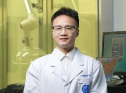 Sheng-Long Ding合作论文数Fudan University7
 Tao Yi (易涛)合作论文数College of Chemistry and Chemical Engineering, Donghua University7Dong Zhirui (董智瑞)合作论文数6
 Qian Chen合作论文数Zhongshan Hospital, Fudan University6Peng Liu合作论文数6Lu Cao (曹璐)合作论文数6
 Xue Fengfeng合作论文数6Liu Shu-Hao合作论文数6Fu Tengfei合作论文数6Jiapeng Zou合作论文数5
 Qintong Xu合作论文数5Ya Yuan合作论文数5




