During World War II, rampant caries disqualified nearly 20% of military recruits from service. To address this issue, President Truman signed legislation on June 24, 1948, creating what would eventually become the National Institute of Dental and Craniofacial Research (NIDCR). As NIDCR celebrates 75 years, we reflect on past accomplishments and strategize for the future. In this unprecedented era of science and technology, we envision leveraging innovations for development, testing, and implementation of diagnostics and therapeutics to address a broad spectrum of diseases and disorders affecting the dental, oral, and craniofacial complex and systemic health. Dr. D’Souza is the director, National Institute of Dental and Craniofacial Research, Bethesda, MD. Dr. King is a director, Division of Extramural Activities , National Institute of Dental and Craniofacial Research, Bethesda, MD. Dr. Webster-Cyriaque is the deputy director, National Institute of Dental and Craniofacial Research, Bethesda, MD. Dr. Shum is the director, Division of Extramural Research, National Institute of Dental and Craniofacial Research, Bethesda, MD. Dr. Fischer is a branch chief, Clinical Research & Clinical Trials, Mucosal Diseases, National Dental Practice-Based Research Network, National Institute of Dental and Craniofacial Research, Bethesda, MD.
COVID-19 is a new human infectious disease that is caused by a novel coronavirus called severe acute respiratory syndrome coronavirus 2 (SARS-CoV-2). It emerged in December 2019 and owing to its rapid spread worldwide, was declared a pandemic by the World Health Organization in March of 2020. Although the molecular mechanisms of SARS-CoV-2 viral infection and the interactions of the pathogen with host tissues remain unclear today, the crisis has garnered the interest and commitment to biomedical research among communities around the globe.
The promise of tissue engineering and regenerative medicine to reduce the burden of disease and improve quality of life are widely acknowledged. Traditional tissue engineering and regenerative medicine approaches rely on generation of tissue constructs in vitro for subsequent transplantation or injection of exogenously manipulated cells into a host. While promising, few such therapies have succeeded in clinical practice. Here, we propose that recent advances in stem cell and developmental biology, immunology, bioengineering, and material sciences, position us to develop a new generation of in vivo regenerative medicine therapies, which we term autotherapies. Autotherapies are strategies based on optimizing endogenous tissue responses and capitalizing on manipulation of stem cell niches and endogenous tissue microenvironments to enhance tissue healing and regeneration.
Activation of osteoblastic bone anabolism in the calvarial sutures is considered to be the essential pathologic condition underlying mutant FGFR2-related craniofacial dysostosis. However, early clinical investigations indicated that abnormal cartilage development in the cranial base was rather a primary site of abnormal feature in Apert Syndrome (AS). To examine the significance of cartilaginous growth of the cranial base in AS, we generated a transgenic mouse bearing AS-type mutant Fgfr2IIIc under the control of the Col2a1 promoter-enhancer (Fgfr2IIIc(P253R) mouse). Despite the lacking expression of Fgfr2IIIc(P253R) in osteoblasts, exclusive disruption of chondrocytic differentiation and growth reproduced AS-like acrocephaly accompanied by short anterior cranial base with fusion of the cranial base synchondroses, maxillary hypoplasia and synostosis of the calvarial sutures with no significant abnormalities in the trunk and extremities. Gene expression analyses demonstrated upregulation of p21, Ihh and Mmp-13 accompanied by modest increase in expression of Sox9 and Runx2, indicating acceleration of chondrocytic maturation and hypertrophy in the cranial base of the Fgfr2IIIc(P253R) mice. Furthermore, an acquired affinity and specificity of mutant FGFR2IIIc(P253R) receptor with FGF2 and FGF10 is suggested as a mechanism of activation of FGFR2 signaling selectively in the cranial base. In this report, we strongly suggest that the acrocephalic feature of AS is not alone a result of the coronal suture synostosis, but is a result of the primary disturbance in growth of the cranial base with precocious endochondral ossification.
Amelogenin and ameloblastin, the major enamel matrix proteins, are important for enamel mineralization. To identify their synergistic roles in enamel development, we generated Amel X −/− /Ambn −/− mice. These mice showed additional enamel defects in comparison with Amel X −/− or Ambn −/− mice. In 7-day-old Amel X −/− /Ambn −/− mice, not only was the ameloblast layer irregular and detached from the enamel surface, as in Ambn −/− , but also, the enamel width was significantly reduced in the double-null mice as compared with Amel X −/− or Ambn −/− mice. Proteomic analysis of the double-null teeth revealed increased levels of RhoGDI (Arhgdia), a Rho-family-specific guanine nucleotide dissociation inhibitor, which is involved in important cellular processes, such as cell attachment. Both Amel X −/− /Ambn −/− mice and Ambn −/− mice displayed positive staining with RhoGDI antibody in the irregularly shaped ameloblasts detached from the matrix. Ameloblastin-regulated expression of RhoGDI suggests that Rho-mediated signaling pathway might play a role in enamel formation.
An Inhibitor of O-Glycosylation Induces Apoptosis in NIH3T3 Cells and Developing Mouse Embryonic Mandibular TissuesJournal of Biological ChemistryVol. 279Issue 48PreviewThe family of UDP-GalNAc:polypeptide N-acetylgalactosaminyltransferases (ppGaNTases) is responsible for initiating mucin-type O-linked glycosylation in higher eukaryotes. To begin to examine the biological role of O-linked glycosylation, mammalian cells were treated with a small molecule inhibitor (designated 1–68A, Ref. 15) of ppGaNTase activity. NIH3T3 cells exposed to the inhibitor were shown to undergo a significant reduction in cell surface O-glycosylation as detected by staining with jacalin and peanut agglutinin lectins after 30 min of treatment; no reduction in staining using antibodies to O-linked N-acetylglucosamine or the lectin concanavalin A was detected. Full-Text PDF Open Access 50382-50390 This article has been withdrawn by the authors for the following reasons. In this work, we employed a small molecule (designated 1-68A) that is an inhibitor of the UDP-GalNAc polypeptide:N-acetylgalactosaminyltransferases to probe the functions of O-glycosylation. Follow-up work by our laboratories indicates that this compound (1-68A) is not stable under the cell culture conditions used in this work; it decomposes by an oxidative mechanism that has not yet been fully elucidated, producing H2O2 in the process. The compound is unstable at physiological pH (decomposing in phosphate-buffered saline, pH 7.4, or either RPMI 1640 or F-12 medium) and is stable at acidic pH (phosphate-buffered saline, pH 4.5). We have since demonstrated that H2O2 can induce cell death in several cell lines, including NIH3T3 cells, under the conditions used in this work. Thus, although we have confirmed that the central observations reported are accurate, we can no longer attribute the finding of apoptosis to inhibition of O-glycosylation per se. To avoid any future ambiguity about these findings, we withdraw the publication.
Introduction: The increasing recognition that use of bisphosphonates may be associated with osteonecrosis of the jaw (ONJ) led the leadership of the American Society for Bone and Mineral Research (ASBMR) to appoint a task force to address a number of key questions related to this disorder.Materials and Methods: A multidisciplinary expert group reviewed all pertinent published data on bisphosphonate-associated ONJ. Food and Drug Administration drug adverse event reports were also reviewed.Results and Conclusions: A case definition was developed so that subsequent studies could report on the same condition. The task force defined ONJ as the presence of exposed bone in the maxillofacial region that did not heal within 8 wk after identification by a health care provider. Based on review of both published and unpublished data, the risk of ONJ associated with oral bisphosphonate therapy for osteoporosis seems to be low, estimated between I in 10,000 and <1 in 100,000 patient-treatment years. However, the task force recognized that information on incidence of ONJ is rapidly evolving and that the true incidence may be higher. The risk of ONJ in patients with cancer treated with high doses of intravenous bisphosphonates is clearly higher, in the range of 1-10 per 100 patients (depending on duration of therapy). In the future, improved diagnostic imaging modalities, such as optical coherence tomography or MRI combined with contrast agents and the manipulation of image planes, may identify patients at preclinical or early stages of the disease. Management is largely supportive. A research agenda aimed at filling the considerable gaps in knowledge regarding this disorder was also outlined.
We previously reported that amelogenin isoforms M180 and leucine-rich amelogenin peptide (LRAP) are expressed in the periodontal region, and that their absence is associated with increased cementum defects in amelogenin-knockout (KO) mice. The aim of the present study was to characterize the functions of these isoforms in osteoclastogenesis and in the proliferation and migration of cementoblast/periodontal ligament cells. The co-cultures of wild-type (WT) osteoclast progenitor and KO cementoblast/periodontal ligament cells displayed more tartrate-resistant acid phosphatase (TRAP)-positive cells than the co-cultures of WT cells. The addition of LRAP to both co-cultures significantly reduced RANKL expression and the TRAP-positive cells. Proliferation and migration rates of the KO cementoblast/periodontal ligament cells were lower than those of WT cells and increased with the addition of either LRAP or P172 (a porcine homolog of mouse M180). Thus, we demonstrate the regulation of osteoclastogenesis by LRAP, and the proliferation and migration of cementoblast/periodontal ligament cells by LRAP and P172.
Currently, much remains unknown of the genes that mediate the biological events during growth and fusion of the midfacial region, and the possible pathways through which these genes function. We took advantage of high-throughput microarray analysis to search for genes that may play a critical role in the growth and fusion of the midfacial region to become the primary palate. We identified several genes that were potentially expressed at different levels between tail somite (TS) 6-8 (pre-fusion) and TS 12-14 (fusion) in the 3 midfacial processes. Expression of 4 of these genes ( Tbx14/15, Dickkopf-1, Fibroblast Growth Factor 8, and Keratin-18) was further verified by reverse-transcription/quantitative PCR and in situ hybridization at the 2 stages of midfacial development. With the identification of these genes, and possibly others, functional analyses can be conducted to improve our understanding of the mechanisms and pathways by which the midface forms.
The molecular mechanisms by which the primordia of the midface grow and fuse to form the primary palate are not well characterized. This is in spite of the fact that failure of growth and/or fusion of these facial primordia leads to the common human craniofacial birth defects, clefts of the lip with or without clefts of the palate. Members of the transforming growth factor beta (Tgfbeta) superfamily have been shown to play critical roles during craniofacial development. Specifically, the role of Tgfbeta-3 in mediating the fusion of the embryonic secondary palatal shelves is well documented. In a screen for genes expressed during fusion of the murine midfacial processes, Tgfbeta2 was identified as a gene differentially expressed during fusion of the lateral and medial nasal processes. The objective of our study was to analyze the spatial and temporal expression of Tgfbeta2 during critical stages of midfacial morphogenesis at both the transcript and protein levels. We also compared the pattern of expression of Tgfbeta2 with that of Bmp4, a gene shown previously to be involved in mediating the fusion process in the midface. Our results showed Tgfbeta2 expression in a very restrictive area of the epithelial layer along the borders of the midfacial primordia, in a pattern very similar to that of Bmp4. The highly restrictive and spatial and temporal pattern of expression of Tgfbeta2 implicates its role in mediating the fusion of the midfacial processes, possibly through interacting with Bmp4 in the regulation of apoptosis and/or epithelial-mesenchymal transformation. A greater understanding of the role of this gene will clarify how the normal midface grows and the mechanisms behind cleft development.
BACKGROUND:Cleft lip with or without cleft palate is the most common congenital anomaly in the craniofacial region. Knowledge of the molecular mechanisms behind normal lip fusion can contribute to better intervention and improved functional clinical outcome. Transforming growth factor-beta3 (TGF-beta3) has been implicated in lip morphogenesis. Therefore, we hypothesized that TGF-beta3 functions during lip fusion through regulation of angiogenesis and mesenchymal cell cycle progression during early developmental stages.METHODS:To test this hypothesis we used the CL/Fraser mouse model, which has a high incidence of cleft lip. Lips isolated from embryonic day (ED) 11.5 mouse embryos were allowed to develop in serum-free organ cultures in the presence or absence of TGF-beta3. The lips that developed in these cultures fused in 2 days.RESULTS:During normal development, we detected positive immunoreactions for TGF-beta3 at the site of fusion. We also detected mesenchymal cells that were immunopositive for Flk-1 and CD31, which are markers for endothelial cell precursors. Exogenous TGF-beta3 accelerated lip fusion in culture. This enhancement was associated with an increase in the number of capillary blood vessels in the lips cultured in the presence of TGF-beta3, in comparison with controls. In tandem, TGF-beta3 increased the level of expression of both Flk-1 and CD31. Our data suggest that an elevated level of TGF-beta3 may promote angiogenesis in developing lips that is mediated by increased Flk-1 and CD31 expression. We also detected increased cyclin D1 expression (a marker for cell proliferation) in the presence of TGF-beta3, which suggests that TGF-beta3 promoted cell proliferation.CONCLUSIONS:TGF-beta3 promoted cell proliferation and angiogenesis in lip mesenchymal tissues. These events led to enhanced lip fusion in the presence of TGF-beta3.
The family of UDP-GalNAc:polypeptide N-acetylgalactosaminyltransferases (ppGaNTases) is responsible for initiating mucin-type O-linked glycosylation in higher eukaryotes. To begin to examine the biological role of O-linked glycosylation, mammalian cells were treated with a small molecule inhibitor (designated 1-68A, Ref. 15) of ppGaNTase activity. NIH3T3 cells exposed to the inhibitor were shown to undergo a significant reduction in cell surface O-glycosylation as detected by staining with jacalin and peanut agglutinin lectins after 30 min of treatment; no reduction in staining using antibodies to O-linked N-acetylglucosamine or the lectin concanavalin A was detected. Apoptosis was also observed in treated cells after 45 min of exposure, ostensibly following the O-glycosylation reduction. Overexpression of several different ppGaNTase isoforms restored cell surface O-glycosylation and rescued inhibitor-induced apoptosis. Additionally, mouse embryonic mandibular organ cultures exposed to 1-68A developed abnormally, presumably because of epithelial and mesenchymal apoptosis that followed a reduction in jacalin and peanut agglutinin staining. Our studies suggest that mucin-type O-linked glycosylation may be required for normal development and that ppGaNTases may play a role in the regulation of apoptosis.
Bone morphogenetic protein 4 (BMP4) and growth/differentiation factor 5 (GDF5) are closely related protein family members and regulate early cartilage patterning and differentiation. In this study, we compared the functional outcome of their actions systematically at various stages of chondrogenesis in mouse embryonic limb bud mesenchyme grown in micromass cultures. Overall, both growth factors enhanced cartilage growth and differentiation in these cultures. Uniquely, BMP4 not only accelerated the formation and maturation of cartilaginous nodules, but also induced internodular mesenchymal cells to express cartilage differentiation markers. On the other hand, GDF5 increased the number of prechondrogenic mesenchymal cell condensation and cartilaginous nodules, without altering the overall pattern of differentiation. In addition, GDF5 caused a more sustained elevated expression level of Sox9 relative to that associated with BMP4. BMP4 accelerated chondrocyte maturation throughout the cultures and sustained an elevated level of Col10 expression, whereas GDF5 caused a transient increase in Col10 expression. Taken together, we conclude that BMP4 is instructive to chondrogenesis and induces mesenchymal cells toward the chondrogenic lineage. Furthermore, BMP4 accelerates the progression of cartilage differentiation to maturation. GDF5 enhances cartilage formation by promoting chondroprogenitor cell aggregation, and amplifying the responses of cartilage differentiation markers. These differences may serve to fine‐tune the normal cartilage differentiation program, and can be exploited for the molecular manipulation in biomimetics. Published 2004 Wiley‐Liss, Inc.
ObjectiveTo understand the molecular mechanisms that lead to increased MMP-13 expression and cartilage degeneration during the progression of osteoarthritis (OA), we have investigated the expression of the transcription factor RUNX2 in OA cartilage and the regulation of MMP-13 expression by RUNX2 and FGF2 in articular chondrocytes.DesignRUNX2 and MMP-13 expression in human OA and control cartilage was analyzed by immunohistochemistry. The effects of RUNX2 over-expression, with or without FGF2 treatment, on MMP-13 promoter activity and enzyme accumulation were measured in articular chondrocytes. Inhibitors of MEK/ERK were assayed for their ability to block FGF2 and RUNX2 up-regulation of the MMP-13 promoter. We analyzed RUNX2 phosphorylation in response to FGF2.ResultsFibrillated OA cartilage exhibited increased RUNX2 immunoreactivity when compared to control cartilage. RUNX2 co-localized with MMP-13 in clusters of chondrocytes in fibrillated OA cartilage. RUNX2 over-expression in cultured chondrocytes increased their responsiveness to FGF2 treatment, which led to increased MMP-13 expression. Inhibitors of MEK/ERK signaling blocked up-regulation of the MMP-13 promoter by RUNX2 and FGF2, and also blocked the activation of RUNX2 by FGF2. FGF2 treatment of articular chondrocytes increased RUNX2 phosphorylation approximately 2-fold.ConclusionsIncreased expression of RUNX2 in OA cartilage may contribute to increased expression of MMP-13. FGF2, which is present in OA synovial fluid, activated RUNX2 via the MEK/ERK pathway and increased MMP-13 expression. However, it is unlikely that RUNX2 is a substrate of ERK1/2. RUNX2 expression and activation may be a significant step in the progression of OA by promoting changes in gene expression and chondrocyte differentiation.
Cleft lip is a common congenital malformation, and labioplasty performed on infants to repair such defects often results in severe scar formation. Since TGF-beta3 has been implicated in wound healing, we therefore hypothesized that TGF-beta3 functions to reduce scarring after cleft lip repair. In this investigation, we demonstrated that exogenous TGF-beta3 reduced scar formation in an incised and sutured mouse lip in vivo. During labioplasty, endogenous TGF-beta3 expression was also elevated. In vitro experiments showed that exogenous TGF-beta3 reduced type I collagen accumulation. Furthermore, TGF-beta3 inhibited alpha-smooth-muscle actin expression, a marker for myofibroblasts. In tandem, TGF-beta3 induced the expression and activity of MMP-9. Analysis of our data suggests that TGF-beta3 is normally secreted following labioplastic wound healing. An elevated level of TGF-beta3 reduces type I collagen deposition by restricting myofibroblast differentiation and thereby collagen synthesis, and by promoting collagen degradation by MMP-9. In combination, these events lead to TGF-beta3-mediated reduced scar formation.
Amelogenins, major components of developing enamel, are predominantly involved in the formation of tooth enamel. Although amelogenins are also implicated in cementogenesis, their precise spatial expression pattern and molecular role are not clearly understood. Here, we report for the first time the expression of two alternate splice forms of amelogenins, M180 and the leucine-rich amelogenin peptide ( LRAP), in the periodontal region of mouse tooth roots. Lack of M180 and LRAP mRNA expression correlated with cementum defects observed in the amelogenin-null mice. The cementum defects were characterized by an increased presence of multinucleated cells, osteoclasts, and cementicles. These defects were associated with an increased expression of the receptor activator of the nuclear factor-kappaB ligand ( RANKL), a critical regulator of osteoclastogenesis. These findings indicate that the amelogenin splice variants, M180 and LRAP, are critical in preventing abnormal resorption of cementum.
Background: Bone morphogenetic proteins (BMPs) are pleiotropic differentiation factors that regulate cell fate determination by orchestrating the activities of downstream signal transducers. Although BMP ligands can elicit signal transduction from heterodimeric combinations of several type-I and type-II receptors, cytoplasmic transducers of the BMP signal include only three known BMP-specific regulatory Smad proteins: Smad1, 5, and 8. In order to determine the combination of signals that regulate chondrogenesis by BMPs, we analyzed the functions of BMP Smad subtypes. Methods: Multipotential mesenchymal C3H10T1/2 cells and monopotential chondroprogenitor MC615 cells were placed in micromass culture in the presence or absence of BMP4. Chondrogenic differentiation was assayed by measuring Sox9 and type-II collagen gene expression and by alcian blue staining. Transactivation of type-II collagen by regulatory Smads singly, or in combination with Smad4, which partners with regulatory Smads, was assayed by luciferase activity. Results: In the absence of BMP4, mesenchymal cells did not exhibit chondrogenic differentiation, whereas chondroprogenitor cells showed increased cartilage marker expression. In the presence of BMP4, the rate and extent of chondrogenesis increased in a dose-dependent manner for both cell types. We further determined that Smad1 or Smad5, but not Smad8, synergized with Smad4 in the transactivation of the type-II collagen promoter in chondroprogenitor cells. In contrast, Smad8 and Smad4 presented modest synergy in mesenchymal cells. Conclusions: Taken together, our data suggest that uncommitted mesenchymal cells do not have the cellular competence to respond to the rate-limiting chondroinductive factor BMP. However, in chondroprogenitor cells, BMP stimulates differentiation through mechanisms mediated by Smad1 or Smad5 in combination with Smad4. Clinical Relevance: Our functional studies of these mesenchymal and chondroprogenitor cells will establish the mechanisms of lineage commitment and provide a platform for molecular manipulations with predictable lineage outcome. Therefore, our knowledge base can provide the molecular basis for stem/progenitor cell differentiation and a paradigm for tissue engineering.
 Hc Slavkin合作论文数35GH Nuckolls合作论文数24
 Pablo Bringas合作论文数12K Takahashi合作论文数11R Dashner合作论文数11I Takahashi合作论文数10Yuji Hatakeyama合作论文数8Ichiro Semba合作论文数8
 Ashok Kulkarni合作论文数National Institute of Dental and Craniofacial Research7O Tanaka合作论文数6
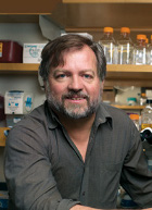 Rik Derynck合作论文数Department of Cell & Tissue Biology, University of California San Francisco5
 Taduru L Sreenath合作论文数Rockville5
 Zena Werb合作论文数Department of Anatomy, University of California, San Francisco5Rajeev Seth合作论文数5
 Harold C. Slavkin合作论文数University of Southern California5ML Mayo合作论文数4T Ikura合作论文数4Glen H. Nuckolls合作论文数4
 David Warburton合作论文数Keck School of Medicine, University of Southern California;Ostrow School of Dentistry, University of Southern California;Department of Surgery, Children’s Hospital Los Angeles4Kazuaki Nonaka合作论文数4
 Junko Hatakeyama合作论文数4A Yamane合作论文数3Y Sakakura合作论文数3Alan Horner合作论文数3M Mayo合作论文数3
 Simeon Buckley合作论文数3
 Michio Nagata (長田道夫)合作论文数Faculty of Medicine, University of Tsukuba3
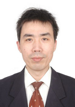 Xibin Wang (王锡斌)合作论文数Department of Automotive Engineering, School of Energy and Power Engineering, Xi'an Jiaotong University3
 Sung Chi Rocky Tuan (段崇智)合作论文数School of Biomedical Sciences, The Chinese University of Hong Kong;Institute for Tissue Engineering and Regenerative Medicine, The Chinese University of Hong Kong3
 Koichi Takahashi合作论文数3K Nonaka合作论文数2N Haruyama合作论文数2C Crohin合作论文数2T Thyagarajan合作论文数2
 Rena D’Souza合作论文数National Institute of Dental and Craniofacial Research, National Institutes of Health;University of Utah;“Craniofacial and Dental Disorders” Research Laboratory, National Institute of Child Health and Human Development, National Institutes of Health2Joan McGowan合作论文数2Yoannis Imbert合作论文数2
 Kazuhito Satomura合作论文数2CW Gibson合作论文数2Satoshi Fukumoto合作论文数2Jm Levsky合作论文数2V Santos合作论文数2Gj Wistow合作论文数2S Kobayashi合作论文数2
 Laurie Kay McCauley合作论文数Department of Periodontics and Oral Medicine, School of Dentistry, University of Michigan;Center for Cell Plasticity and Organ Design, Medical School, University of Michigan;School of Dentistry, University of Michigan2
 Koka Sreenivas合作论文数Mayo Clin, Dept Dent Specialties, Rochester, MN 55905 USA2Van Poznak C合作论文数2
 Ruggiero Salvatore L合作论文数New York Center for Oral and Maxillofacial Surgery2




