Hepatocellular carcinoma (HCC) is a challenging disease to evaluate in terms of prognosis, requiring close attention to the prognosis of HCC patients. Exosomes have been shown to play an important role in HCC development and have significant potential in managing HCC patient prognosis, as they are detectable in patients' blood. By using small extracellular vesicular RNA, liquid biopsies can reflect the underlying physiological and pathological status of the originating cells, providing a valuable assessment of human health. No study has explored the diagnostic value of mRNA expression changes in exosomes for liver cancer. The present study investigated establishing a risk prognosis model based on mRNA expression levels in exosomes from blood samples of liver cancer patients and evaluated its diagnostic and prognostic value, providing new targets for liver cancer detection. We obtained mRNA data from HCC patients and normal controls from the TCGA and exoRBase 2.0 databases and established a risk prognostic assessment model using exosomes-related risk genes selected through prognostic analysis and Lasso Cox analysis. The patients were divided into high-risk and low-risk groups based on median risk score values to validate the independence and evaluability of the risk score. The clinical value of the model was further analyzed using a nomograph model, and the efficacy of immunotherapy and cell-origin types of prognostic risk genes were further assessed in the high- and low-risk groups by immune checkpoint and single-cell sequencing. A total of 44 genes were found to be significantly associated with the prognosis of HCC patients. From this group, we selected six genes (CLEC3B, CYP2C9, GNA14, NQO1, NT5DC2, and S100A9) as exosomal risk genes and used them as a basis for the risk prognosis model. The clinical information of HCC patients from the TCGA and ICGC databases demonstrated that the risk prognostic score of the model established in this study was an independent prognostic factor with good robustness. When pathological stage and risk prognostic score were incorporated into the model to predict clinical outcomes, the nomograph model had the best clinical benefit. Furthermore, immune checkpoint assays and single-cell sequencing analysis suggested that exosomal risk genes were derived from different cell types and that immunotherapy in the high-risk groups could be beneficial. Our study demonstrated that the prognostic scoring model based on exosomal mRNA was highly effective. The six genes selected using the scoring model have been previously reported to be associated with the occurrence and development of liver cancer. However, this study is the first to confirm that these related genes existed in the blood exosomes, which could be used for liquid biopsy of patients with liver cancer, thereby avoiding the need for puncture diagnosis. This approach has a high value in clinical application. Through single-cell sequencing, we found that the six genes in the risk model originate from multiple cell types. This finding suggests that the exosomal characteristic molecules secreted by different types of cells in the microenvironment of liver cancer may serve as diagnostic markers.
Glycated low-density lipoprotein (G-LDL) is an established proatherosclerotic factor, but the mechanism is not completely understood. In vitro, we evaluated the uptake and transcytosis rates of N-LDL and G-LDL in endothelial cells and the uptake and transcytosis rates of G-LDL were much higher than those of N-LDL. Then, using small interfering RNAs, the receptor mediating G-LDL uptake and transcytosis was screened among eight candidate receptors, and the mechanism of the receptor regulation was thoroughly examined. We discovered that scavenger receptor A (SR-A) knockdown dramatically decreased the uptake and transcytosis rates of G-LDL. Additionally, endothelial cells with overexpressed SR-A had enhanced G-LDL uptake and transcytosis. In vivo, G-LDL was injected in the tail vein of ApoE-/- mice to investigate whether G-LDL affects atherosclerotic plaque formation. Compared with the injection of N-LDL, the injection of G-LDL accelerated atherosclerotic plaque formation in ApoE-/- mice, which was ameliorated by endothelial cells specific SR-A knockdown. Together, our results provide the first demonstration that the transcytosis of G-LDL across endothelial cells is much faster than that of N-LDL and SR-A is the major type of receptor responsible for G-LDL binding and transcytosis across endothelial cells.
Abstract Objective To elucidate the mechanism whereby advanced glycation end products (AGEs) accelerate atherosclerosis (AS) and to explore novel therapeutic strategies for atherosclerotic cardiovascular disease. Methods and results The effect of AGEs on low-density lipoprotein (LDL) transcytosis across endothelial cells (ECs) was assessed using an in vitro model of LDL transcytosis. We observed that AGEs activated the receptor for advanced glycation end products (RAGE) on the surface of ECs and consequently upregulated Caveolin-1, which in turn increased caveolae-mediated LDL transcytosis and accelerated AS progression. Our molecular assessment revealed that AGEs activate the RAGE-NF-κB signaling, which then recruits the NF-κB subunit p65 to the RAGE promoter and consequently enhances RAGE transcription, thereby forming a positive feedback loop between the NF-κB signaling and RAGE expression. Increased NF-κB signaling ultimately upregulated Caveolin-1, promoting LDL transcytosis, and inhibition of RAGE suppressed AGE-induced LDL transcytosis. In ApoE −/− mice on a high-fat diet, atherosclerotic plaque formation was accelerated by AGEs but suppressed by EC-specific knockdown of RAGE. Conclusion AGEs accelerate the development of diabetes-related AS by increasing the LDL transcytosis in ECs through the activation of the RAGE/NF-κB/Caveolin-1 axis, which may be targeted to prevent or treat diabetic macrovascular complications.
Background: Activation of NLRP3 inflammasome accelerates the formation of atherosclerotic plaques. Here, we evaluated the effects of inflammation on the expression of the NLRP3 inflammasome in endothelial cells (ECs). Methods: The effect of TNF-alpha on transcytosis of LDL was measured. VCAM-1 binding peptide targeting cationic liposomes (PCLs) were prepared as siRNA vectors. Methylated NLRP3 siRNA was encapsulated into the PCLs to knock down NLRP3 in vitro and in vivo. In rats with partial carotid ligation, TNF-alpha-induced LDL retention in the carotid artery endothelium was observed. In ApoE(-/-) mice, NLRP3 siRNA-PCLs were injected intravenously to observe their effect on the formation of atherosclerosis. Results: Our results showed that TNF-alpha upregulated NLRP3 in ECs, promoting the assembly of the NLRP3 inflammasome and processing of pro-IL-1 beta into IL-1 beta. Moreover, TNF-alpha accelerated LDL transcytosis in ECs. Knockdown of NLRP3 prevented TNF-alpha-induced NLPR3 inflammasome/IL-1 beta signaling and LDL transcytosis. Using optimized cationic liposomes to encapsulate methylated NLRP3 siRNA, resulting in targeting of VCAM-1-expressing ECs, to knockdown NLRP3, TNF-alpha-induced NLRP3 inflammasome activation and LDL transcytosis were prevented. Using the partial carotid ligation as an atherosclerosis rat model, we found that local administration of NLRP3 siRNA-PCLs efficiently knocked down NLPR3 expression in the carotid endothelium and dramatically attenuated the deposition of atherogenic LDL in carotid ECs in TNF-alpha-challenged rats. Furthermore, NLRP3 siRNA-PCLs were injected intravenously in ApoE(-/-) mice, resulting in reduced plaque formation. Conclusion: These findings established a novel strategy for targeting the NLRP3 inflammasome using NLRP3 siRNA-PCLs to interrupt LDL transcytosis, representing a potential novel therapy for atherosclerosis.
Background: Atherosclerosis (AS) is the basis of diabetic macrovascular complications. The plasma low-density lipoprotein (LDL) particles transcytosis across endothelial cells (ECs) and deposition under the endothelium is the initiation step of AS. We previously reported that high glucose inhibits the autophagic degradation of Caveolin-1 and promote LDL transcytosis across ECs, which in turn accelerates atherosclerotic progression. Since Sirt6 is a chromatin-associated protein with deacetylation activity, whether it can regulate Caveolin-1 acetylation and regulating the autophagic degradation of Caveolin-1 remains elusive. Methods: Autophagy and histone acetylation were assessed in the umbilical cords of patients with gestational diabetes mellitus (GDM) by immunohistochemistry. An in vitro model of LDL transcytosis was established, and the role of Sirt6 in LDL transcytosis across endothelial cells was clarified. The effect of Sirt6 on the autophagic degradation of Caveolin-1 under hyperglycemic conditions was explored in a streptozotocin (STZ)-induced diabetic AS model established using the ApoE / mice. Results: Caveolin-1 and acetylated histone H3 levels were significantly increased, while LC3B and Sirt6 were downregulated in the monolayer of the vascular wall from GDM and type 2 diabetes mellitus (T2DM) patients. Immunoprecipitation assays showed that Sirt6 interacts with Caveolin-1 and specifically mediated its acetylation levels. Immuno-electron microscopy (EM) further indicated that Sirt6 overexpression triggered the autophagic lysosomal degradation of Caveolin-1. ECs-specific overexpression of Sirt6 by adeno-associated viral vector serotype 9 (AAV9) induced autophagy, reduced Caveolin-1 expression, and ameliorated atherosclerotic plaque formation in STZ-induced diabetic ApoE / mice. Conclusion: Sirt6-mediated acetylation of Caveolin-1 activates its autophagic degradation and inhibits high glucose-stimulated LDL transcytosis. Thus, the Sirt6/Caveolin-1 autophagic pathway plays a crucial role in diabetic AS, and the overexpression or activation of Sirt6 is a novel therapeutic strategy.
Background Aging is a natural life process and with an aging population, age-related diseases (e.g. type 2 diabetes mellitus (T2DM), atherosclerosis-based cardiovascular diseases) are the primary mortality cause in older adults. Telomerase is often used as an aging biomarker. Detection and characterization of novel biomarkers can help in a more specific and sensitive identification of a person’s aging status. Also, this could help in age-related diseases early prevent, ultimately prolonging the population’s life span. Sirtuin 6 (Sirt6) - a member of the Sirtuins NAD + -dependent histone deacetylases family - is mainly intracellularly expressed, and is reported to be involved in the regulation of aging and aging-related diseases. Whether serum Sirt6 is correlated with aging and could be used as an aging biomarker is unknown. In the present study, we aimed to investigate the age-related Sirt6 changes in the serum of human adults. Methods Participants were divided into three groups according to age: 20–30 years (Young); 45–55 years (Middle-aged); and ≥ 70 years (Old). The Sirt6 and telomerase serum concentrations were determined by ELISA. The Sirt6 and human telomerase reverse transcriptase (hTERT) expression in vessels from amputated human lower limbs were analyzed using real-time quantitative PCR (RT-qPCR) and immunohistochemical staining. The relationships between variables were evaluated by Pearson correlation analysis. Results The Sirt6 and telomerase serum levels reduced with an increase in age. A similar tendency was observed for Sirt6 and hTERT in the vessel. Serum levels of Sirt6 were higher in females compared with males. Pearson’s regression analysis revealed that the Sirt6 serum level positively correlated with telomerase ( r = 0.5743) and both were significantly negatively correlated with age ( r = − 0.5830 and r = − 0.5993, respectively). Conclusions We reported a negative correlation between serum Sirt6 concentration and aging in human beings. Therefore, the Sirt6 serum level is a potential sex-specific aging marker.
Subendothelial retention of apolipoprotein B100-containing lipoprotein, such as low-density lipoprotein (LDL), is the initial step of atherogenesis. Activation of autophagy exhibits beneficial effects for the treatment of atherosclerosis. In our previous study, we demonstrated that hyperglycemia suppressed autophagic degradation of caveolin-1, which in turn resulted in acceleration of caveolae-mediated LDL transcytosis across endothelial cells and lipid retention. Therefore, targeting the crossed pathway in autophagy activation and LDL transcytosis interruption may be a promising antiatherosclerotic strategy. In metabolic diseases, including atherosclerosis, salidroside, a phenylpropanoid glycoside compound (3,5-dimethoxyphenyl) methyl-β-glucopyranoside), is the most important compound responsible for the therapeutic activities of Rhodiola. However, whether salidroside suppresses LDL transcytosis to alleviate atherosclerosis has not yet been elucidated. In the present study, we demonstrated that salidroside significantly decreased LDL transcytosis across endothelial cells. Salidroside-induced effects were dramatically blocked by AMPK (adenosine monophosphate-activated protein kinase) inhibitor (compound c, AMPKα siRNA) and by overexpression of exogenous tyrosine-phosphorylated caveolin-1 using transfected cells with phosphomimicking caveolin-1 on tyrosine 14 mutant plasmids (Y14D). Furthermore, we observed that salidroside promoted autophagosome formation via activating AMPK. Meanwhile, the interaction between caveolin-1 and LC3B-II, as well as the interaction between active Src (indicated by the phosphorylation of Src on tyrosine 416) and LC3B-II, was significantly increased, upon stimulation with salidroside. In addition, both bafilomycin A1 (a lysosome inhibitor) and an AMPK inhibitor (compound c) markedly prevented salidroside-induced autophagic degradation of p-Src and caveolin-1. Moreover, the phosphorylation of caveolin-1 on tyrosine 14 was disrupted due to the downregulation of p-Src and caveolin-1, thereby directly decreasing LDL transcytosis by attenuating the number of caveolae on the cell membrane and by preventing caveolae-mediated LDL endocytosis released from the cell membrane. In ApoE-/- mice, salidroside significantly delayed the formation of atherosclerotic lesions. Meanwhile, a significant increase in LC3B, accompanied by attenuated accumulation of the autophagy substrate SQSTM1, was observed in aortic endothelium of ApoE-/- mice. Taken together, our findings demonstrated that salidroside protected against atherosclerosis by inhibiting LDL transcytosis through enhancing the autophagic degradation of active Src and caveolin-1.
Diabetes is one of the most common comorbidities of COVID-19. We aimed to conduct a multidimensional analysis of risk factors associated with the severity and mortality of patients with COVID-19 and diabetes. In this retrospective study involving 1443 patients with COVID-19, we analyzed the clinical and laboratory characteristics and risk factors associated with disease severity in patients with COVID-19 with and without diabetes. Binary logistic regression analyses were performed to identify the risk factors associated with mortality in patients with COVID-19 and diabetes. The 84-day survival duration for critical patients with COVID-19 and diabetes who had different levels of leukocytes and neutrophils, or treated with immunoglobulin or not, was conducted using Kaplan–Meier survival curves. Of the 1443 patients with COVID-19, 256 (17.7%) had diabetes, had a median age of 66.0 [IQR 58.0–73.8] years, and were more likely to develop severe (41.8% vs. 35.6%) and critical disease (34.0% vs. 14.9%), followed by higher mortality (21.1% vs. 7.0%), than those without diabetes. Higher levels of leukocytes (> 5.37 × 10⁹/L), older age, and comorbid cerebrovascular disease and chronic renal disease independently contributed to in-hospital death of patients with COVID-19 and diabetes. Leukocytes > 5.37 × 10⁹/L and the application of immunoglobulin were associated with shorter survival duration and lower mortality, respectively, in critical patients with COVID-19 and diabetes. More attention should be paid to patients with COVID-19 and diabetes, especially when they have high leukocyte counts (> 5.37 × 10⁹/L). Timely and adequate intravenous immunoglobulin (IVIG) use may reduce the mortality of critical patients with COVID-19 and diabetes.
Elevated plasma low density lipoprotein (LDL) is an established risk factor for cardiovascular disease. In addition to being able to cross the endothelial barrier to become accumulated in subendothelial space and thereby initiate atherosclerosis, LDL may exert a direct effect on vascular endothelial cells through activation of LDL receptor and its downstream signaling. Whether LDL can modulate the signaling for autophagy in endothelial cells is not clear. The present study firstly demonstrated that LDL can suppress endothelial autophagy through activation of the PI3K/Akt/mTOR signaling pathway and can promote glucose uptake by translocating glucose transporter 1 (GLUT1) from cytoplasm to cell membrane, actions similar to those of insulin. A co-immunoprecipitation assay found that LDL receptor (LDLR) and insulin receptor (IR) formed a complex in HUVECs. Knock down of the insulin receptor by small interfering RNA blocked the suppression of autophagy by LDL, as well as the signaling pathway involved. We conclude that LDL may mimic the action of insulin in endothelial cells, which might partly explain the increased incidence of diabetes in patients receiving some LDL-lowering therapy.
Critical Limb Ischemia (CLI) is common but uncommonly diagnosed. Improved recognition and early diagnostic markers for CLI are needed. Therefore, the aim of our study was to identify plasma biomarkers of CLI in patients with type 2 diabetes mellitus (T2DM). In this study, antibody-coated glass slide arrays were used to determine the plasma levels of 274 human cytokines in four matched cases of diabetes with and without CLI. Potential biomarkers were confirmed in an independent cohort by ELISA. After adjusting for confounding risk factors, only plasma level of Siglec-5 remained significantly associated with an increased odds ratio (OR) for diabetes with CLI by binary logistic regression analysis. Receiver operating characteristic (ROC) curve analysis revealed the optimal cut-off points for Siglec-5 was 153.1 ng/ml. After entering Siglec-5, the AUC was 0.99, which was higher than that of confounding risk factors only (AUC = 0.97, P < 0.05). Siglec-5 was expressed in plaques, but not in healthy artery wall in T2DM patients. Elevated plasma Siglec-5 was independently associated with CLI in T2DM. Plasma Siglec-5 levels are implicated as an early diagnostic marker of CLI in T2DM patients and it may become a target for the prevention or treatment of CLI in diabetes.
Persistent elevation of plasma TNF-α is a marker of low grade systemic inflammation. Palmitic acid (PA) is the most abundant type of saturated fatty acid in human body. PA is bound with albumin in plasma and could not pass through endothelial barrier freely. Albumin-bound PA has to be transported across monolayer endothelial cells through intracellular transcytosis, but not intercellular diffusion. In the present study, we discovered that TNF-α might stimulate PA transcytosis across cardiac microvascular endothelial cells, which further impaired the insulin-stimulated glucose uptake by cardiomyocytes and promoted insulin resistance. In this process, TNF-α-stimulated endothelial autophagy and NF-κB signaling crosstalk with each other and orchestrate the whole event, ultimately result in increased expression of fatty acid transporter protein 4 (FATP4) in endothelial cells and mediate the increased PA transcytosis across microvascular endothelial cells. Hopefully the present study discovered a novel missing link between low grade systemic inflammation and insulin resistance.
Aim:The clock gene mPer2 controls circadian periods and plays a critical role in clock resetting and responses to drugs of abuse. Mice deficient in mPer2 exhibit a marked susceptibility to acute liver injury. Clinical observations have demonstrated the existence of a relationship between circadian rhythm and liver cirrhosis. Here, we sought direct evidence for clock function to liver fibrosis using mPer2-deficient mice.Methods:Hepatic fibrosis was induced in wild-type (WT) and mPer2-/- mice by repetitive intraperitoneal carbon tetrachloride (CCl(4)) injection. Masson trichrome staining and analysis of alpha-smooth muscle actin (alpha-SMA) immunohistochemistry were performed to show the collagen accumulation and the hepatic stellate cell (HSC) activation, respectively. The mRNA levels of fibrosis-related genes were monitored by quantitative real-time polymerase chain reaction. The protein level of TIMP-1 was determined by immunohistochemistry. Transferase deoxytidyl uridine end labeling, alpha-SMA double staining and 4',6'-diamidino-2-phenylindole dihydrochloride staining were performed to show HSC apoptosis in vivo and in vitro, respectively.Results:CCl(4) caused much more severe liver fibrosis and activated more HSC in mPer2 null mice as compared to WT animals. Meanwhile, mPer2 null mice exhibited less efficiency in fibrosis resolution. Apoptotic HSC were significantly fewer in mPer2 null mice compared with WT mice after CCl(4); transfected Per2 cDNA into cultured HSC resulted in more HSC apoptosis with upregulation of TRAIL-R2/DR5 expression.Conclusion:Loss of clock gene mPer2 predisposes liver fibrosis by increasing HSC activation and inhibiting HSC apoptosis.
Mice lacking A(1) adenosine receptors (A(1)AR) were thought to be protected from developing fatty liver; however, the contribution of A(1)AR to hepatic fibrosis has not been explored. Here we found that the expression of A(1)AR was decreased in fibrotic liver induced by chronic carbon tetrachloride (CCl4) but increased in that induced by bile duct ligation (BDL). Therefore, we examined whether A(1)AR contributes to hepatic fibrosis in CCl4 and BDL animal models using A(1)AR knockout mice. Compared with wild-type (WT) mice, hepatic fibrosis resulting from chronic CCl4 exposure was attenuated in A(1)AR(-/-) mice with markedly decreased collagen deposition and reduced hepatic stellate cell activation, whereas bile duct-ligated A(1)AR(-/-) mice displayed a significant increase in hepatic fibrosis. Hepa-tocyte damage was reduced in A(1)AR(-/-) mice after a single injection of CCl4, with down-regulation of CYP2E1 and UCP2 gene expression in livers, which resulted in impaired liver sensitivity to CCl4. However, BDL caused severe bile infarcts in livers of A(1)AR(-/-) mice, with significantly elevated levels of bile acid compared with those in WT mice. CCl4 and BDL resulted in different expression patterns of genes involved in fibrogenesis in A(1)AR(-/-) mice. These results indicate that A(1)AR participates in the pathogenesis of hepatic fibrosis with a complex mechanism, and the effect of targeting adenosine and its receptors in the prevention of hepatic fibrosis should be cautiously evaluated.
 Xiong Jia合作论文数10
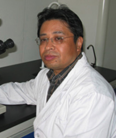 Baixiang Li (李百祥)合作论文数Public Health College of Harbin Medical University10
 Si Jin (金肆)合作论文数Liyuan Hospital, Tongji Medical College, Huazhong University of Science and Technology10Yajing Lu合作论文数7Wenzhuo Cheng合作论文数7
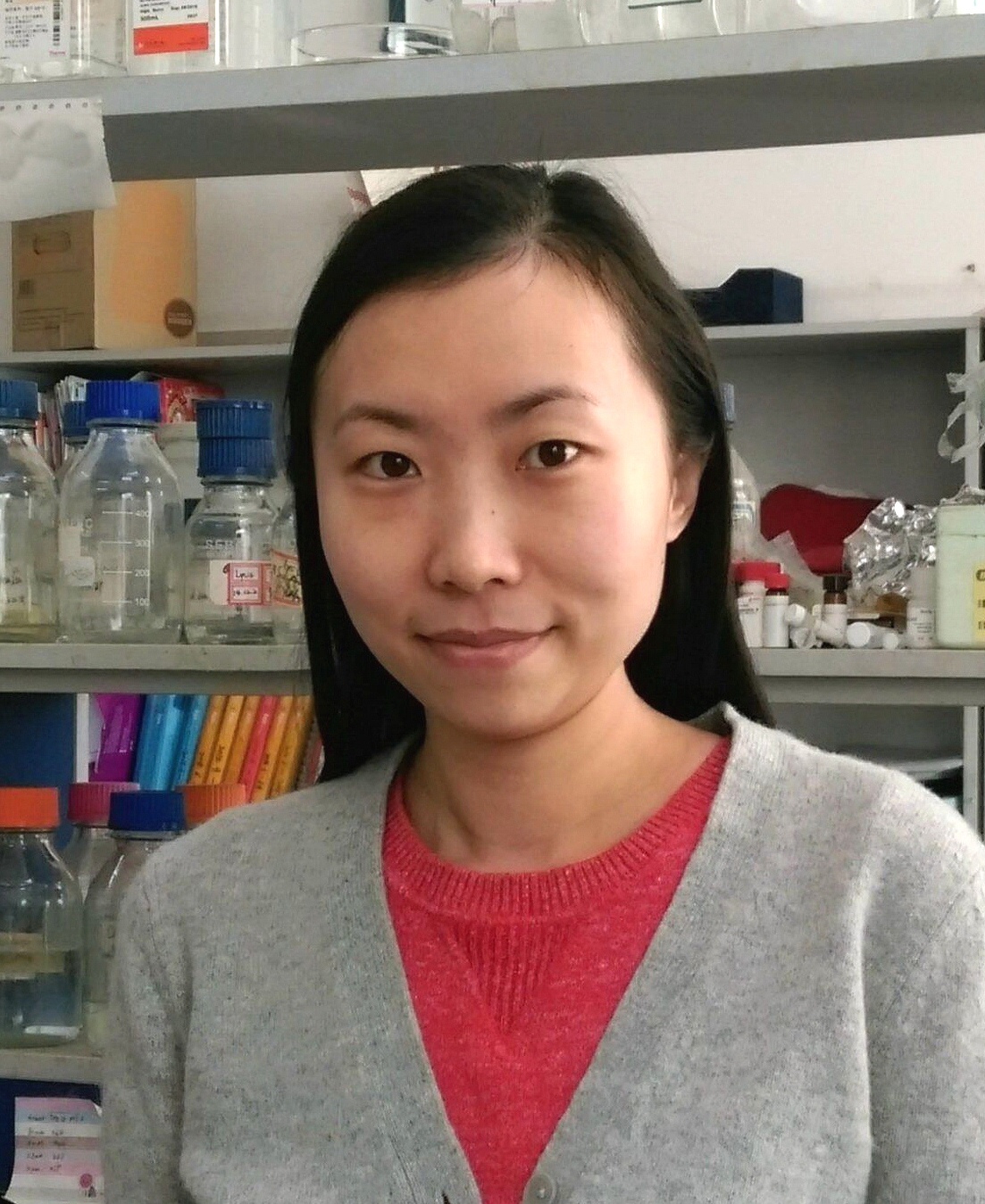 Ying Zhao (赵颖)合作论文数Department of Biochemistry and Biophysics, School of Basic Medical Sciences, Peking University7
 Xiaoyan Yang (杨晓燕)合作论文数School of Basic Medicine, Tongji Medical College, Huazhong University of Science and Technology5Meng Shu合作论文数4Yan Shu合作论文数4Yan Zhu合作论文数4Song Yi合作论文数4Li Wang合作论文数4Juyi Li (李居怡)合作论文数4Ling Wang合作论文数3
 Ye Li合作论文数3Wenjing Li合作论文数2Aiping Deng合作论文数2Wu Guangjie合作论文数2Peng Chen合作论文数2
 Ping Yang (杨平)合作论文数College of Architecture and Environment, Sichuan University2
 Zichun Hua (华子春)合作论文数Xinxiang Medical University;School of Life Sciences, Nanjing University2
 Shu Meng (孟舒)合作论文数Xinhua Hospital, School of Medicine, Shanghai Jiao Tong University2Zheyi Han合作论文数2
 Jianfa Zhang (张建法)合作论文数School of Chemistry and Chemical Engineering, Nanjing University of Science and Technology2Tengjiao Wang合作论文数1Yaogong Wu合作论文数1Liwen Kuang (邝莉雯)合作论文数1Shuli Liu合作论文数1Qiyu Xiao (肖棋予)合作论文数1Yan Lou合作论文数1Xiufang Wang合作论文数1ZhongJing Wang (王中京)合作论文数1Shiming Wang (王世明)合作论文数1Xing Shasha合作论文数1Tao Zheng合作论文数1
 Binghui Li (李炳辉)合作论文数Liyuan Hospital, Tongji Medical College, Huazhong University of Science and Technology1Dan Wu (吴丹)合作论文数Huazhong University of Science and Technology,Wuhan,China1
 Fang Bian合作论文数1Qiyong Mei (梅其勇)合作论文数1
 Xiaolong Du合作论文数Department of Physics and Astronomy, College of Physical Sciences, University of California, Los Angeles1
 Zibo Feng (冯自波)合作论文数Lyuan Hospital, Tongji Medical College, Huazhong University of Science & Technology1




