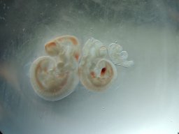
Pre-patterning of the embryo, driven by spatially localized factors, is a common feature across several non-mammalian species 1-4 . However, mammals display regulative development and thus it was thought that blastomeres of the embryo do not show such pre-patterning, contributing randomly to the three lineages of the blastocyst: the epiblast, primitive endoderm and trophectoderm that will generate the new organism, the yolk sac and placenta respectively 4-6 . Unexpectedly, early blastomeres of mouse and human embryos have been reported to have distinct developmental fates, potential and heterogeneous abundance of certain transcripts 7-12 . Nevertheless, the extent of the earliest intra-embryo differences remains unclear and controversial. Here, by utilizing multiplexed and label-free single-cell proteomics by mass-spectrometry 13 , we show that 2-cell mouse and human embryos contain an alpha and a beta blastomere as defined by differential abundance of hundreds of proteins exhibiting strong functional enrichment for protein synthesis, transport, and degradation. Such asymmetrically distributed proteins include Gps1 and Nedd8, depletion or overexpression of which in one blastomere of the 2-cell embryo impacts lineage segregation. These protein asymmetries increase at 4-cell stage. Intriguingly, halved mouse zygotes display asymmetric protein abundance that resembles alpha and beta blastomeres, suggesting differential proteome localization already within zygotes. We find that beta blastomeres give rise to a blastocyst with a higher proportion of epiblast cells than alpha blastomeres and that vegetal blastomeres, which are known to have a reduced developmental potential, are more likely to be alpha. Human 2-cell blastomeres also partition into two clusters sharing strong concordance with clusters found in mouse, in terms of differentially abundant proteins and functional enrichment. To our knowledge, this is the first demonstration of intra-zygotic and inter-blastomere proteomic asymmetry in mammals that has a role in lineage segregation.
Background: The combination of immune checkpoint modulators with chemotherapy improves efficacy compared with chemotherapy alone in PD-L1+ advanced TNBC (IMpassion130; KEYNOTE-355). The addition of IPAT to paclitaxel (PAC) improved efficacy in a phase 2 trial in advanced TNBC (LOTUS). Preliminary overall response rate (ORR) data from a multicenter phase 1b study (NCT03800836) evaluating a triplet combination of IPAT, atezolizumab, and taxane chemotherapy showed promising anti-tumor activity in a similar patient population, irrespective of PD-L1 status [Schmid, AACR 2019]. Here, we report follow-up results including progression-free survival (PFS) from this study. Patients and Methods: Eligible patients had measurable unresectable locally advanced/metastatic TNBC, ECOG performance status 0/1, and had received no prior systemic therapy for advanced disease (prior [neo]adjuvant chemotherapy and/or radiation permitted if all chemotherapy was completed ≥12 months before first dose). Patients with brain metastases were excluded. Patients received oral IPAT 400 mg/day on days 1–21 and IV atezolizumab 840 mg on days 1 & 15 in combination with PAC 80 mg/m2 (Arm A) or nab-PAC 100 mg/m2 (Arm B) on days 1, 8, & 15. Cycles were repeated every 28 days until loss of clinical benefit, unacceptable toxicity, or consent withdrawal. Arms C and D evaluated sequential regimens comprising a doublet induction therapy with the third agent added on day 15 (Arm C: IPAT + PAC, then + atezolizumab; Arm D: atezolizumab + PAC, then + IPAT). Tumors were assessed every 8 weeks. Key endpoints were confirmed ORR (per RECIST v1.1), duration of response (DoR), PFS, and safety. Results: At the data cut-off (26 Jul 2020), results were available from 114 patients (Arm A n=70, Arm B n=20, Arm C n=12, Arm D n=12). Median duration of follow-up was 11.1 months. Efficacy results are summarized in the table. Safety of the combination appeared to be consistent with the known safety profile of the individual drugs. Grade ≥3 adverse events (AEs) occurred in 55% of patients (including rash [13%], diarrhea [12%], and neutropenia [10%]) and serious AEs in 34%. AEs led to discontinuation of IPAT in 6% of patients and atezolizumab in 4%. No new safety signals were identified. Conclusions: Updated results demonstrate a lower ORR than in the preliminary report of the first 26 patients. Subgroup analyses according to PD-L1 or PIK3CA/AKT1/PTEN alteration status or taxane backbone show no consistent trend across endpoints, although small sample sizes limit interpretation. Further biomarker analyses focusing on subgroups and biology may identify subsets of patients deriving a benefit. Citation Format: Peter Schmid, Peter Savas, Enrique Espinosa, Valentina Boni, Antoine Italiano, Shane White, Karen Cheng, Lisa Lam, Lidia Robert, Victor Laliman, Kalpit Shah, Marie-Paule Sablin. Phase 1b study evaluating a triplet combination of ipatasertib (IPAT), atezolizumab, and a taxane as first-line therapy for locally advanced/metastatic triple-negative breast cancer (TNBC) [abstract]. In: Proceedings of the 2020 San Antonio Breast Cancer Virtual Symposium; 2020 Dec 8-11; San Antonio, TX. Philadelphia (PA): AACR; Cancer Res 2021;81(4 Suppl):Abstract nr PS12-28.
This study describes a technical breakthrough in endolymphatic sac research, made possible by the use of the recently generated Prox1-GFP transgenic mouse model. Whole-mount imaging techniques through the decalcified temporal bone and three-dimensional observations of Prox1-GFP mouse tissue revealed the positive labeling of the endolymphatic sac in adult stage, and allowed, for the first time, the GFP-based identification of endolymphatic sac epithelial cells. Prox1 expression was observed in all parts of the endolymphatic sac epithelia. In intermediate portion of the endolymphatic sac, mitochondria-rich cells did not express Prox1, although ribosome-rich cells showed strong GFP labeling. The anatomical relationship between the endolymphatic sac and the surrounding vasculature was directly observed. In the endolymphatic sac, expression of Prox1 may suggest progenitor cell-like pluripotency or developmental similarity to systemic lymphatic vessels in other organs. This whole-mount imaging technique of the endolymphatic sac can be combined with other conventional histological, sectioning, and labeling techniques and will be very useful for future endolymphatic sac research.
Intracellular calcium ([Ca²⁺]i) signaling mediates physiological and pathological processes in multiple organs, including the renal podocyte; however, in vivo podocyte [Ca²⁺]i dynamics are not fully understood. Here we developed an imaging approach that uses multiphoton microscopy (MPM) to directly visualize podocyte [Ca²⁺]i dynamics within the intact kidneys of live mice expressing a fluorescent calcium indicator only in these cells. [Ca²⁺]i was at a low steady-state level in control podocytes, while Ang II infusion caused a minor elevation. Experimental focal podocyte injury triggered a robust and sustained elevation of podocyte [Ca²⁺]i around the injury site and promoted cell-to-cell propagating podocyte [Ca²⁺]i waves along capillary loops. [Ca²⁺]i wave propagation was ameliorated by inhibitors of purinergic [Ca²⁺]i signaling as well as in animals lacking the P2Y2 purinergic receptor. Increased podocyte [Ca²⁺]i resulted in contraction of the glomerular tuft and increased capillary albumin permeability. In preclinical models of renal fibrosis and glomerulosclerosis, high podocyte [Ca²⁺]i correlated with increased cell motility. Our findings provide a visual demonstration of the in vivo importance of podocyte [Ca²⁺]i in glomerular pathology and suggest that purinergic [Ca²⁺]i signaling is a robust and key pathogenic mechanism in podocyte injury. This in vivo imaging approach will allow future detailed investigation of the molecular and cellular mechanisms of glomerular disease in the intact living kidney.
Podocytes are critical in the maintenance of a healthy glomerular filter; however, they have been difficult to study in the intact kidney because of technical limitations. Here we report the development of serial multiphoton microscopy (MPM) of the same glomeruli over several days to visualize the motility of podocytes and parietal epithelial cells (PECs) in vivo. In podocin-GFP mice, podocytes formed sporadic multicellular clusters after unilateral ureteral ligation and migrated into the parietal Bowman's capsule. The tracking of single cells in podocin-confetti mice featuring cell-specific expression of CFP, GFP, YFP or RFP revealed the simultaneous migration of multiple podocytes. In phosphoenolpyruvate carboxykinase (PEPCK)-GFP mice, serial MPM found PEC-to-podocyte migration and nanotubule connections. Our data support a highly dynamic rather than a static nature of the glomerular environment and cellular composition. Future application of this new approach should advance our understanding of the mechanisms of glomerular injury and regeneration.
Budding yeast septins assemble into hetero-octamers and filaments required for cytokinesis. Solvent-exposed cysteine (Cys) residues provide sites for attaching substituents useful in assessing assembly kinetics and protein interactions. To introduce Cys at defined locations, site-directed mutagenesis was used, first, to replace the native Cys residues in Cdc3 (C124 C253 C279), Cdc10 (C266), Cdc11 (C43 C137 C138), Cdc12 (C40 C278), and Shs1 (C29 C148) with Ala, Ser, Val, or Phe. When plasmid-expressed, each Cys-less septin mutant rescued the cytokinesis defects caused by absence of the corresponding chromosomal gene. When integrated and expressed from its endogenous promoter, the same mutants were fully functional, except Cys-less Cdc12 mutants (which were viable, but exhibited slow growth and aberrant morphology) and Cdc3(C124V C253V C279V) (which was inviable). No adverse phenotypes were observed when certain pairs of Cys-less septins were co-expressed as the sole source of these proteins. Cells grew less well when three Cys-less septins were co-expressed, suggesting some reduction in fitness. Nonetheless, cells chromosomally expressing Cys-less Cdc10, Cdc11, and Cdc12, and expressing Cys-less Cdc3 from a plasmid, grew well at 30°C. Moreover, recombinant Cys-less septins—or where one of the Cys-less septins contained a single Cys introduced at a new site—displayed assembly properties in vitro indistinguishable from wild-type. Proteins 2013; 81:1964–1979. © 2013 Wiley Periodicals, Inc.
Activation of angiotensin receptor type 1 (AT1) contributes to NADPH oxidase (Nox)-derived oxidative stress during metabolic syndrome. However, the specific role of AT1 in modulating redox signaling, mitochondrial function, and oxidative stress in the heart remains more elusive. To test the hypothesis that AT1 activation increases oxidative stress while impairing redox signaling and mitochondrial function in the heart during diet-induced insulin resistance in obese animals, Otsuka Long Evans Tokushima Fatty (OLETF) rats (n = 8/group) were treated with the AT1 blocker (ARB) olmesartan for 6 wk. Cardiac Nox2 protein expression increased 40% in OLETF compared with age-matched, lean, strain-control Long Evans Tokushima Otsuka (LETO) rats, while mRNA and protein expression of the H₂O₂-producing Nox4 increased 40-100%. ARB treatment prevented the increase in Nox2 without altering Nox4. ARB treatment also normalized the increased levels of protein and lipid oxidation (nitrotyrosine, 4-hydroxynonenal) and increased the redox-sensitive transcription factor Nrf2 by 30% and the activity of antioxidant enzymes (SOD, catalase, GPx) by 50-70%. Citrate synthase (CS) and succinate dehydrogenase (SDH) activities decreased 60-70%, whereas cardiac succinate levels decreased 35% in OLETF compared with LETO, suggesting that mitochondrial function in the heart is impaired during obesity-induced insulin resistance. ARB treatment normalized CS and SDH activities, as well as succinate levels, while increasing AMPK and normalizing Akt, suggesting that AT1 activation also impairs cellular metabolism in the diabetic heart. These data suggest that the cardiovascular complications associated with metabolic syndrome may result from AT1 receptor-mediated Nox2 activation leading to impaired redox signaling, mitochondrial activity, and dysregulation of cellular metabolism in the heart.
One emerging topic in renin–angiotensin system (RAS) research is the direct local control of renin synthesis and release by endogenous metabolic intermediates. During the past few years, our laboratory has characterized the localization and signaling of the novel metabolic receptor GPR91 in the normal and diabetic kidney and established GPR91 as a new, direct link between high glucose and RAS activation in diabetes. GPR91 (also called SUCNR1) binds tricarboxylic acid (TCA) cycle intermediate succinate which can rapidly accumulate in the local tissue environment when energy supply and demand are out of balance. In a variety of physiological and pathological conditions associated with metabolic stress, succinate signaling via GPR91 appears to be an important mediator or modulator of renin secretion. This review summarizes our current knowledge on the control of renin release by molecules of endogenous metabolic pathways with the main focus on succinate/GPR91.
We clarified the localization of lymphatic vessels in the tympanic membrane and proliferation of lymphatic vessels during regeneration after perforation of the tympanic membrane by using whole-mount imaging of the tympanic membrane of Prox1 GFP mice. In the pars tensa, lymphatic vessel loops surrounded the malleus handle and annulus tympanicus. Apart from these locations, lymphatic vessel loops were not observed in the pars tensa in the normal tympanic membrane. Lymphatic vessel loops surrounding the malleus handle were connected to the lymphatic vessel loops in the pars flaccida and around the tensor tympani muscle. Many lymphatic vessel loops were detected in the pars flaccida. After perforation of the tympanic membrane, abundant lymphatic regeneration was observed in the pars tensa, and these regenerated lymphatic vessels extended from the lymphatic vessels surrounding the malleus at day 7. These results suggest that site-specific lymphatic vessels play an important role in the tympanic membrane.
The contributions of angiotensin receptor type 1 (AT1) activation to NADPH oxidase (Nox)‐derived oxidative stress during hypertension and diabetes are well established. The role of AT1 in modulating redox signaling, mitochondrial function and oxidative stress in the heart during metabolic syndrome, however, remains elusive. To test the hypothesis that AT1 activation increases oxidative stress while impairing mitochondrial function and redox signaling in the heart during metabolic syndrome, diet‐induced obese, insulin resistant (OLETF) rats were treated with the AT1 blocker (ARB) Olmesartan for 6 weeks. Cardiac Nox2 expression increased 40% in OLETF compared to age‐matched lean (LETO) rats while expression of the H2O2‐producing Nox4 increased 40%. ARB treatment prevented the increase in Nox2 without altering Nox4. ARB treatment also normalized increased protein and lipid oxidation and increased the redox‐sensitive transcription factor Nrf2 30%, and antioxidant enzymes by 50–70%. Mitochondrial aconitase activity increased 60% in OLETF compared to LETO whereas succinate decreased 35%; ARB treatment normalized both. These data demonstrate that AT1 activation contributes to increased Nox2‐derived oxidative stress and to impaired redox signaling and mitochondrial activity, and suggests that, besides its anti‐hypertensive effects, ARB treatment may improve myocardial function during metabolic syndrome.
Metabolic syndrome (MetS) is commonly associated with elevated renin-angiotensin system, oxidative stress, and steatohepatitis with down-regulation of uncoupling proteins (UCPs). However, the mechanisms linking renin-angiotensin system, steatosis, and UCP2 to hepatic oxidative damage during insulin resistance are not described. To test the hypothesis that angiotensin receptor activation contributes to decreased hepatic UCP2 expression and aconitase activity and to increased oxidative damage after increased glucose intake in a model of MetS, lean and obese Long Evans rats (n = 10/group) were randomly assigned to the following groups: 1) untreated Long Evans Tokushima Otsuka (lean, strain control), 2) untreated Otsuka Long Evans Tokushima Fatty (OLETF) (MetS model), 3) OLETF + angiotensin receptor blocker (ARB) (10 mg olmesartan/kg·d × 6 wk), 4) OLETF + high glucose (HG) (5% in drinking water × 6 wk), and 5) OLETF + ARB + HG (ARB/HG × 6 wk). HG increased body mass (37%), plasma triglycerides (TGs) (35%), plasma glycerol (87%), plasma free fatty acids (28%), and hepatic nitrotyrosine (74%). ARB treatment in HG decreased body mass (12%), plasma TG (15%), plasma glycerol (23%), plasma free fatty acids (14%), and hepatic TG content (42%), suggesting that angiotensin receptor type 1 (AT1) activation and increased adiposity contribute to the development of obesity-related dyslipidemia. ARB in HG also decreased hepatic nitrotyrosine and increased hepatic UCP2 expression (59%) and aconitase activity (40%), as well as antioxidant enzyme activities (50-120%), suggesting that AT1 activation also contributes to protein oxidation, impaired lipid metabolism, and antioxidant metabolism in the liver. Thus, in addition to promoting obesity-related hypertension, AT1 activation may also impair lipid metabolism and antioxidant capacity, resulting in steatosis via decreased UCP2 and tricarboxylic acid cycle activity.
Lithium (Li) is a commonly used treatment for bipolar disorders. However, the use of Li is complicated by its renal side effect causing nephrogenic diabetes insipidus (NDI). The mechanism by which Li causes NDI is incompletely understood but cyclooxygenase 2 (COX‐2) and prostaglandin E2 (PGE2) are primary players. It has been established that Li inhibits luminal uptake of dicarboxylates including succinate resulting in their increased urinary excretion. Succinate is the ligand of the newly discovered metabolic receptor GPR91 that is highly expressed in the collecting ducts and its signaling involves COX‐2 and PGE2. Therefore, we tested the hypothesis that GPR91 plays an important role in Li‐induced NDI. Wild type (WT) and GPR91 −/− mice (n=5 each) were fed normal and Li‐added diets for 14 days and then euthanized to collect kidney tissues. Li‐induced polyuria was significantly less in GPR91 −/− mice (osmolality=604.85 ± 56.96 mOsm, urine output=10.96 ± 1.07 ml/24h) compared to WT mice (osmolality=394.60 ± 29.81 mOsm, urine output=19.37 ± 1.30 ml/24h) with no significant difference in Li intake. AQP2 protein abundance in the medulla was 1.5 fold higher in Li‐treated GPR91 −/− versus WT mice. Also, urinary succinate excretion progressively increased during Li treatment. These data suggest that deletion of GPR91 leads to a significant resistance to Li‐induced polyuria and may provide a novel therapeutic strategy.
The importance of local tissue metabolism, succinate accumulation and signaling via its receptor GPR91 in the activation of the intra‐renal renin‐angiotensin system (RAS) in DN has been established by our laboratory using the streptozotocin (STZ) model. However, the genetic Akita (A) mouse model of type I diabetes is an excellent alternative to study DN due to lack of the toxic side effects of STZ. In this study we used wild type A (n=6) and GPR91−/− A (A−/−, n=5) mice at 18‐wks of age. Although both groups develop robust hyperglycemia, we observed noticeable decreases in water intake, food intake and urine volume in A−/− mice. Urinary albumin excretion showed 1.69±0.7 fold increase in A group compared to WT and only 1.04±0.2 increase in in A−/− mice. Immunohistochemical analyses of TGFβ found strong glomerular labeling in A but not A−/− kidneys. Whole kidney Western blots demonstrated higher TGFβ content in A mice (1.27±0.3) than A−/− (0.68±0.1). A mice revealed higher cortical renin content (1.37±0.2) than A−/− (1.0±0.1). A significant difference in medullary renin was observed between A (1.34±0.2) and A−/− mice (0.8±0.1). We concluded that GPR91 plays an important role in the pathogenesis of DN.
In the renal tubules, ATP released from epithelial cells stimulates purinergic receptors, regulating salt and water reabsorption. However, the mechanisms by which ATP is released into the tubular lumen are multifaceted. Pannexin1 (Panx1) is a newly identified. ubiquitously expressed protein that forms connexin-like channels in the plasma membrane, which have been demonstrated to function as a mechanosensitive ATP conduit. Here, we report on the localization of Panx1 in the mouse kidney. Using immunofluorescence, strong Panx1 expression was observed in renal tubules, including proximal tubules, thin descending limbs, and collecting ducts, along their apical cell membranes. In the renal vasculature, Panx1 expression was localized to vascular smooth muscle cells in renal arteries, including the afferent and efferent arterioles. Additionally, we tested whether Panx1 channels expressed in renal epithelial cells facilitate luminal ATP release by measuring the ATP content of urine samples freshly collected from wild-type and Panx1(-/-) mice. Urinary ATP levels were reduced by 30% in Panx1(-/-) compared with wild-type mice. These results suggest that Panx1 channels in the kidney may regulate ATP release and via purinergic signaling may participate in the control of renal epithelial fluid and electrolyte transport and vascular functions.
Septins are GTP-binding proteins that form ordered, rod-like multimeric complexes and polymerize into filaments, but how such supramolecular structure is related to septin function was unclear. In Saccharomyces cerevisiae, four septins form an apolar hetero-octamer (Cdc11-Cdc12-Cdc3-Cdc10-Cdc10-Cdc3-Cdc12-Cdc11) that associates end-to-end to form filaments. We show that septin filament assembly displays previously unanticipated plasticity. Cells lacking Cdc10 or Cdc11 are able to divide because the now-exposed subunits (Cdc3 or Cdc12, respectively) retain an ability to homodimerize via their so-called G interface, thereby allowing for filament assembly. In such cdc10Δ and cdc11Δ cells, the remaining septins, like wild-type complexes, localize to the cortex at the bud neck and compartmentalize nonseptin factors, consistent with a diffusion barrier composed of continuous filaments in intimate contact with the plasma membrane. Conversely, Cdc10 or Cdc11 mutants that cannot self-associate, but "cap" Cdc3 or Cdc12, respectively, prevent filament formation, block cortical localization, and kill cells.
 Janos Peti-Peterdi合作论文数10Gevorgyan Haykanush合作论文数5
 Anne D Riquier-Brison合作论文数4
 Burford James L合作论文数4
 Rudy M Ortiz合作论文数Dept Mol & Cell Biol, Univ Calif Merced3
 José Pablo Vázquez-Medina合作论文数Department of Integrative Biology, University of California, Berkeley3
 Akira Nishiyama (西山成)合作论文数Graduate School of Medicine, Faculty of Medicine, Kagawa University3
 Jose Abraham Viscarra合作论文数School of Natural Sciences, University of California3
 Ruben Rodriguez合作论文数3
 Thorwald Max A合作论文数3Daisuke Nakano合作论文数3Irina Popovich合作论文数2
 Eva Nogales合作论文数Department of Molecular and Cell Biology, College of Letters & Science, University of California, Berkeley;Howard Hughes Medical Institute;Molecular Biophysics and Integrative Bioimaging Division, Lawrence Berkeley National Laboratory2
 Aurélie Bertin合作论文数CNRS, Université Paris-Sud Laboratoire de Physique des Solides2Michael A. Mcmurray合作论文数Milliman & Robertson2Takenori Miyashita合作论文数2
 Young-Kwon Hong合作论文数Cutaneous Biology Research Center, Massachusetts General Hospital and Harvard Medical School2
 Nozomu Mori合作论文数2
 Jeremy Thorner合作论文数Division of Biochemistry, Biophysics and Structural Biology, Department of Molecular and Cell Biology, College of Letters and Science, University of California, Berkeley2
 Matthias Hackl合作论文数2Maciej Meglicki合作论文数1Priscilla Montez合作论文数1Nikolai Slavov合作论文数1Lisa K Iwamoto-Stohl合作论文数1Victor Laliman合作论文数1R. Gray Huffman合作论文数Barnett Inst, Northeastern Univ1
 Valentina Boni合作论文数1Jason Derks合作论文数Barnett Inst, Northeastern Univ1
 Harrison Specht合作论文数Broad Institute of MIT and Harvard1Aleksandra A. Petelski合作论文数Barnett Inst, Northeastern Univ1Victoria Jorgensen合作论文数1Rachel Mandelbaum合作论文数1Karen Cheng合作论文数1Saad Khan合作论文数1
 Bailey A T Weatherbee合作论文数Department of Biological Sciences, University of Delaware, Newark, DE, 19716, USA1Ali Ahmady合作论文数1
 Antonia Weberling合作论文数Univ Cambridge, Mammalian Embryo & Stem Cell Grp, Dept Physiol Dev & Neurosci, Downing St, Cambridge CB2 3EG, England1Carlos W Gantner合作论文数Univ Melbourne, Florey Inst Neurosci & Mental Hlth, Melbourne, Vic, Australia1
 Katalin Susztak合作论文数Department of Genetics, Perelman School of Medicine, University of Pennsylvania1
 Lidia Robert合作论文数1
 Richard J Paulson合作论文数Keck School of Medicine, University of Southern California;Department of Obstetrics and Gynecology, University of Southern California1
 Sánchez-Vásquez Estefania合作论文数Laboratorio de biología del desarrollo, Instituto de Investigaciones Biotecnológicas-Instituto Tecnológico de Chascomús (CONICET-UNSAM), Chascomús, 7130 Buenos Aires, Argentina.1
 Natalia De Val合作论文数Department of Integrative Structural and Computational Biology, The Scripps Research Institute;Center for HIV/AIDS Vaccine Immunology and Immunogen Discovery, The Scripps Research Institute;The Scripps Research Institute, The Scripps Research Institute1Villanueva Karie合作论文数1
 Shah Kalpit合作论文数The Chicago Medical School, Rosalind Franklin University;The Chicago Medical School, Rosalind Franklin University;Rosalind Franklin University, Rosalind Franklin University1
 Hsiung Chris C-S合作论文数Division of Hematology, The Children's Hospital of Philadelphia;Perelman School of Medicine, University of Pennsylvania;Perelman School of Medicine, University of Pennsylvania1
 Savas Peter合作论文数Translational Breast Cancer Genomics Laboratory, Peter MacCallum Cancer Centre1
 Nguyen Mien T X合作论文数Keck School of Medicine, USC1
 Enrique Espinosa合作论文数1




