OBJECTIVES:To evaluate basal-luminal cell of origin subtyping using Prediction Analysis of Microarray of 50 genes (PAM50) genomic classification profiles for predicting disease progression in men undergoing active surveillance (AS) for prostate cancer (PCa). PATIENTS AND METHODS:In the prospective Miami Active Surveillance Trial (MAST) trial, 205 men undergoing AS received serial multiparametric magnetic resonance imaging (MRI) and biopsies, including MRI-targeted and systematic sampling. The highest-grade core from each biopsy was sent for expression profiling using Decipher, a clinical-grade transcriptome assay (Veracyte Inc., San Diego, CA, USA). Basal-luminal subtyping was evaluated using PAM50 molecular subtype models. PCa grade progression was compared across subtypes, as were gene mutation signatures, prognostic indices, and pathway activities. Kaplan-Meier curves, log-rank test, and multivariable Cox regression were used to assess association between PAM50 and grade progression. Heatmaps and volcano plots were rendered to illustrate potential mechanistical differences between PAM50 subtypes. RESULTS:Of the 205 patients, 128 had transcriptome data for baseline basal-luminal classification. PAM50 identified 46 Luminal A (LA), 26 Luminal B (LB), and 56 Basal subtypes. Decipher scores were lowest in LA, followed by Basal, and highest in LB. Grade progression-free survival was worse in patients with the LB subtype (median 1.7 years) compared to those with LA and Basal subtypes (median 2.9 years; log-rank P = 0.005); LB patients had grade progression-free survival of 34% by 24 months of AS, compared to 63% for Basal or 68% for LA. Transcriptome analysis showed distinct enrichment profiles for each subtype, with LB strongly associated with SPOP and CHD1 mutations. Limitations include small sample size and single-institution setting. CONCLUSION:The PAM50 basal-luminal subtyping shows promise as a molecular classification tool for predicting progression risk in PCa. This is one of the only prospective studies evaluating PAM50 subtyping for predicting cancer progression in a cohort of men undergoing AS for PCa.
Radiomics extracted from cone beam computed tomography (CBCT) can be assessed at time points during treatment and may provide an advantage over assessments in a pre-treatment setting using diagnostic images, like magnetic resonance imaging (MRI) or computed tomography (CT), for prostate cancer (pCa) patients receiving radiotherapy (RT). The purpose of this study was to analyze correlations between prostate radiomic features (RFs) derived from T2-weighted (T2w) MRI, CT, and first fraction CBCT for patients receiving RT for pCa. Forty-seven patients were analyzed. The prostate volumes were manually segmented, and 42 radiomic features were extracted, of which seven volume-normalized RFs were considered. The absolute Spearman correlation was calculated among the RFs of the aforementioned imaging modalities (RM) and prostate volume (RV) since the motivation of this paper was to analyze the strength of the correlation. The Benjamini-Hochberg adjustment was applied to p-values to account for multiple comparisons. No high correlations were found between CT/CBCT vs. T2w. The intramodality RM demonstrated that CT RFs were much higher than the other modalities. For example, intramodality RM≥0.95 the percentage of RFs was 17% for CT, 9% for CBCT, and 4.5% for T2w. The differences in RFs across different modalities can be viewed positively: the lack of correlation between RFs across T2w and CT/CBCT could indicate a fundamental difference in the extractable image information. It could also indicate that some RFs did not have any extractable information. A future study will include evaluating the predictive performance of patient outcomes using radiomic features from CT, CBCT, and T2w, which could help in answering such questions.
Outcomes in glioblastoma are improved by surgical resection and adjuvant radiation (RT). In primary GBM (pGBM), large clinical target volume (CTV) margins typically cover occult invasion. In recurrent GBM (rGBM), RT often uses tiny CTV margins that likely omit occult invasion due to re-RT radiation necrosis concerns. Whole-brain spectroscopic MRI (sMRI) is an emerging technique with similar resolution to PET that may help define the CTV for rGBM. Patients with pGBM (n = 18) and rGBM (n = 19) underwent sMRI with RT simulation. T1-post contrast (T1PC) and T2/FLAIR MRI volumes were contoured. sMRI generated choline/N-acetylaspartate > 2x (Cho/NAA > 2x) volumes are known to correlate with high-risk invasion. Hausdorff distances were calculated to define the margin necessary to cover Cho/NAA > 2x in pGBM and rGBM. In rGBM, mock CTV expansions from T1PC volumes were created to determine non-selective CTV expansion sizes needed to cover Cho/NAA > 2x volumes. For pGBM, the median T1PC, Cho/NAA > 2x, and T2/FLAIR volumes were 32.3 cc, 45.0 cc, and 74.8 cc respectively. For rGBM, the median T1PC, Cho/NAA > 2x, and T2/FLAIR volumes were 21.7 cc, 58.9 cc, and 118.3 cc, respectively. T2/FLAIR volumes increased more relative to T1PC volumes in rGBM than pGBM (p ≤ 0.001). Meanwhile, the median Hausdorff distance between T1PC and Cho/NAA > 2x was 22.9 mm in pGBM and 25.7 mm in rGBM, suggesting that the high-risk volume does not significantly change. In rGBM, it is common to use no CTV expansion from the T1PC volume which only included 61
TPS283 Background: Some patients with oligometastases may have the potential for long-term disease-free survival with just aggressive local therapy as shown by randomized trials for total consolidation of macroscopic metastases using metastasis-directed therapy (MDT). Long-term outcomes of pooled STOMP and ORIOLE trials in oligorecurrent metastatic castration-sensitive prostate cancer (omCSPC) demonstrated MDT improved progression free survival. However, men with high-risk mutations, including pathogenic alterations in ATM , BRCA1/2 , Rb1 , and TP53 , did poorly. Additional data suggests men with metastatic castration-resistant prostate cancer and similar mutations are sensitive to PARP inhibition (PARPi) with niraparib. We are launching a first-in-man biomarker-driven trial in omCSPC patients with high-risk mutations to evaluate the efficacy of MDT + androgen deprivation therapy (ADT) versus MDT + ADT + niraparib/abiraterone acetate plus prednisone (nira/AAP). Methods: This study is a multi-site, non-blinded, randomized phase II trial in patients with omCSPC. Men with histologically confirmed (at any site) omCSPC (≤3 metastases on standard imaging or ≤5 on Axumin/Choline/PSMA-PET/CT) and germ-line/somatic high-risk mutations ( TP53, BRCA1/2, PALB2, ATM, BRIP1, CHEK2, FANCA, RAD51B, RAD54L, MUTYH ) will be randomized (1:1) to MDT + 6- months ADT versus MDT + 6-months ADT + 6-months nira/AAP. A range of MDT radiation fractionation regimens are permitted. Subjects who meet eligibility criteria and qualify for enrollment will be stratified according to: (i) institution; (ii) conventional/enhanced imaging, (iii) PSADT <6-months, (iv) initial surgery/radiation, and (v) BRCA1/2 status. This study has been IRB approved (NCT06212583). We assume an accrual time of 24 months, with 18 months of additional follow-up time, and will randomize a total of 88 patients (44 patients in each arm). The primary endpoint will be to assess frequency of PSA failure (> 0.2 ng/mL post primary surgery or nadir + 2 post definitive radiation) with testosterone >100 ng/dl at 18-months after randomization (powered for 20% improvement over control arm by Fisher’s exact test). Secondary endpoints will include toxicity, health-related quality of life (HRQoL), time to locoregional progression, time to distant progression, time to new metastasis, radiographic progression-free survival, and duration of response. Discovery correlatives associated with clinical outcome will be assessed by collection of including, but not limited to, cell free DNA, circulating-tumor cells, immunologic biomarkers, microbiota and radiomics. Clinical trial information: NCT06212583 .
Androgen deprivation therapy (ADT) significantly alters metabolism in prostate cancer patients. Serum metabolomics are the gold standard for studying these effects; however, saliva offers a non-invasive alternative for biomarker assessment. This study evaluates the impact of ADT on serum and saliva metabolites and the correlation between the two matrices. By evaluating saliva as a potential surrogate for serum, this work explores its application in biomarker analysis of chronic risk factors within community health programs. Blood and saliva were collected from 20 eugonadal men and 20 men receiving hormonal therapy for prostate cancer, and were processed per standard protocols and stored at -80°C. Untargeted metabolomics using UPLC-MS analysis of polar and non-polar metabolites were performed; and glucocorticoids were quantified using targeted LC-MS/MS. Data were analyzed using MassHunter, METLIN database, and MetaboAnalyst, with log-transformation and by Mann-Whitney tests (False Discovery Rate (FDR)-adjusted P-values < 0.05). In untargeted metabolomics, 1, 198 metabolites were identified across serum and saliva. Comparing ADT to eugonadal serum, 35 metabolites had fold-changes (FC)>2 and 35 had FC<0.5, with 248 metabolites reaching statistical significance (adjusted-P<0.05). Similarly, in saliva, 54 metabolites had FC>2 and 69 had FC<0.5, with 82 metabolites showing significant differences (P<0.05). Comparing serum to saliva across all participants, 248 metabolites had FC>2 and 268 had FC<0.5, with 697 reaching statistical significance (P<0.05). When integrating fold changes (FC>2 or FC<0.5) and significance thresholds (adjusted-p<0.1), we identified 14 upregulated and 20 downregulated metabolites in serum, and 14 upregulated and 28 downregulated metabolites in saliva of ADT patients compared to eugonadal patients; and 247 upregulated and 268 downregulated metabolites in saliva compared to serum among participants. Targeted metabolomics also confirmed strong correlations between serum and salivary cortisol (r=0.612, p<0.001) and cortisone (r=0.618, p<0.001). These findings underscore the potential of glucocorticoids as biomarkers for stress-related metabolic alterations, suggesting that saliva potentially reflects the systemic metabolomic profile. This highlights saliva’s promise for non-invasive biomarker studies in stress and broader community health applications. ADT is associated with significant changes in serum and salivary metabolome, highlighting its systemic and local impact on metabolism. Moreover, targeted metabolomic analysis revealed strong correlations between glucocorticoids in serum and saliva, suggesting saliva may serve as a non-invasive surrogate for serum in biomarker studies to facilitate research in community health programs. Alireza Abdshah, Mohammad Alyamani, Marijo Bilusic, Dipen J. Parekh, Benjamin Spieler, Rakesh Singal, Brandon A. Mahal, Erin Kobetz, Sanoj Punnen, Matthew C. Abramowitz, Jaime R. Merchan, Nima Sharifi. Metabolomic insights into androgen deprivation therapy, and saliva as a non-invasive surrogate for serum to enable community-based biomarker studies [abstract]. In: Proceedings of the American Association for Cancer Research Annual Meeting 2025; Part 1 (Regular Abstracts); 2025 Apr 25-30; Chicago, IL. Philadelphia (PA): AACR; Cancer Res 2025;85(8_Suppl_1):Abstract nr 1929.
Extracting longitudinal image quantitative data, known as delta-radiomics, has the potential to capture changes in a patient’s anatomy throughout the course of radiation treatment for prostate cancer. Some of the major challenges of delta-radiomics studies are contouring the structures for individual fractions and accruing patients’ data in an efficient manner. The manual contouring process is often time consuming and would limit the efficiency of accruing larger sample sizes for future studies. The problem is amplified because the contours are often made by highly trained radiation oncologists with limited time to dedicate to research studies of this nature. This work compares the use of automated prostate contours generated using a deformable image-based algorithm to make predictive models of genitourinary and changes in total international prostate symptom score in comparison to manually contours for a cohort of fifty patients. Area under the curve of manual and automated models were compared using the Delong test. This study demonstrated that the delta-radiomics models were similar for both automated and manual delta-radiomics models.
Purpose:The aim of this study is to develop a framework for quantitative analysis of longitudinal T2-weighted MRIs (T2w) following radiotherapy (RT) for prostate cancer. Materials and methods:The developed methodology includes: (i) deformable image registration of longitudinal series to pre-RT T2w for automated detection of prostate, peripheral zone (PZ), and gross tumor volume (GTV); and (ii) T2w signal-intensity harmonization based on three reference tissues. The REgistration and HARMonization (REHARM) framework was applied on T2w acquired in a clinical trial consisting of two pre-RT and three post-RT MRI exams. Image registration was assessed by the DICE coefficient between automatic and manual contours, and intensity normalization via inter-patient histogram intersection. Longitudinal consistency was evaluated by the repeatability coefficient and Pearson correlation (r) between the two T2w exams before RT. Results:T2w from 107 MRI exams (23 patients) were utilized. Following REHARM, the histogram intersections for prostate, PZ and GTV increased from median = 0.43/0.16/0.13 to 0.66/0.44/0.46. The repeatability in T2w intensity estimation was better for the automatic than the manual contours for all three regions of interest (r = 0.9, p < 0.0001, for GTV). The changes in the tissues' T2w values pre- and post-RT became significant, indicating the measurable quantitative signal related to radiation. Conclusions:The developed methodology allows to automate longitudinal analysis reducing data acquisition-related variation and improving consistency. The quantitative characterization of RT-induced changes in T2w will lead to new understanding of radiation effects enabling prediction modeling of RT response.
BACKGROUND:4Kscore is used to aid the decision for prostate biopsy, however its role in active surveillance (AS) has not been investigated in a magnetic resonance imaging (MRI)-based protocol. Our objective was to assess the association between 4Kscore and progression in men undergoing AS on a prospective MRI-based protocol. METHODS:This was a single-institution, single-arm, non-therapeutic, interventional trial of 166 men with biopsy-confirmed prostate cancer enrolled between 2014-2020. Patients were placed on a trial-mandated AS protocol including yearly multiparametric (mp)MRI, prostate biopsy, and 4Kscore followed for 48 months after diagnosis. We analyzed protocol-defined and grade progression at confirmatory and subsequent surveillance biopsies. RESULTS:Out of 166 patients, 83 (50%) men progressed per protocol and of them 41 (24.7% of whole cohort) progressed by grade. At confirmatory biopsy, men with a baseline 4Kscore ≥ 20% had a higher risk of grade progression compared to those with 4Kscore < 20% (OR = 4.04, 95% CI: 1.05-15.59, p = 0.043) after adjusting for National Comprehensive Cancer Network (NCCN) risk and baseline PIRADS score. At surveillance biopsies, most recent 4Kscore ≥ 20% significantly predicted per protocol (OR = 2.61, 95% CI: 1.03-6.63, p = 0.044) and grade progression (OR = 5.13, 95% CI: 1.63-16.11, p = 0.005). CONCLUSIONS:For patients on AS, baseline 4Kscore predicted grade progression at confirmatory biopsy, and most recent 4Kscore predicted per-protocol and grade progression at surveillance biopsy.
RATIONALE AND OBJECTIVES:Active surveillance (AS) is the preferred management strategy for low-risk prostate cancer. This study aimed to evaluate the impact of Habitat Risk Score (HRS), an automated approach for mpMRI analysis, for early detection of progressors in a prospective AS clinical trial (MAST NCT02242773). MATERIALS AND METHODS:The MAST protocol includes Confirmatory mpMRI ultrasound fusion (MRI-US) biopsy and yearly surveillance MRI-US biopsies for up to 3 years. Clinical and mpMRI data from patients that progressed based on protocol criteria at years 1-3 were reviewed. Patients were classified as "MRI/HRS Progressors" if the PI-RADS lesion(s) had been targeted throughout the surveillance and resulted in positive biopsies, or as "Missed Progressors" if the lesion(s) were not identified by PI-RADS ("PI-RADS Miss") or were missed by the biopsy ("Needle Miss"). HRS maps were generated for each patient and evaluated for association with histopathological progression. RESULTS:Of the 34 patients, 15 were classified as "MRI/HRS Progressors" and 19 as "Missed Progressors" (12 "PI-RADS Miss", seven "Needle Miss"). In all cases, HRS confirmed the PI-RADS assessment. In the "PI-RADS Miss" group, HRS identified the lesions in all patients that were not targeted by biopsy and resulted in patient reclassification. HRS volumes showed clear association with tumor evolution both in terms of volume and aggressiveness over time. CONCLUSION:HRS volumes can serve as a quantitative biomarker for early detection of progression and lead to timely conversion to treatment, thereby improving patient outcomes and reducing the burden of unnecessary surveillance.
Purpose/Objective(s) Overall survival is improved in glioblastoma (GBM) by multimodality therapy including surgical resection and adjuvant radiation (RT). In primary GBM (pGBM), a classic problem is the delineation of the infiltrating microscopic tumor margin for focal therapies. In recurrent GBM (rGBM), the imaging is further confounded by changes from the pGBM treatments. As a result, rGBM is often not given RT due to confusing imaging or delivered with a tiny clinical target volume (CTV) that is likely missing high-risk non-enhancing tumor due to fears about re-RT radiation necrosis. Whole-brain spectroscopic MRI (sMRI) is an emerging MRI technique with similar resolution to brain PET that can detect native metabolites and has been applied to define CTV in pGBM. We wished to determine whether it may also have usefulness in defining the CTV for rGBM. Materials/Methods Patients with pGBM (n=21) and rGBM (n=21) were prospectively consented for sMRI. T1-post contrast (T1PC) and T2/FLAIR MRI volumes were contoured. sMRIs were used to generate relative choline:N-acetylaspartate>2 (rCN>2) maps normalized to contralateral normal appearing white matter, which have been validated to correlate strongly with the presence of occult tumor invasion. sMRI-defined volumes were contoured using the rCN>2 maps. Hausdorff distances were calculated to define the margin necessary to cover rCN>2 in both pGBM and rGBM. In rGBM, mock CTV expansions from the T1PC volume were created to determine non-selective CTV expansions needed to cover occult tumor invasion. Results For pGBM, the mean T1PC volume was 49.9 cc, mean T2/FLAIR volume was 108.5 cc, and mean rCN>2 was 72.5 cc. For rGBM, the mean T1PC volume was 28.6 cc, mean T2/FLAIR volume was 140.7 cc, and mean rCN>2 was 51.8 cc. The ratio of T1PC to T2/FLAIR volumes was 0.53 in pGBM and 0.25 in rGBM (p<0.001). The mean Hausdorff distance indicating the maximum additional distance between T1PC and rCN>2 was 22.9 mm in pGBM and 28.3 mm in rGBM. Our findings support ∼2 cm expansions in pGBM as per clinical standards. But, in rGBM, no CTV expansion from the T1PC volume (i.e., per RTOG 1205 trial protocol) resulted in only 54% of total disease coverage. With 10-, 15- and 20-mm CTV expansions from the T1PC volume, 94%, 98%, and 99% of total disease was covered, respectively. Conclusion sMRIs rCN>2 maps define occult disease in both pGBM and rGBM. These volumes extend approximately 2-3 cm beyond the disease seen on T1PC MRI. In rGBM, CTV expansions based on rCN>2 maps could allow for increased coverage of occult disease typically missed with standard radiation therapy volumes.
Purpose/Objective(s) The STOMP and ORIOLE randomized clinical trials (RCTs) showed progression-free survival (PFS) benefits of metastasis-directed therapy (MDT) alone without androgen-deprivation therapy (ADT) for oligometastatic hormone-sensitive prostate cancer (omHSPC). However, most patients with bone metastatic (BM) omHSPC recur with additional BM following MDT alone. We hypothesized the addition of BM-targeting alpha-emitter radium-223 (Ra223), approved for treatment of BM castration-resistant prostate cancer (mCRPC), could delay progression of disease. In addition, biomarkers to determine patients who benefit most from MDT are still poorly defined. We report on the first RCT to examine Ra223 and evaluate novel biomarkers in BM omHSPC. Materials/Methods In this phase 2 multi-institutional RCT (NCT04037358), men with recurrent omHSPC (≥1 BM & ≤5 radiation fields) were: i) stratified by institution, primary management (radiotherapy vs surgery), PSA doubling time and prior ADT, and ii) randomized 1:1 to stereotactic ablative radiation (SABR) MDT or SABR and 6 monthly cycles of Ra223. The primary endpoint was composite PFS [(PSA ≥ 25% increase and ≥ nadir + 2 ng/mL) and/or (RECIST 1.1 or new lesion on bone scan) and/or (ADT initiation or symptomatic decline)]. Tissue, liquid and imaging correlative studies were obtained and analyzed as biomarkers. Results From 8/2019-3/2023, 64 patients were randomized (33 to SABR and 31 to SABR/Ra223). Arms were balanced for key covariates. 26 (87%) patients received the full 6 planned cycles of Ra223. Median PFS was 10.2 months with SABR and 10.4 months with SABR/Ra223 (stratified HR 1.7, 95% CI, 0.78-3.69, p = 0.18). 13 patients (20%) experienced grade 3 toxicity (no grade 4 or 5), of which 6 were non-treatment-related, 2 related to SABR, and 5 related to SABR/Ra223 (lymphopenia, back pain). Baseline circulating tumor cells (CTCs) were detected in 55% of patients, and prostate-specific membrane antigen (PSMA)+ CTCs at baseline were prognostic for PFS (p = 0.029). Compared with the observation arm from the ORIOLE RCT, T-cell receptor deep sequencing (TCR-seq) identified significantly more clonotypic expansions between baseline and day 90 with SABR (p = 0.009) and SABR/Ra223 (p = 0.0007). Greater unique productive TCR rearrangements (UPRs) were prognostic for PFS independent of treatment arm (aHR, 0.45; 95% CI, 0.21-0.96; p = 0.04). UPRs were also associated with PFS in the ORIOLE RCT (HR, 0.46; 95% CI, 0.19-1.05; p = 0.07). Conclusion SABR alone for omHSPC affords PFS benefits, but emergence of additional BM in most patients remains a challenge. We report for the first time that the addition of Ra223 to SABR MDT in this low volume BM state does not delay progression of disease. CTCs are present in omHSPC, and SABR MDT induces a systemic adaptive immune response. We validate the TCR repertoire as a prognostic biomarker in omHSPC treated with SABR MDT. These results underline the importance of RCTs in omHSPC with concurrent collection of biological correlates.
Purpose/Objective(s)Human-papilloma virus (HPV)-positive oropharyngeal cancer (OPC) is a potentially curable disease with a rising incidence in the United States. Despite improved radiation therapy (RT) techniques, toxicities remain a concern. Adaptive RT (ART) can reduce radiation to organs-at-risk (OARs) without sacrificing tumoricidal dose to target volumes, but there are barriers to routine implementation. These include (1) technical barriers to ART and (2) timing of ART for shrinking tumor volumes or changes in body contours (such as weight loss or contracture of OARs). Magnetic resonance (MR)-Linacs allow for frequent, online, and standardized plan adaption, made possible by improved visualization of tumors and OARs as well as an integrated planning system. This study aims to: (1) develop an ART workflow protocol using an online adapt-to-shape approach on our MR-Linac while (2) identifying barriers to performance (such as treatment times and the influence to quality of life) and (3) characterizing quantitative imaging biomarkers during RT.Materials/MethodsSixteen eligible participants with non-metastatic HPV+ OPC will be enrolled. All participants will undergo simulation on our institution's MR-Linac. Participants will receive concurrent chemoRT with a dose prescription of 70 Gy in 35 fractions on the MR-Linac. ART planning will occur every 5th fraction of RT (i.e. the 6th, 11th, 16th, 21st, 26th, and 31st fractions). At 3-months follow-up, participants will undergo another MRI scan on the MR-Linac. The primary endpoint is to assess the accuracy of ART by (1) calculating the percent difference between initial and weekly ART plans and (2) confirming accuracy of deformable imaging registration. Secondary endpoints include (1) treatment times, calculated from guidelines set by the MR-Linac Consortium Head and Neck Tumor Site, as well as (2) quality of life measurements using study questionnaires (i.e. EORTC QLQs Core 30 and H&N43) at all study timepoints. As an exploratory endpoint, quantitative imaging biomarkers of tumor and nodal volume regression will be descriptively characterized.ResultsThis study is a trial-in-progress and has not reached pre-specified endpoints for analysis.ConclusionMR-Linacs are underutilized in the treatment of OPC patients, despite previous validation of clinically acceptable head and neck RT plans. For OPC patients, replanning is often static (i.e. at one timepoint), offline, and limited in standardization. This work will help clarify the role of MR-guided ART in HPV+ OPC: its feasibility, quality, and overall patient experience.
Background Glioblastoma (GBM) cellularity correlates with whole brain spectroscopic MRI (sMRI) generated relative choline to N -Acetyl-Aspartate ratio (rChoNAA) mapping. In recurrent GBM (rGBM), tumor volume (TV) delineation is challenging and rChoNAA maps may assist with re-RT targeting. Methods Fourteen rGBM patients underwent sMRI in a prospective study. Whole brain sMRI was performed to generate rChoNAA maps. TVs were delineated by the union of rChoNAA ratio over 2 (rChoNAA > 2) on sMRI and T1PC. rChoNAA > 2 volumes were compared with multiparametric MRI sequences including T1PC, T2/FLAIR, diffusion-restriction on apparent diffusion coefficient (ADC) maps, and perfusion relative cerebral blood volume (rCBV). Results rChoNAA > 2 (mean 27.6 cc, range 6.6–79.1 cc) was different from other imaging modalities ( P ≤ 0.05). Mean T1PC volumes were 10.7 cc (range 1.2–31.4 cc). The mean non-overlapping volume of rChoNAA > 2 and T1PC was 29.2 cm 3 . rChoNAA > 2 was 287% larger (range 23% smaller–873% larger) than T1PC. T2/FLAIR volumes (mean 111.7 cc, range 19.0–232.7 cc) were much larger than other modalities. rCBV volumes (mean 6.2 cc, range 0.2–19.1 cc) and ADC volumes were tiny (mean 0.8 cc, range 0–3.7 cc). Eight in-field failures were observed. Three patients failed outside T1PC but within rChoNAA > 2. No grade 3 toxicities attributable to re-RT were observed. Median progression-free and overall survival for re-RT patients were 6.5 and 7.1 months, respectively. Conclusions Treatment of rGBM may be optimized by sMRI, and failure patterns suggest benefit for dose-escalation within sMRI-delineated volumes. Dose-escalation and radiologic-pathologic studies are underway to confirm the utility of sMRI in rGBM.
Quantitative T2-weighted MRI (T2W) interpretation is impeded by the variability of acquisition-related features, such as field strength, coil type, signal amplification, and pulse sequence parameters. The main purpose of this work is to develop an automated method for prostate T2W intensity normalization. The procedure includes the following: (i) a deep learning-based network utilizing MASK R-CNN for automatic segmentation of three reference tissues: gluteus maximus muscle, femur, and bladder; (ii) fitting a spline function between average intensities in these structures and reference values; and (iii) using the function to transform all T2W intensities. The T2W distributions in the prostate cancer regions of interest (ROIs) and normal appearing prostate tissue (NAT) were compared before and after normalization using Student's t-test. The ROIs' T2W associations with the Gleason Score (GS), Decipher genomic score, and a three-tier prostate cancer risk were evaluated with Spearman's correlation coefficient (rS ). T2W differences in indolent and aggressive prostate cancer lesions were also assessed. The MASK R-CNN was trained with manual contours from 32 patients. The normalization procedure was applied to an independent MRI dataset from 83 patients. T2W differences between ROIs and NAT significantly increased after normalization. T2W intensities in 231 biopsy ROIs were significantly negatively correlated with GS (rS = -0.21, p = 0.001), Decipher (rS = -0.193, p = 0.003), and three-tier risk (rS = -0.235, p < 0.001). The average T2W intensities in the aggressive ROIs were significantly lower than in the indolent ROIs after normalization. In conclusion, the automated triple-reference tissue normalization method significantly improved the discrimination between prostate cancer and normal prostate tissue. In addition, the normalized T2W intensities of cancer exhibited a significant association with tumor aggressiveness. By improving the quantitative utilization of the T2W in the assessment of prostate cancer on MRI, the new normalization method represents an important advance over clinical protocols that do not include sequences for the measurement of T2 relaxation times.
This work represents the largest study of T kinetics in patients treated with salvage radiation and ADT. Approximately half of patients did not normalize their T levels by 2 years. Our data validate an incremental and meaningful FFP benefit of adding short-term ADT and PLNRT to PBRT independent of T recovery.
The m_CTV plans had significantly less dose to the bladder, no increased dose to the rectum and significantly lower predicted rates of early and late GU toxicity. Using a m_CTV encompassing observed local failures from PSMA PET could improve the therapeutic ratio for patients undergoing sRT. Further clinical validation is warranted.
 Alan Pollack合作论文数Sylvester Comprehensive Cancer Center, Miller School of Medicine, University of Miami Health Systems;Department of Radiation Oncology, Miller School of Medicine, University of Miami92
 Radka S. Stoyanova合作论文数Radiation Oncology Department, University of Miami55
 Dal Pra Alan合作论文数Princess Margaret Cancer Centre, University Health Network36
 Kwon Deukwoo合作论文数34
 Sanoj Punnen合作论文数Sylvester Comprehensive Cancer Center, University of Miami32
 Matthew T Studenski合作论文数23
 Padgett Kyle R合作论文数McKnight Brain Institute, University of Florida20Rodrigo Delgadillo合作论文数Departments of Radiation Oncology18N. Dogan合作论文数18Kryvenko Oleksandr N合作论文数17
 Felix M. Chinea合作论文数Hlth Equ & Inclus Strategy, Doxim14
 Adrian Lazaro Breto合作论文数University of Miami14
 Nagy Elsayyad合作论文数13
 Isildinha Reis合作论文数Sylvester Comprehensive Cancer Center, University of Miami13
 Elizabeth Bossart合作论文数12Samuels Michael Andrew合作论文数Department of Radiation Oncology, University of Miami School of Medicine11
 Jeff M. Michalski合作论文数Department of Radiation Oncology, School of Medicine, Washington University in St. Louis;Division of Clinical Research, School of Medicine, Washington University in St. Louis;Institute of Clinical and Translational Sciences, School of Medicine, Washington University in St. Louis;Siteman Cancer Center, School of Medicine, Washington University in St. Louis11
 Brandon A. Mahal合作论文数Sylvester Comprehensive Cancer Center, Miller School of Medicine, University of Miami;Sylvester Comprehensive Cancer Center;Department of Radiation Oncology, Miller School of Medicine, University of Miami11Benjamin Spieler合作论文数10
 Laura Freedman合作论文数Department of Radiation Oncology, University of Miami10
 Elai Davicioni合作论文数Veracyte, Inc.10Samuels Stuart E合作论文数Neuroscience Program, University of Miami School of Medicine10
 Adrian Ishkanian合作论文数10
 Nesrin Dogan合作论文数Radiation Oncology Department, Loyola University Medical Center10Benjamin O Spieler合作论文数9G. Azzam合作论文数Sylvester Comprehensive Cancer Center, University of Miami Miller School of Medicine9
 John Ford合作论文数9
 Merce Jorda合作论文数Miami9
 Jason Efstathiou合作论文数Mass General Research Institute;Harvard Medical School9
 Rahul Tendulkar合作论文数Department of Radiation Oncology, Cleveland Clinic Cancer Center;Cleveland Clinic Lerner College of Medicine, Case Western Reserve University9
 Bridget Fey Koontz合作论文数GenesisCare/The Brody School of Medicine, East Carolina University8Mitchell S. Anscher合作论文数8
 Stanley Liauw合作论文数Department of Radiation Oncology, The University of Chicago8
 Sandra Gaston合作论文数8Benjamin Rich合作论文数Sylvester Comprehensive Cancer Center, University of Miami Miller School of Medicine7
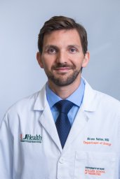 Bruno Nahar合作论文数Desai Sethi Urology Institute, University of Miami Miller School of Medicine7F. Yang合作论文数7Patel Vivek合作论文数7
 Andrew Stephenson合作论文数Rush University Medical Center7
 Thomas M. Pisansky合作论文数Mayo Clinic-Rochester7
 Robert B. Den合作论文数Department of Radiation Oncology, Thomas Jefferson University;Alpha Tau Medical7
 Daniel Spratt合作论文数Department of Radiation Oncology, University Hospitals Cleveland Medical Center;Case Western Reserve University School of Medicine7
 Dipen J. Parekh合作论文数Department of Urology, Miller School of Medicine, University of Miami7
 Drew Moghanaki合作论文数Virginia Commonwealth University7
 William Jin合作论文数6J. Ford合作论文数6Nachiketh Soodana Prakash合作论文数Dept Urol, Univ Miami6
 Chad Ritch合作论文数6
 Rosa Patricia Castillo合作论文数Fundación Santa Fe de Bogota6




