Cognitive impairment is a common comorbidity of epilepsy and adversely impacts people with both frontal lobe (FLE) and temporal lobe (TLE) epilepsy. While its neural substrates have been investigated extensively in TLE, functional imaging studies in FLE are scarce. In this study, we profiled the neural processes underlying cognitive impairment in FLE and directly compared FLE and TLE to establish commonalities and differences. We investigated 172 adult participants (56 with FLE, 64 with TLE and 52 controls) using neuropsychological tests and four functional MRI tasks probing expressive language (verbal fluency, verb generation) and working memory (verbal and visuo-spatial). Patient groups were comparable in disease duration and anti-seizure medication load. We devised a multiscale approach to map brain activation and deactivation during cognition and track reorganization in FLE and TLE. Voxel-based analyses were complemented with profiling of task effects across established motifs of functional brain organization: (i) canonical resting-state functional systems; and (ii) the principal functional connectivity gradient, which encodes a continuous transition of regional connectivity profiles, anchoring lower-level sensory and transmodal brain areas at the opposite ends of a spectrum. We show that cognitive impairment in FLE is associated with reduced activation across attentional and executive systems, as well as reduced deactivation of the default mode system, indicative of a large-scale disorganization of task-related recruitment. The imaging signatures of dysfunction in FLE are broadly similar to those in TLE, but some patterns are syndrome-specific: altered default-mode deactivation is more prominent in FLE, while impaired recruitment of posterior language areas during a task with semantic demands is more marked in TLE. Functional abnormalities in FLE and TLE appear overall modulated by disease load. On balance, our study elucidates neural processes underlying language and working memory impairment in FLE, identifies shared and syndrome-specific alterations in the two most common focal epilepsies and sheds light on system behaviour that may be amenable to future remediation strategies.
The adapted Rey Auditory Verbal Learning Test paradigm fMRI might support verbal memory lateralization. Temporal lobe epilepsy laterality influences hippocampal memory laterality indexes. Left temporal lobe epilepsy has shown a higher proportion of atypical verbal memory compared with language, potentially secondary to memory functional reorganization. BACKGROUND AND PURPOSE: fMRI is a noninvasive tool for predicting postsurgical deficits in candidates with pharmacoresistant temporal lobe epilepsy. We aimed to test an adapted paradigm of the Rey Auditory Verbal Learning Test to evaluate differences in memory laterality indexes between patients and healthy controls and its association with neuropsychological scores. MATERIALS AND METHODS: We performed a prospective study of 50 patients with temporal lobe epilepsy and 22 healthy controls. Participants underwent a block design language and memory fMRI. Laterality indexes and the hippocampal anterior-posterior index were calculated. Language and memory lateralization was organized into typical and atypical on the basis of laterality indexes. A neuropsychological assessment was performed with a median time from fMRI of 8 months and was compared with fMRI performance. RESULTS: We studied 40 patients with left temporal lobe epilepsy and 10 with right temporal lobe epilepsy. Typical language occurred in 65.3% of patients and 90.9% of healthy controls (P = .04). The memory fMRI laterality index was obtained in all healthy controls and 92% of patients. The verbal memory laterality index was bilateral (24.3%) more frequently than the language laterality index (7.69%) in patients with left temporal lobe epilepsy. Atypical verbal memory was greater in patients with left temporal lobe epilepsy (56.8%) than in healthy controls (36.4%), and the proportion of bilateral laterality indexes (53.3%) was larger than right laterality indexes (46.7%). Atypical verbal memory might be associated with higher cognitive scores in patients. No relevant differences were seen in the hippocampal anterior-posterior index according to memory impairment. CONCLUSIONS: The adapted Rey Auditory Verbal Learning Test paradigm fMRI might support verbal memory lateralization. Temporal lobe epilepsy laterality influences hippocampal memory laterality indexes. Left temporal lobe epilepsy has shown a higher proportion of atypical verbal memory compared with language, potentially to memory functional reorganization.
Epileptic networks, defined as brain regions involved in epileptic brain activity, have been mapped by functional connectivity in simultaneous electroencephalography and functional magnetic resonance imaging (EEG-fMRI) recordings. This technique allows to define brain hemodynamic changes, measured by the Blood Oxygen Level Dependent (BOLD) signal, associated to the interictal epileptic discharges (IED), which together with ictal events constitute a signature of epileptic disease. Given the highly time-varying nature of epileptic activity, a dynamic functional connectivity (dFC) analysis of EEG-fMRI data appears particularly suitable, having the potential to identify transitory features of specific connections in epileptic networks. In the present study, we propose a novel method, defined dFC-EEG, that integrates dFC assessed by fMRI with the information recorded by simultaneous scalp EEG, in order to identify the connections characterised by a dynamic profile correlated with the occurrence of IED, forming the dynamic epileptic subnetwork. Ten patients with drug-resistant focal epilepsy were included, with different aetiology and showing a widespread (or multilobar) BOLD activation, defined as involving at least two distinct clusters, located in two different lobes and/or extended to the hemisphere contralateral to the epileptic focus. The epileptic focus was defined from the IED-related BOLD map. Regions involved in the occurrence of interictal epileptic activity; i.e., forming the epileptic network, were identified by a general linear model considering the timecourse of the fMRI-defined focus as main regressor. dFC between these regions was assessed with a sliding-window approach. dFC timecourses were then correlated with the sliding-window variance of the IED signal (VarIED), to identify connections whose dynamics related to the epileptic activity; i.e., the dynamic epileptic subnetwork. As expected, given the very different clinical picture of each individual, the extent of this subnetwork was highly variable across patients, but was but was reduced of at least 30% with respect to the initially identified epileptic network in 9/10 patients. The connections of the dynamic subnetwork were most commonly close to the epileptic focus, as reflected by the laterality index of the subnetwork connections, reported higher than the one within the original epileptic network. Moreover, the correlation between dFC timecourses and VarIED was predominantly positive, suggesting a strengthening of the dynamic subnetwork associated to the occurrence of IED. The integration of dFC and scalp IED offers a more specific description of the epileptic network, identifying connections strongly influenced by IED. These findings could be relevant in the pre-surgical evaluation for the resection or disconnection of the epileptogenic zone and help in reaching a better post-surgical outcome. This would be particularly important for patients characterised by a widespread pathological brain activity which challenges the surgical intervention.
Epileptic activity arises from dysfunction of pathological neuronal network (epileptic network). Aim: to assess maintenance of epileptic network in between IEDs occurrence. We acquired simultaneous EEG-fMRI resting state in 10 drug-resistant focal epilepsy patients, having multifocal BOLD response and maximal t-value in the IED field. We compared results of functional connectivity (FC) before and after removing IEDs effect for two seed types: a 10mm-diameter sphere drawn in the BOLD map maximum and the epileptic component obtained by Indipendent component analysis. We evaluated the results in terms of mean t-value and the percentage of overlap between the FC maps. Mean t-value was significant lower after regressing out the IEDs at group level (p = 0.008 for sphere; p = 0.0004 for epileptic component). Overlap extent was 85% ± 12% and 87% ± 12% for the sphere and the epileptic component, respectively. Regions involved in a specific epileptic network show coherent oscillation independent of the occurrence of interictal activity. This could indicate the presence of epileptic activity invisible to the scalp-EEG or the presence of new and pathological resting state networks. Regions involved in a specific epileptic network show functional coupling independently of the occurrence of interictal activity as revealed on scalp EEG.
ObjectiveAssessment of language dominance using functional magnetic resonance imaging (fMRI) is a standard tool to estimate the risk of language function decline after epilepsy surgery. Although there has been considerable research in the characterization of language networks in bilingual individuals; little is known about the clinical usefulness of language mapping in a secondary language in patients with epilepsy, and how language lateralization assessed by fMRI may differ by the use of native or a secondary language paradigms. In this study we investigate language representation in a population of nonnative English speakers to assess differences in fMRI language lateralization between the first (native) and second language (English).MethodsSixteen nonnative English-speaking patients with focal drug-resistant epilepsy underwent language fMRI in their first (native) language (L1) and in English (L2). Differences between language maps using L1 and L2 paradigms were examined at the single subject level by comparing within-subject lateralization indexes obtained for each language. Differences at the group level were examined for each of the tasks and languages.ResultsGroup maps for the second language (English) showed overlapping areas of activation with the native language, but with larger clusters, and more bilaterally distributed than for the first language. However, at the individual level, lateralization indexes were concordant between the two languages, except for one patient with bilateral hippocampal sclerosis who was left dominant in English and showed bilateral dominance for verb generation and right dominance for verbal fluency in his native tongue.SignificanceLanguage lateralization can generally be reliably derived from fMRI tasks in a second language provided that the subject can follow the task. Subjects with greater likelihood of atypical language representation should be evaluated more carefully, using more than one language paradigm.
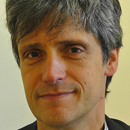 Matthias Koepp合作论文数Department of Clinical and Experimental Epilepsy, Queen Square Institute of Neurology, University College London17
 Christian Vollmar合作论文数Klinikum Grosshadern, University of Munich;Department of Neurology, Germany;Department of Neurology, University of Munich16
 John Duncan合作论文数Department of Clinical and Experimental Epilepsy, University College London15
 Mark R. Symms合作论文数Institute of Neurology, NSE Epilepsy Research MRI Unit12
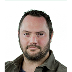 Jason Stretton合作论文数MRC Cognition and Brain Sciences Unit, University of Cambridge11
 Pamela J. Thompson合作论文数11
 David W Carmichael合作论文数MRI Unit, National Society for Epilepsy9
 Mark Richardson合作论文数Centre for Developmental Neurobiology, School of Neuroscience, Institute of Psychiatry, Psychology & Neuroscience, King’s College London7Bonelli Silvia B合作论文数NSE MRI Unit, National Society for Epilepsy6
 Helen Cross合作论文数Great Ormond Street Institute of Child Health, University College London6
 Britta Wandschneider合作论文数für Neurologie, Charité-Universitätsmedizin6
 Meneka K Sidhu合作论文数6
 Ronit Pressler合作论文数Developmental Neurosciences Dept, University College London;Telemetry Unit, Great Ormond Street Hospital for Children6A Donaire合作论文数5M. Carreno合作论文数5
 Gavin Winston合作论文数Department of Medicine, Queen's University;School of Computing, Queen's University5
 Paul M. Thompson合作论文数Imaging Genetics Center, Lab of Neuro Imaging, Institute for Neuroimaging and Informatics, Keck School of Medicine, University of Southern California5
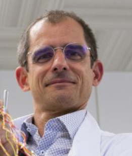 Serge Vulliémoz合作论文数Department of Clinical Neurosciences, Faculty of Medicine, Geneva University Hospitals;Division of Neurology, Geneva University Hospitals5
 Nuria Bargallo合作论文数Neuroradiology Section, Radiology Department, Hospital Clinic de Barcelona;The Lysholm Department of Neuroradiology, National Hospital for Neurology and Neurosurgery;Neuroradiology Section, Centre Medic Alomar;Magnetic Resonance Image Core Facility, IDIBAPS5J Duncan合作论文数4
 Suejen Perani合作论文数Kings Coll London, Inst Psychiat Psychol & Neurosci, Dept Basic & Clin Neurosci, London, England4Isabel Manzanares合作论文数Hosp Clin Barcelona, EpiCARE Network4
 Gareth Barker合作论文数Department of Neuroimaging, School of Neuroscience, King’s College London;Institute of Psychiatry, Psychology & Neuroscience, King’s College London4Xavier Setoain合作论文数4
 Chris Clark合作论文数Department of Developmental Neurosciences, University College London4Teresa Boget合作论文数4
 Luis Pintor合作论文数4Jonathan O'Muircheartaigh合作论文数4M C Edo合作论文数3E Conde Blanco合作论文数3K. St Pier合作论文数3E Santamarina合作论文数3
 Tim M Tierney合作论文数UCL Queen Square Institute of Neurology, University College London3Manuel Toledo合作论文数3
 Caciagli Lorenzo合作论文数Montreal Neurological Institute and Hospital, McGill University Montreal3
 Beate Diehl合作论文数University College London;National Hospital for Neurology and Neurosurgery3
 Louis Lemieux合作论文数Queen Square Institute of Neurology, University College London3Iannotti Giannina R合作论文数3
 Margitta Seeck合作论文数Département des Neurosciences Cliniques, Hôpitaux Universitaires de Genève3
 Francesca Pittau合作论文数Department of Neurological Sciences, University of Bologna3
 Frederic Grouiller合作论文数3Manuel Quintana合作论文数3
 Jordi Rumia合作论文数Hospital Clínic, Universitat de Barcelona3E. Conde合作论文数2J Alvarez Sabín合作论文数2
 Elhum A Shamshiri合作论文数University Hospitals and Faculty of Medicine of Geneva, University of Geneva2Veena Kumari H M合作论文数Channabasaveswara Institute of Technology2de Francisco J合作论文数Universidad Autónoma de Barcelona, Hospital Universitario Vall d'Hebron2Sanjay Sisodiya合作论文数Department of Clinical and Experimental Epilepsy, Queen Square Institute of Neurology, Faculty of Brain Sciences, University College London2




