Although women with prior LEEP history are generally considered as having higher risk of preterm birth, recent meta-analysis indicated that the absolute risk of preterm birth is less than 10%. Recently, cervical elastography has emerged as an ancillary tool to cervical length (CL) for prediction of preterm birth. Here, we aimed to determine if a previous LEEP before pregnancy is associated with the change of cervical strain as well as shortening of CL. This data is a part of multicentre prospective observational study about prediction of preterm birth using cervical elastography (E-cervix study) in Korea between March 2018 and February 2019. The cervical elastography was performed at the time of CL measurement in midtrimester of asymptomatic gravidas who underwent a LEEP prior to pregnancy (n = 30) or who had not had this procedure (n = 34). Cervical elastography was obtained at midsagittal plane same as for CL measurement using E-cervixTM (WS80A, Samsung Medison, Seoul, Korea) and mean value of elastographic parameters including contrast index (ECI), hardness ratio (HR), mean strain level in internal os (IOS) and external os (EOS) was obtained by three repeated examination. Statistical analysis was done using Mann-Whitney U test. There was no difference in gestational age at examination between LEEP and control group (median:16.5 vs 18.1 weeks, p = 0.188]. CL was significantly shorter in LEEP group (3.1 vs 3.8 cm, p < 0.001). The ECI, IOS and EOS was higher and HR was lower in LEEP group but neither was statistically significant significant (2.74 vs 2.36, p = 0.477; 0.19 vs 0.18, p = 0.220; 0.24 vs 0.23, p = 0.562; 76.8 vs 85.4, p = 0.101). LEEP prior to pregnancy results in cervical shortening in midtrimester ultrasonography but seems not to cause significant change in cervical strain. Supported by Korea Health Technology R&D Project through the Korea Health Industry Development Institute (KHIDI), funded by the Ministry of Health & Welfare, Republic of Korea (grant number: HI18C1696). Please note: The publisher is not responsible for the content or functionality of any supporting information supplied by the authors. Any queries (other than missing content) should be directed to the corresponding author for the article.
To determine whether change in cervical elastographic parameters following progesterone treatment in pregnant women with short cervix is associated with a higher prevalence of adverse pregnancy outcome. Of multicentre prospective cohort including 1033 consecutive pregnant women, 80 were given progesterone for short cervical length (CL) of leqq; 25 mm. Cervical elastography was performed pre-(baseline) and 1,2, and 4 weeks post-progesterone administration using E-cervixTM (WS80A, Samsung Medison). Adverse pregnancy outcome included delivery< 37 weeks of gestation, treatment for threatened labour, or requiring cervical cerclage. Difference and % change from baseline in cervical elastography parameters (contrast index (ECI), hardness ratio (HR), mean strain level of internal (IOS), and external OS (EOS)) between pre- and post-progesterone treatment were compared between the normal and adverse outcome groups. Diagnostic performance for prediction of adverse pregnancy outcome was assessed using ROC curve analysis. Of 80 patients, 60 were eligible for analysis where 35 delivered at term and 25 had adverse pregnancy outcome. Between pregnant women who had adverse pregnancy outcome vs normal group, HR and IOS at were significantly different at 1 week (61 vs 73%, 0.29 vs 0.29) whereas CL demonstrated no difference. HR % change at 1 week from the baseline in normal group showed a positive trend whereas it was negative in adverse outcome group (+17.5 vs -4.5%, p = 0.09). Parameters at 2 and 4 weeks were not statistically significant. Area under curve (AUC) of IOC % change at 1 week in prediction of adverse outcome was 0.84(0.71, 0.97). Performing cervical elastography before and after 1 week of progesterone administration may be of help to predict progesterone treatment efficacy. Supported by Korea Health Technology R&D Project through the Korea Health Industry Development Institute (KHIDI), funded by the Ministry of Health & Welfare, Republic of Korea (grant number: HI18C1696). Please note: The publisher is not responsible for the content or functionality of any supporting information supplied by the authors. Any queries (other than missing content) should be directed to the corresponding author for the article.
Sphingosine-1-phosphate (S1P), which is formed by phosphorylation of sphingosine through a process catalysed by sphingosine kinase (SK), is a multifunctional mediator of a variety of cellular responses including proliferation, differentiation, motility, and survival. K6PC-5, which was recently synthesized as a novel SK activator, is expected to increase S1P levels. Indeed studies have already demonstrated that K6PC-5 exhibits anti-aging effects on intrinsic aged murine skin by increasing fibroblasts, collagen synthesis, dermal thickness, and epidermal differentiation. However, photoaging and intrinsic aging have highly different clinical and histopathological properties. In this study, we developed a photoaged murine model by exposing mice that were 56 weeks old to ultraviolet (UV)B and UVA radiation for 8 weeks. We then investigated whether K6PC-5, as an SK activator, had anti-aging effects on photoaged murine skin in addition to its effects on intrinsic aged murine skin and determined the mechanism. K6PC-5 increased dermal collagen density in photoaged skin through increases in fibroblasts and collagen production. Photoaged murine skin treated with K6PC-5 showed an increase in stratum corneum (SC) integrity with increased corneodesmosome density and an improvement in barrier recovery rate. Matrix metalloproteinase 13 remained unchanged. These results indicate that topical application of K6PC-5 improves photoaged skin by improving skin barrier and increasing fibroblast count and function. In conclusion, K6PC-5, as an S1P activator, improves long-term UV-exposed aged skin as well as intrinsic aged skin.
 S. Jeong合作论文数12B. Park合作论文数12S. Lee合作论文数9Hyungkeun Kim合作论文数Yonsei University8Jae Boum Youm合作论文数Mitochondrial Signaling Laboratory, Inje University7
 Jae-Hoon Bae合作论文数6E. Choi合作论文数5
 Jeonghoon Jeon合作论文数4Kwak Hyun Jung合作论文数4H. Park合作论文数2H. Kwon合作论文数2H. Shim合作论文数2
 Hee-Dong Park合作论文数2Seol H J合作论文数2
 Hanel Hwang合作论文数2
 Joseph Sung (沈祖堯)合作论文数Lee Kong Chian School of Medicine, Nanyang Technological University2
 Junehee Kwon合作论文数Department of Retail, Hospitality, and Tourism Management, College of Education, Health, and Human Sciences, University of Tennessee, Knoxville2S. Oh合作论文数2
 Ih Bae合作论文数2Won Joon Seong合作论文数School of Medicine, Kyungpook National University Hospital2sughun hong合作论文数1H. Kim合作论文数1Y. Kim合作论文数1Eunkyung Lee合作论文数1H. Gwak合作论文数1Moo Gak Hwang合作论文数1T. Mauro合作论文数1C. Chung合作论文数1M. Kim合作论文数1
 Inkyung Jung合作论文数1Chang Soochul合作论文数1
 Mankil Jung合作论文数Department of Chemistry, Yonsei University1Y. Lee合作论文数1
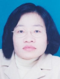 Sai Peck Lee合作论文数Faculty of Computer Science and Information Technology, University of Malaya1
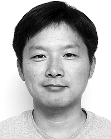 Dongwan Shin合作论文数Dept Comp Sci, New Mexico Inst Min & Technol1Seihyun Ahn合作论文数1
 Yang-Han Lee合作论文数Department of Electrical Engineering;Tamkang University;Department of Electrical Engineering, Tamkang University1Kyung-Hwan Kim合作论文数Sogang University1
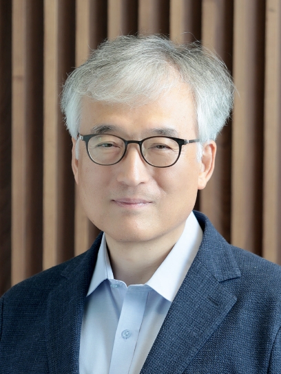 Hyeongtag Jeon合作论文数Division of Materials Science and Engineering, Hanyang University1Sukyoung Yi合作论文数1Myeon Haeng Cho合作论文数Department of Biology;Yonsei University;Department of Biology, Yonsei University1
 Yong Sam Jung合作论文数Department of Bioscience and Biotechnology, Chungnam National University1
 Jeong Hee Hong合作论文数Department of Physiology, University of Texas Southwestern Medical Center1Hochul Yi合作论文数1
 Hyun Jik Kim合作论文数Department of Otolaryngology Head and Neck Surgery 134, Yonsei University College of Medicine1Wen-Bin Yeh合作论文数1
 Jae-Heung Ko合作论文数1




