Recurrence risk estimates underpin meningioma research, including molecular classification and clinical trial benchmarking, yet are often based on retrospective or historical data. The aim of this study was to assess the variation of recurrence risk estimates across calendar periods, WHO classification editions, geographical settings, and healthcare systems.thetermine We analyzed 4,111 patients with primary WHO-1/-2 meningiomas from 31 centers in 15 countries (1990–2019). Recurrence was defined according to local radiological assessment. The 5- and 10-year recurrence risks were estimated using regression standardization with inverse probability of censoring weights, adjusting for key clinical, surgical, and histopathological variables. Recurrence risk estimates varied across all domains examined. More recent calendar periods were associated with higher predicted recurrence risk, particularly for WHO-2 at 5 years (e.g., ≥ 2013 vs. ≤ 2007: RR 1.60, 95
Chordomas involving the cervical spine are best treated by surgical resection and post-operative radiotherapy. Excision of these tumors can be complicated by injury to surrounding soft tissues and neurovascular structures. We report a case of a cervical chordoma resection during which time the left vertebral artery was injured, requiring aneurysm clip sacrifice and the modification of the vertebral body implant to accommodate the applier end of the clips. The implant was modified with the removal of a small portion of one side to accommodate the proximal ends of aneurysm clips. The patient remained neurologically intact without cerebrovascular insult. Modification of the implant did not affect the structural integrity of the corpectomy implant, the planning of radiation therapy, or the final clinical result. At 2.5 years from surgery, the patient remains clinically well with no evidence of recurrence and no structural failure of the implant.
TPS2089 Background: The efficacy of systemic therapies for brain metastases (BM) is hindered by the blood-brain barrier (BBB) and brain-tumor barrier. Transcranial low-intensity focused ultrasound combined with IV microbubble oscillators (MB-FUS), allows for localized, controlled, non-invasive and temporary BBB opening, which has been shown to enhance tumor drug delivery of systemic therapies, as well as impro efficacy of immunotherapies. Non-small cell lung cancer (NSCLC) is the most common cause of BM, and this randomized controlled trial (RCT) aims to evaluate the safety and efficacy of MB-FUS-mediated BBB opening combined with standard of care (SOC) systemic therapy versus systemic therapy alone for patients with NSCLC BM. Methods: LIMITLESS is prospective, multicenter, parallel-arm, RCT, ongoing at up to 30 centers, that randomizes patients with NSCLC BM, in a 2:1 ratio to either: (i) Arm 1: MR-guided MB-FUS plus all FDA approved on-label use of immune checkpoint inhibitors (ICIs) with or without chemotherapy regimen (SOC systemic therapy), or (ii) Arm 2: SOC systemic therapy alone. Included patients are ≥18 years aged, with normal organ function, KPS ≥70, and have ≥0.5 cm size BM meeting measurable disease criteria as per RANO-BM. Patients on both arms receive standard-of-care therapy, while those on arm 1 also undergo MB-FUS. Patients undergo pre-treatment brain MRI, followed by IV administration of microbubbles for enhanced sonication effects. BBB opening is performed using a transcranial 220 kHz device with 1024-element phased array transducer with real-time acoustic feedback-based power control for maintaining effective microbubble activation. The primary study outcome is the overall objective response rate (ORR) at 6 months as assessed using RANO-BM criteria. Using a Bayesian design for power analysis, a superior ORR of 60% is assumed for MB-FUS arm versus 30% in the control arm for a total sample size of N = 96, 64 participants in MB-FUS and 32 in control arm, for 80% power using a two-sided chi-square test with an alpha of 0.05. For the upper-bound estimate, ORR of 45% in MB-FUS arm and 30% in the control arm, the study needs N = 369 participants: 246 in LIFU arm and 123 in control arm. The secondary outcomes are best objective response rate and median time-to-response per treatment arm. Exploratory outcomes are median progression-free survival (PFS), overall survival (OS), median intracranial PFS, median extracranial PFS, and quality of life. Patient enrollment commenced in 2022 and is ongoing (ClinicalTrials.Gov Registration: NCT05317858). Clinical trial information: NCT05317858 .
Background/Objectives: Connectomics is an evolving branch of neuroscience that determines structural and functional connectivity in the brain. The objective of this prospective imaging study is to evaluate the effect of whole brain radiotherapy (WBRT) on the connectome. Methods: A combination of diffusion tensor imaging (DTI) and functional magnetic resonance imaging (fMRI) was used to study the structural and functional connectivity of the brain, and a machine learning algorithm trained to analyze subject-specific data was applied to create individualized brain maps with 15 neuronal networks for each patient. These brain maps were compared to normal brains from the human connectome project, producing an anomaly matrix. Connectome analysis and multi-dimensional neurocognitive testing on a web-based platform were performed at baseline and 3 months post-WBRT. The change in anomaly frequency was co-related with neurocognitive outcomes. Results: At baseline, connectome analysis revealed that the multiple demand network had the most anomalies (46%). Pre- and post-WBRT comparison revealed increases in proportional anomaly frequency across multiple networks. Pearson correlation showed correlation between neurocognitive domain decline and anomaly changes: learning and memory domain with subcortical network [Verbal recall (Pearson coefficient −0.94; p < 0.01), verbal revision (Pearson coefficient −0.89; p = 0.01), and verbal recognition (Pearson coefficient −0.94; p < 0.01)]. Conclusions: This proof-of-concept study integrated data from DTI and fMRI in the form of connectome and revealed significant changes in brain connectivity, with WBRT that also correlated with neurocognitive outcomes. Further studies in a larger cohort are underway, and correlations with white matter changes and tumor locations/numbers will be performed.
TPS2094 Background: Liquid biopsy in glioblastoma (GBM) is hindered by a lack of requisite circulating tumor (ct) and cell-free (cf) DNA levels in blood due to the blood-brain barrier (BBB). This limits the identification of blood-based tumor biomarkers along with the development and use of biomarker-driven systemic therapies. Low intensity focused ultrasound combined with intravenously administered microbubble oscillators (MB-FUS), leads to non-invasive BBB opening. This trial aims to evaluate the utility of LIFU for bolstering blood ctDNA and cfDNA for enhance liquid biopsy in patients with GBM. Methods: LIBERATE is an ongoing, prospective, multi-center, self-controlled, pivotal trial evaluating safety and technical efficacy of transcranial MR-guided MB-FUS for increasing blood ctDNA and cfDNA levels in adults, aged 18-80 years with GBM. Patients with suspected GBM planned for tumor biopsy or resection at 17 centers in US and Canada are being enrolled. Patients with multifocal tumors or tumors arising from deep midline, thalamus, cerebellum, or brainstem are excluded. Patients are administered IV microbubbles for enhanced sonication, after which MR-guided BBB opening using a 220 kHz device, with 1024-element phased array transducer, is performed with real-time acoustic feedback control for effective cavitation. Pre- and post-procedure, phlebotomy and MRI brain are done. Patients are offered optional 2 nd procedure during adjuvant chemotherapy phase if willing. Primary efficacy endpoint is correlation between biomarker patterns in tumor tissue collected during surgery/biopsy and blood collected following MB-FUS procedure. Confirmatory secondary efficacy endpoint is ratio between greatest yield of cfDNA in blood post-MB-FUS compared to cfDNA level in blood pre-MB-FUS. The primary study hypothesis is that agreement rate on biomarker pattern between resected/biopsied tumor tissue and blood is > 70%. The secondary hypothesis is that MB-FUS BBBO leads to a ≥2-fold rise in blood cfDNA. Assuming the true agreement rate expected is 89%, a sample of N = 50 patients will provide 90% power to meet the primary endpoint (Exact test, Binomial Proportion, one-sided Alpha = 0.025). Exploratory endpoints include (1) sensitivity of detection of known specific somatic mutations in ctDNA from blood samples collected before and after MB-FUS, (2) estimation of ctDNA levels in samples collected at 30-minutes, 1-hour, 2-hour, and 3-hour post-MB-FUS to determine time of greatest yield, (3) correlation of MRI parameters related to grading of BBB opening and ctDNA-based biomarkers from post-MB-FUS blood samples, (4) biomarker correlation between plasma cfDNA sampled during adjuvant chemotherapy phase and tumor tissue harvested at surgery. Patient enrollment commenced in 2022 and is ongoing (NCT05383872). Clinical trial information: NCT05383872 .
Antibody-drug conjugates (ADCs) have transformed treatment in HER2-positive breast cancer (BC), particularly in patients with brain metastases (BM), due to agents like trastuzumab emtansine (T-DM1) and trastuzumab deruxtecan (T-DXd), both demonstrating intracranial efficacy. Radiotherapy (RT), especially stereotactic radiosurgery (SRS), remains a mainstay in BM management. However, combining ADCs with RT may increase the risk of symptomatic radiation necrosis (SRN), a potentially debilitating complication that can significantly impair neurological function and quality of life. This meta-analysis evaluates SRN outcomes in patients receiving concurrent (C-ADC) versus non-concurrent ADCs with RT (NC-ADC). A systematic search was conducted in January 2025 across PubMed, Cochrane Library, and major oncology conference proceedings (ASCO, SNO, ESMO, SABCS). Eligible studies included randomized controlled trials and cohort studies (CS) that reported SRN or related toxicity outcomes in HER2-positive BC patients with BM treated with either C-ADC or NC-ADC approaches. Pooled SRN proportions and risk ratios (RR) with 95% confidence intervals (CIs) were calculated using a random-effects model. Heterogeneity was assessed using the I² statistic. Of 884 studies screened, 9 CS (N=421) met inclusion criteria. The median patient age was 56.3 years (IQR: 49.8–57). Prior intracranial RT was reported in 41.3%, with 19.3% receiving SRS and 19.6% undergoing whole-brain RT. Median time to C-ADC was 8.75 days (IQR: 8.0–18.0), and to NC-ADC was 273.5 days (IQR: 225.8–327.8). SRN occurred in 19.5% (95% CI: 9.2%–29.8%; I²=39.2%) of C-ADC patients versus 6.9% (95% CI: 2.5%–11.2%; I²=52.0%) in NC-ADC. C-ADC was associated with significantly increased SRN risk (RR=2.73, 95% CI: 1.45–5.11, P=0.002; I²=0.0%), with consistent findings across included studies. Concurrent ADC-RT treatment is linked to a significantly higher SRN risk. Careful patient selection and prospective studies are essential to optimize treatment sequencing strategies that balance efficacy with safety and minimize toxicity.
e14011 Background: Adolescents and young adults (AYA), aged 15 to 39 at diagnosis, account for 2% of invasive cancers in US. Despite significant advances in cancer treatment, brain metastases (BM) remain an underexplored complication in this population, contributing to significant morbidity and mortality. This study aims to evaluate prognostic factors for BM in AYA patients (pts). Methods: Our study used the National Cancer Database (2010-2021) to analyze AYA patients diagnosed with the 20 most common cancer types. Patients with ≥6 months of follow-up and known survival status were included. Demographic and socioeconomic factors were analyzed by descriptive statistics. Cox analyses were used to identify potential prognostic factors. Kaplan–Meier analysis was conducted to evaluate median overall survival (mOS). Results: Of 382,241 AYA pts, 2,504 (0.65%) were diagnosed with BM with a median age of 34 years (IQR: 28–37). Most were male (51.7%) and White (75.9%). The most common causes of BM were lung (37.5%), testicular (15.5%), breast (14.9%), melanoma (13.8%), lymphoma (4.3%), sarcoma (3.3%) and colorectal (2.5%). Pts with BM were significantly more likely to have bone (40.1% vs 2.2%, P < 0.001) and lung metastases (44% vs 2.6%, P < 0.001), but were less likely to have liver metastases (26.2% vs 73.8%, P < 0.001) compared to those without BM. The mOS for pts with BM was 27.4 months (95% CI: 25.43–29.44), with 1-, 3-, 5- and 10-year overall survival (OS) rates of 75.6%, 43.5%, 34.8%, and 24.7%, respectively. When stratified by cancer group, testicular cancer (TC) had the best outcomes (mOS: NR, 5-y OS: 58.1%), while lung cancer (mOS: 22.7 months, 5-y OS: 25.8%) and melanoma (mOS: 22.6 months, 5-y OS: 35.1%) had the worst outcomes. In multivariate analysis, pts with TC had significantly better outcomes compared to other cancers (HR: 0.3, 95% CI: 0.14–0.72, p = 0.006). Treatment with stereotactic radiosurgery (SRS) significantly improved survival compared to whole-brain radiation therapy (HR: 0.8, 95% CI: 0.71–0.94, p = 0.004). Liver metastases (HR: 1.3, 95% CI: 1.18–1.50, p < 0.001) and lung metastases (HR: 1.2, 95% CI: 1.08–1.36, p = 0.001) were associated with worse survival. Conclusions: BMs in AYA pts are rare and outcomes appear more favorable than older populations. SRS use is associated with the best survival rates. Further research on tumor biology including genetic and epigenetic drivers, and strategies for early detection and prevention is necessary to improve outcomes in this unique population. Survival outcomes by cancer. All Testicular Breast Melanoma Lung Survival Rate%, (95% CI)1-Year3-Year 75.6(74-77.3)43.5(41.6-45.6) 81.7(77.9-85.7)60(55.1-65.2) 80.1(76.1-84.2)44.9(39.9-50.5) 68.6(63.9–73.7)40.4(35.4-46.1) 74.5(71.8-77.4)37.5(34.5-40.9) mOS (months)(95% CI) 27.4(25.4–29.4) NA(96.9-NA) 29.1(26.4-36.3) 22.6(18.1-29.4) 22.7(20.7-25.8)
Background and objectives Intraoperative magnetic resonance imaging (iMRI) is valuable for assessing the extent of brain tumor resections and preventing repeat procedures for unexpected residual tumors. However, prolonged procedure times and restrictions on ferromagnetic materials deter widespread iMRI use. This study explored two methods to increase iMRI utilization, both involving transferring patients to the magnet after wound closure rather than moving the magnet into the operating room (OR). Methods A process improvement database of 40 consecutive patients undergoing iMRI for brain tumor surgery was analyzed. Phase-1, conducted between November 2020 and February 2021, involved transporting patients to the magnet and returning them to the OR for extubation. Phase-2, conducted between August 2021 and November 2021, designated an extubation area in the recovery room, eliminating the need to return patients to the OR. Diagnosis was used to match Phase-1 and Phase-2 cohorts with a retrospective comparative cohort (Phase-0) collected between June 2019 and August 2019. Several time intervals were recorded for analysis. Results A total of 57 cases were analyzed; 56% of patients were male, with a mean age of 56 years. iMRI volumes significantly increased in Phase-1 (100%, n = 20) and Phase-2 (100%, n = 19) compared to Phase-0 (22%, n = 4, p < 0.001). OR occupancy (p = 0.18) and anesthesia duration (p = 0.23) were statistically similar between groups, but the Phase-2 group demonstrated a clinically relevant median decrease of 78 minutes in case duration and a 41-minute reduction in anesthesia duration compared to Phase-0. Both Phase-1 and Phase-2 showed a significant 11-hour decrease in post-surgical MRI acquisition times compared to Phase-0 (p < 0.001). Conclusion Transferring patients to the magnet significantly increased iMRI utilization and facilitated immediate postoperative imaging without negatively impacting OR efficiency or anesthetic safety. Establishing an extubation site in the recovery room saved valuable OR time and reduced patient intubation time. The practice of "moving the patient, not the magnet," combined with recovery room extubation, is now routine in our neurosurgery service.
TPS2089 Background: 20-40% of HER-2-positive breast cancer patients develop brain metastases (BMs), for which stereotactic radiosurgery (SRS) is used for local disease control. Tucatinib, an oral HER-2-selective tyrosine kinase inhibitor (TKI), has been demonstrated to be safe and efficacious in HER-2-positive breast cancer in combination with capecitabine and trastuzumab. The synergism between these three drugs and radiation may potentially enhance DNA damage, causing increased apoptosis. We hypothesize that this 3-drug therapy, in combination with SRS in patients with HER-2 positive breast cancer BM (BCBM), may be safe and provide superior long-term control of intracranial and systemic disease. Methods: This is a prospective, single-arm, multicenter, ongoing, phase 1 clinical trial. Patients aged 18-80 years, ECOG score 0-2, normal organ function, and up to 10 newly-diagnosed BMs is planned to enroll at six centers in the US. Any number of prior systemic therapies are allowed, except tucatinib and capecitabine. Key exclusion criteria include leptomeningeal metastases, evidence of intra-tumoral or peri-tumoral hemorrhage, and BMs within 5 mm of the optic chiasm/nerve. Patients receive oral tucatinib alongside SRS, followed by a two-week dose-limiting toxicity (DLT) period, and subsequently continue the regimen until disease progression or intolerable toxicity. DLTs are defined by Grade 3 or 4 thrombocytopenia, grade 4 anemia, grade 4 neutropenia lasting more than seven days, febrile neutropenia, and grade 3 non-hematologic toxicity. Primary study endpoint is maximum tolerated dose (MTD) of tucatinib in combinatorial therapy. This de-escalation study starts at 300 mg dose level (DL), with DL-1 250 mg, and DL-2 200 mg, based on a 3 + 3 design. Cohort expansion at MTD is planned to a total of 40 patients. Secondary endpoints include efficacy as determined by response rate, intracranial progression-free survival (PFS), extracranial PFS, overall survival, quality of life as assessed by FACT-Br, toxicity, and neurocognitive function. The trial has been approved by the Institutional Review Board (IRB) and currently is enrolling patients. Clinical trial information: NCT05553522 .
This study evaluates the clinical utility of 3D turbo spin echo (3D-TSE) imaging in stereotactic radiosurgery (SRS) planning for brain metastases, examining the value of adding 3D-TSE to MPRAGE. Dual-sequence MRI (MPRAGE + 3D-TSE) significantly prolonged time to distant intracranial failure (DIF) in whole brain radiotherapy (WBRT)-naïve patients (n = 308, 11.4 vs. 6.8 months, p = 0.03), but not in patients previously treated with WBRT and salvaged with SRS (n = 39, 6.5 vs. 5.6 months, p = 0.76). Additional lesion detection attributed to 3D-TSE imaging was greater in WBRT-naïve (19.6 %) than in post-WBRT patients (12.0 %) (p < 0.001). The dual-sequence imaging may improve time to distant failure in the modern era for most patients treated with primary stereotactic radiosurgery and should be integrated into the SRS workflow.
Low-grade gliomas (LGGs) exhibit low overall mutational burden and an immunosuppressive microenvironment, contributing to immunotherapy resistance. Intratumoral heterogeneity (ITH) further complicates the targeting of tumor-specific antigens (TSAs), yet its influence on the antigenic and immune landscapes in LGG remains understudied, particularly due to reliance on single-biopsy analyses. We performed exome and transcriptome sequencing on 70 spatially mapped biopsies from seven IDH-mutant Grade II astrocytoma patients. Tumor purity, immune deconvolution, and neoantigen prediction were conducted to assess regional immune variation and antigenic burden. In one patient (P375), neoantigen-specific CD8+ T cells were isolated using barcoded peptide-HLA multimers. Reactive T-cell receptor (TCR) clonotypes were identified via single-cell TCR sequencing and functionally validated in Jurkat76 and CD8+ T cells. Spatial profiling of whole tumors revealed most mutations were biopsy-specific, with a steep drop in mutations shared across multiple regions. RNA-seq–based unsupervised clustering with xCell and DESeq2 revealed two distinct immune microenvironments: immune-hot regions with elevated immune infiltration, and immune-cold regions with minimal immune presence. Notably, mutation-derived n-mer peptides from immune-hot regions showed enhanced predicted immunogenicity relative to those from immune-cold areas. In one patient (P375), neoantigen-specific CD8⁺ TCR clonotypes were identified targeting PRMT5 mutations found in immune-hot regions. A mutant PRMT5-specific TCR demonstrated robust, antigen-dependent activation and dose-dependent cytotoxicity in vitro. This TCR showed high specificity toward the mutant peptide with minimal cross-reactivity to the wild-type sequence. Furthermore, engineered CD8⁺ T cells effectively killed glioma cells endogenously expressing the full-length mutant PRMT5, supporting its candidacy as a viable immunotherapeutic target. Our findings reveal significant 3D spatial and immunologic heterogeneity in low-grade gliomas and demonstrate that neoantigens such as mutant PRMT5 can be selectively targeted by highly specific, cytotoxic TCR-engineered CD8⁺ T cells. These results support the potential of personalized TCR-based immunotherapy for low-grade glioma.
e14006 Background: Brain metastases (BM) from neuroendocrine neoplasms (NENs) are rare and associated with poor prognosis. Limited data exists in the literature regarding the impact of different treatment (Tx) modalities on survival outcomes in NENs with BM, particularly in relation to primary tumor sites. Methods: NCDB data (2010–2021) was analyzed to assess Tx modalities and their association with primary tumor sites, focusing on NEN histological subtypes including small cell cancer (SCC). Kaplan-Meier analysis estimated median overall survival (OS) in months (mo). Multivariate analyses evaluated the impact of primary sites and Tx combinations, including systemic therapy (Sys) with stereotactic radiosurgery (SRS), whole-brain radiotherapy (WBRT), and standalone therapies. Results: A total of 42,291 patients were included with a median age of 66 years (IQR: 59–72), 50.5% were males, and 87.8% white. OS was highest with Sys+SRS (OS: 10.3 mo) and Sys+WBRT (OS: 8.7 mo); Sys-only Tx showed OS of 7.8 mo, while SRS-only (OS: 2.9 mo) and WBRT-only (OS: 2 mo) had poorer outcomes (P < 0.0001). Compared to Sys+SRS, multivariate analysis showed increased risk of death for Sys+WBRT (HR = 1.22, P < 0.001), Sys only (HR = 1.33, P < 0.001), SRS only (HR = 2.19, P < 0.001), and WBRT only (HR = 3.13, P < 0.001). Breast cancer had similar OS compared to lung primaries (HR = 1.15, 95% CI: 0.77-1.71, P = 0.500). Gastrointestinal cancers (large intestine/rectum and small intestine) showed no differences in OS (HR = 1.02, 95% CI: 0.92-1.14, P = 0.667). Genitourinary cancers, comprising male and female sites, had a modestly higher but non-significant HR (HR = 1.13, 95% CI: 0.97-1.31, P = 0.123). Other systems, including the endocrine system, oral cavity/pharynx, and soft tissues, showed no difference (HR = 1.00, P = 0.948). Conclusions: Sys+SRS was associated with the best OS in NEN-BM, whereas single modality approaches alone had significantly worse outcomes. After adjusting for confounders, primary tumor site did not emerge as a significant determinant of survival. Primary sites and Tx modalities. Primary Site All, N (%) Sys + SRS, n (%) Sys + WBRT, n (%) Sys only, n (%) SRS only, n (%) WBRT only, n (%) Breast & Endocrine System 39 (0.2) 6 (0.2) 11 (0.2) 8 (0.1) 0 (0.0) 4 (0.1) Esophagus and Stomach 110 (0.4) 11 (0.6) 22 (0.3) 38 (0.5) 3 (0.8) 12 (0.5) Female Genitourinary System 105 (0.4) 9 (0.5) 29 (0.3) 30 (0.4) 1 (0.3) 11 (0.5) Large Intestine and Rectum 132 (0.5) 9 (0.5) 26 (0.3) 52 (0.7) 4 (1.1) 6 (0.3) Liver, Biliary System & Pancreas 158 (0.7) 12 (0.7) 41 (0.4) 43 (0.6) 0 (0.0) 18 (0.8) Male Genitourinary System 87 (0.4) 7 (0.4) 21 (0.2) 32 (0.4) 3 (0.8) 7 (0.3) Lung 24103 (97.1) 1713 (96.6) 8516 (98.1) 7313 (96.9) 354 (96.2) 2144 (97.2) Small Intestine 10 (0.0) 0 (0.0) 2 (0.0) 3 (0.0) 1 (0.3) 1 (0.0) Soft Tissues, Oral Cavity & Pharynx 70 (0.2) 6 (0.3) 12 (0.1) 27 (0.3) 3 (0.5) 6 (0.1)
1039 Background: Antibody-drug conjugates (ADCs) have transformed the outcomes of HER2-positive breast cancer (BC), particularly in patients with brain metastases (BM) due to the use of ADCs like trastuzumab emtansine (T-DM1) and trastuzumab deruxtecan (T-DXd), which have demonstrated intracranial efficacy. Radiotherapy (RT), especially stereotactic radiosurgery (SRS), remains a cornerstone for BM management. However, combining ADCs with RT may increase the risk of symptomatic radiation necrosis (SRN). This meta-analysis evaluates SRN outcomes in patients receiving concurrent (C-ADC) versus non-concurrent ADCs with RT (NC-ADC). Methods: A systematic search was performed in January 2025 across PubMed, Cochrane, and conference proceedings from ASCO, SNO, ESMO, and SABCS. Eligible studies included randomized controlled trials and cohort studies (CS) of C-ADC and NC-ADC in HER2-positive BC patients with BM. Studies reporting SRN rates or related outcomes were included. A random-effects model was used to calculate pooled proportions and risk ratios (RR) with 95% confidence intervals (CIs). Heterogeneity across studies was assessed using the I² statistic, with values of 0–25% considered low, 26–50% moderate, and greater than 50% high. Results: Out of 884 studies screened, 9 CS (N = 421) were included. The median age was 56.3 years (IQR: 49.8–57). Patients receiving prior intracranial RT was 41.28%, 19.28% underwent SRS, and 19.58% received prior whole brain radiotherapy (WBRT). The median time of C-ADC was 8.75 days (IQR: 8.0–18.0) and NC-ADC was 273.5 days (IQR: 225.75–327.75). The pooled proportion of SRN in the C-ADC group was 19.5% (95% CI: 9.2%–29.8%; I² = 39.19%, τ² = 0.0061, P = 0.1382) indicating moderate heterogeneity. NC-ADC group experienced a pooled SRN proportion of 6.9% (95% CI: 2.5%–11.2%; I² = 51.98%, τ² = 0.0014, P = 0.0089), signifying high heterogeneity. The pooled RR showed a significantly increased SRN risk in the C-ADC group (RR = 2.726, 95% CI: 1.454–5.109, P = 0.002). Heterogeneity for the pooled RR was negligible (I² = 0.0%, Q = 0.20, P = 0.977), indicating consistent findings across studies. Conclusions: C-ADC is associated with a significantly higher risk of SRN. This risk is concerning but must be balanced against potential improvements in local control and efficacy outcomes. Prospective studies are needed to optimize treatment schedule and sequences to minimize toxicity and optimize survival. Meta-analytical findings. Analysis/Measure Effect Size 95% CI I² P-Value Notes Pooled Proportion (Overall) 0.110 0.050–0.169 76.53% 0.0003 High heterogeneity (τ²=0.0047) C-ADC Proportion 0.195 0.092–0.298 39.19% 0.0002 Moderate heterogeneity (τ²=0.0061) NC-ADC Proportion 0.069 0.025–0.112 51.98% 0.0021 High heterogeneity (τ²=0.0014) Risk Ratio (C-ADC vs. NC-ADC) 2.726 1.454–5.109 0.0% 0.002 No heterogeneity (τ²=0.0000)
12095 Background: Palliative care (PC) improves the quality of life for patients with advanced cancer, including those with brain metastases (BM). Despite nearly 170,000–200,000 cases of BM annually in the United States, evidence supporting utilization of early PC in malignant brain tumors remains limited. This study aimed to identify factors influencing access to PC for patients with BM from lung, breast, and colorectal cancer to delineate patient uptake of PC. Methods: This retrospective cohort study analyzed the National Cancer Database (NCDB) data (2010-2021) for patients with BM from lung, breast, and colorectal cancer. Variables analyzed included age, sex, race, ethnicity, facility type, insurance status, socioeconomic factors, and primary cancer site. The primary outcome was receipt of PC. Descriptive statistics summarized patient characteristics, and logistic regression identified predictors of access to PC, reporting odds ratios (OR) with 95% confidence intervals (CIs). Results: Of 214,940 patients with BM, 94% had lung cancer, 4.2% had breast cancer, and 1.7% had colorectal cancer. The cohort was 50.9% female, 82.6% White, 12.2% Black, 3.8% Asian, 96.4% non-Hispanic, and 3.6% Hispanic. In the multivariate analysis, older patients had lower odds of receiving PC compared to patients aged 18-59 (60-69: OR 0.95, 95% CI 0.93-0.98; 70+: OR 0.88, 95% CI 0.86-0.91; P < 0.001), while sex was not associated with PC (OR 0.99, 95% CI 0.97-1.01; P = 0.333). Compared to White patients, Black (OR 0.96, 95% CI 0.93-0.99; P = 0.014), and Asian (OR 0.95, 95% CI 0.91-1.00; P = 0.066) patients had lower likelihood of PC. Hispanics were also less likely to receive PC (OR 0.85, 95% CI 0.80-0.90; P < 0.001). Integrated facilities (i.e., part of a larger network with centralized cancer care) had a lower probability of providing PC than community hospitals (OR 0.97, 95% CI 0.95-0.99; P = 0.002), whereas academic facilities showed no differences (P = 0.105). Medicaid patients had slightly higher odds of receiving PC than those with private insurance (OR 1.04, 95% CI 1.00-1.07; P = 0.026), while Medicare and other government insurance showed no significant differences. Medicaid expansion improved access (January 2014: OR 1.55, 95% CI 1.50-1.60; late expansion: OR 1.55, 95% CI 1.50-1.61; P < 0.001). Patients living > 10 miles from a facility had lower odds of access (OR 0.97, 95% CI 0.94-0.99; P = 0.002). Compared to patients with breast cancer, those with lung cancer had lower odds (OR 0.92, 95% CI 0.87-0.96; P < 0.001), and colorectal cancer patients had the lowest odds (OR 0.73, 95% CI 0.67-0.80; P < 0.001) of accessing PC. Conclusions: Significant disparities in PC access among patients with BM exists. Age, racial and ethnic subgroups, geographic barriers, and primary cancer type impact uptake of PC. Targeted interventions, including strategies to improve access for underserved populations, are needed to increase health equity among BM patients.
 Mitchel Berger合作论文数Brain Tumor Center, University of California, San Francisco;School of Medicine, University of California, San Francisco122
 Philip Theodosopoulos合作论文数Mayfield Clinic79
 Manish Aghi合作论文数Department of Neurological Surgery, School of Medicine, University of California, San Francisco67
 Andrew T. Parsa合作论文数Department of Neurological Surgery, Northwestern University66
 Steve Braunstein合作论文数Department of Radiation Oncology, University of California, San Francisco63
 Stephen T Magill合作论文数60
 Penny K. Sneed合作论文数60
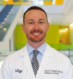 David R Raleigh合作论文数54P Sneed合作论文数52
 Minesh Mehta合作论文数Department of Radiation Oncology, Herbert Wertheim College of Medicine, Florida International University;Miami Cancer Institute52
 Susan M. Chang合作论文数Weill Institute for Neurosciences, University of California, San Francisco;School of Medicine, University of California, San Francisco50
 Michael D. Prados合作论文数Weill Institute for Neurosciences, University of California, San Francisco;Helen Diller Family Comprehensive Cancer Center, University of California, San Francisco;Department of Neurological Surgery, School of Medicine, University of California, San Francisco48Rupesh Kotecha合作论文数48
 Manmeet Singh Ahluwalia合作论文数Miami Cancer Institute, Baptist Health South Florida;Miami Neuroscience Institute, Baptist Health South Florida47
 David Larson合作论文数School of Medicine, University of California, San Francisco46
 Arie Perry合作论文数Division of Neuropathology, Departments of Pathology and Laboratory Medicine, School of Medicine, University of California, San Francisco37Jean L. Nakamura合作论文数Radiation Oncology, University of California36
 Javier Villanueva-Meyer合作论文数34
 Ranjini Tolakanahalli合作论文数Departments of Human Oncology and Medical Physics, University of Wisconsin Comprehensive Cancer Center32
 Lijun Ma合作论文数Department of Radiation Oncology, University of California San Francisco29Nancy Ann Oberheim Bush合作论文数28Vasudevan Harish N合作论文数Department of Developmental and Regenerative Biology, Icahn School of Medicine at Mount Sinai27Nicholas Butowski合作论文数26D. Wieczorek合作论文数25Philip H. Gutin合作论文数Mem Sloan Kettering Canc Ctr25
 Kathleen Lamborn合作论文数Department of Neurological Surgery, Weill Institute for Neurosciences, University of California, San Francisco;School of Medicine, University of California, San Francisco25A GutiéRrez合作论文数25Shannon Fogh合作论文数25
 Yazmin Odia合作论文数24
 Tugce Kutuk合作论文数Department of Radiation Oncology, Miami Cancer Institute23Joanna Phillips合作论文数Department of Neurological Surgery, Helen Diller Research Center, University of California San Francisco, San Francisco, CA, USA23
 Joseph F. Costello合作论文数Helen Diller Family Comprehensive Cancer Center, University of California, San Francisco;Department of Neurological Surgery, University of California, San Francisco23
 Edward Chang (张复伦)合作论文数Weill Institute for Neurosciences, University of California, San Francisco;Department of Neurological Surgery, School of Medicine, University of California, San Francisco22
 William (Chien-Wen) Chen合作论文数Departments of Radiation Oncology and Neurological Surgery, University of California San Francisco21Morshed Ramin A合作论文数The Brain Tumor Center, The University of Chicago Pritzker School of Medicine21
 Soonmee Cha合作论文数Department of Radiology, University of California, San Francisco;School of Medicine, University of California, San Francisco21
 Franco DeMonte合作论文数Department of Neurosurgery, Division of Surgery, The University of Texas MD Anderson Cancer Center21
 William M. Wara合作论文数Department of Radiation Oncology, University of California, San Francisco;Kaiser Permanente21Igor J. Barani合作论文数21
 Shawn Hervey-Jumper合作论文数School of Medicine, University of California, San Francisco;Weill Institute for Neurosciences, University of California, San Francisco;Medical School, University of Michigan21Martin C Tom合作论文数19
 Ahmad Ozair合作论文数19
 Michael E. Sughrue合作论文数Department of Neurological Surgery, Columbia University19
 Tihan Tarik合作论文数Department of Pathology, School of Medicine, The University of California, San Francisco18Hall Matthew D合作论文数Department of Radiation Oncology, City of Hope National Medical Cancer18Arjun Sahgal合作论文数18
 William Dillon合作论文数Department of Radiology and Biomedical Imaging, University of California, San Francisco17
 Michael T. Lawton合作论文数Department of Neurosurgery, Barrow Neurological Institute17R Kotecha合作论文数16




