Laser interstitial thermal therapy (LITT) is a local surgical treatment for brain metastases (BM). Here, we present and discuss the main radiologic findings and challenges related to LITT-treated BM. Prior to LITT, imaging features are useful for prognostic stratification and patient selection, as well as treatment planning. During the LITT session, intra-procedural imaging is acquired immediately at the beginning of the procedure for tumor delineation, during the ablation to monitor treatment using MR thermometry, and immediately post-LITT to evaluate the ablated region. Images obtained during the LITT procedure are useful to estimate extent of ablation, either by analyzing the real-time thermometry data or by comparing pre- and post-ablation images. In the months following LITT, follow-up scans exhibit characteristic imaging patterns for responding versus progressing lesions, which should be accounted for during post-LITT surveillance. In light of these patterns, and considering the strategies adopted by previous LITT studies, we propose dedicated criteria for local radiologic response and local radiologic progression in clinical trials, by adapting the RANO-BM criteria for local therapies to a "dual baseline" framework dedicated to LITT treated BM. Finally, we provide suggestions for the adoption of standardized post-LITT imaging protocols based on the current consensus.
Focused ultrasound (FUS) is an emerging therapeutic and diagnostic technology in neuro-oncology, offering new strategies for molecular diagnosis, drug delivery and tumor ablation across a range of brain tumors, including glioblastoma (GBM), brain metastases, and diffuse intrinsic pontine glioma (DIPG). The prognosis for aggressive brain tumors remains poor, despite advances in surgery, radiation, and chemotherapy. A considerable challenge is the limited ability to deliver therapeutics across the blood-brain barrier (BBB), particularly to infiltrative or non-enhancing tumor regions. FUS introduces an incisionless approach to the molecular subtyping of brain tumors, enhancing therapeutic delivery, and offers novel therapeutic approaches such as sonodynamic therapy (SDT). This review summarizes the FUS mechanisms and highlights the critical role of imaging modalities confirming target engagement, assessing bioeffects and outcomes, and ensuring safety. We also explore future directions, including the integration of liquid biopsy, artificial intelligence, and outpatient-ready FUS platforms, which will position FUS as a promising adjunct to standard neuro-oncologic care.
A major obstacle to identifying effective therapies for the aggressive brain tumor glioblastoma is the lack of human-specific, immunocompetent models that reflect the human tumor microenvironment. To address this, we developed the immune-human organoid tumor transplantation (iHOTT) model, an autologous co-culture platform that integrates patient-derived tumor cells and matched peripheral blood mononuclear cells within human cortical organoids to enable the study of patient-specific immune responses and tumor-immune interactions. This platform preserves tumor and immune populations, immune signaling, and cell-cell interactions observed in patient tumors. Treatment of iHOTT with pembrolizumab, a checkpoint inhibitor, mirrors cell-type shifts and cell-cell interactions observed in patients. T cell receptor (TCR) sequencing further reveals pembrolizumab-driven expansion of stem-like CD4 T cell clonotypes exhibiting patient-specific repertoires. These findings establish iHOTT as a physiologically relevant platform for exploring autologous tumor-immune interactions and underscore the need for antigen-targeted strategies to enhance immunotherapy in glioblastoma.
Background:Multicentric glioma (MCG) are defined radiographically as two or more tumor foci with separation of MRI T2-FLAIR (T2-weighted Fluid-attenuated Inversion Recovery) hyperintensities presenting synchronously (sMCG) and/or metachronously (mMCG). Previous studies have not stratified isocitrate dehydrogenase (IDH) wild-type and mutant gliomas by sMCG and mMCG. In this large, single-institution cohort stratified by IDH status, we evaluated MCG prevalence, prognostic implications, time to mMCG (TtM), location, and pathological concordance. Methods:We identified 836 IDH wild-type and 531 IDH mutant diffuse glioma patients treated at our institution with evaluable MRI. We inspected MRIs at presentation for sMCG and subsequent imaging for mMCG, reviewed radiology and pathology reports, and performed overall survival (OS) and TtM analyses. Results:MCG prevalence was higher in IDH wild-type cases (18%) than in IDH mutant cases (9%) (P < .0001). We found 20 sMCG and 26 mMCG in IDH mutant and 91 sMCG and 54 mMCG in IDH wild-type patients. In 7 wild-type and 1 mutant instances, mMCG arose from sMCG (smMCG). While sMCG and mMCG were associated with lower OS in IDH wild-type cases (sMCG: HR = 1.46, P = .003; mMCG: HR = 1.44, P = .02), only mMCG was associated with lower OS in IDH mutant cases (sMCG: HR = 1.22, P = .7; mMCG: HR = 2.64, P = .001). IDH wild-type cases had shorter TtM than mutant cases (P < .0001). Among double-biopsied MCG, pathology discordance occurred in 5/7 mutant but 0/28 wild-type cases. Conclusion:Results from this cohort of diffuse gliomas stratified by IDH status showed differences between IDH wild-type and mutant cohorts in prevalence, prognosis, TtM, distribution, and pathological concordance.
e14094 Background: Malignant transformation (MT) of IDH mutant gliomas is a poorly characterized process by which a lower grade glioma evolves into a higher grade glioma. We stratified patients based on histology and treatment to examine the prevalence of MT in IDH -mutated patients, the prognostic implications of early and late MT, and the differences between spontaneous, treatment associated, and IDH mutant inhibitor associated MT. Methods: We identified 935 IDH mutant patients seen at UCLA from 1998 to 2025 who provided IRB consent. pMT was defined by a second resection with pathology indicating a worsened grade (2 to 3, 2 to 4, 3 to 4). To increase sample size, new contrast enhancement (CEnew) on MRI scans indicative of true progression (TP) was considered iMT. CEnew identified as pseudoprogression (PsP) was not included as MT. Patients were then stratified by pre-MT treatment (none/spontaneous, radiation and/or chemotherapy, or IDH mutant inhibitor only). We defined early MT as shorter than one standard deviation < median ttMT, and late MT as longer than one standard deviation < median ttMT. Time to MT (ttMT), overall survival (OS), and residual survival (censored patients excluded) were analyzed by Kaplan-Meier and Cox multivariate analyses. Results: In our cohort, 328/935 had MT (pMT + iMT): 88 spontaneous, 234 post chemotherapy and/or radiation, and 6 post IDH mutant inhibitor. 111 were early transformers and 26 were late transformers. Early transformers had shorter median ttMT (early=2.841, late=28.66 years, p<0.0001) as expected, and shorter median OS (early=9.784, late=undefined years, p<0.0001). There was no difference in residual survival after MT. Grade 2 astrocytomas had a median time to transformation of 8.55 years while grade 2 oligodendrogliomas had a median time to MT of 16.42 years. There was no difference between time to iMT versus pMT. Older age predicted shorter ttMT (HR 1.022, p=0.0003) and worse OS (HR 1.026, p<0.0001) while higher KPS predicted increased OS (HR 0.983, p=0.0033). Using grade 3 astrocytoma as reference, lower grade tumors had significantly lower hazard ratios of ttMT (G2O HR 0.05438, p=0.0003; G2A HR 1.118, p=0.4740). Relative to RT/Chemo associated MT, spontaneous (HR 1.407, p=0.0131) and IDH mutant inhibitor MT (HR 2.705, p=0.0202) were associated with shorter ttMT. However, among MT patients only (censored patients excluded), only spontaneous MT was associated with a better OS (HR 0.521, p=0.0013). Residual survival was higher in G2Os (HR 0.4817, p=0.0018), but no differences were observed based on treatment received before MT. Conclusions: Treatment type, along with tumor grade and age at diagnosis, emerged as clinically relevant predictors of transformation risk and survival. Using this clinical cohort, future studies will focus on defining molecular features associated with MT.
Abstract Isocitrate dehydrogenase (IDH) mutant gliomas often transform from low to high grade aggressive tumors. The genetic and molecular drivers of this Malignant Transformation (MT) are poorly understood, and predicting whether a patient will undergo MT is an unmet challenge of high clinical relevance. To stratify patients by MT risk, we applied integrated spatial DNA and RNA profiling to biopsies from 18 retrospective glioma patients which will either remain stable, undergo MT or have already transformed. The resulting dataset consisted of >600,000 single cells, measuring 962 DNA loci, 1150 RNAs, and their spatial locations. Using this dataset, we found that genetic copy number alterations (CNAs), cell types compositions, and cellular neighborhoods did not predict MT. Instead, second order effects i.e. pairwise interactions, are highly predictive of future transformation. First, we identified abnormal chromosomal contact patterns that clearly separate future stable versus future MT samples. Second, we identified 24 ligand-receptors (LR) pairs cross-expressed in neighboring cells as the main molecular factors predictive of transformation and recurrences. We then validated a cross-expressing pair of ENPP2-LPAR1 interactions with a separate cohort of patient samples. In addition, using the LR+ cell pairs as an anchor, we identified cell signaling-specific gene expression programs that can predict from bulk or single cell RNAseq data the time to recurrence. We used a cell-interaction-based foundation model (CIFM) optimized on the spatial RNA data in forward simulations and identified potential myeloid signaling factors involved in MT. Lastly, we analyzed the effect of detection sensitivity on the ability to capture pertinent LR+ neighboring cells by down-sampling transcript and showed that the ability to detect cross-expressing signaling LR transcripts (typically <10 copies per cell) decays rapidly with lower sensitivity, but is more robust to down-sampling of the areas of the tissue imaged. The importance of second-order features suggests that increased depth and dimensionality of data on a smaller quantity of samples can provide valuable insight, and that high-sensitivity and multiple-modalities spatial approaches can help identify markers to risk-stratify patients, aid in therapeutic decision making, and uncover potential therapeutic targets.
We conducted a randomized surgical window-of-opportunity trial (NCT04606316) in recurrent, resectable glioblastoma. Between 2021 and 2024, 71 patients were screened, and 63 were randomized (intention-to-treat [ITT] population), and 58 received study treatment. Patients received pre-surgical immune checkpoint blockade (ICB) with dual anti-PD1 nivolumab + anti-CTLA4 ipilimumab (Arm 1), nivolumab alone (Arm 2), or placebo (Arm 3). Following surgery, Arms 1 and 3 received dual ICB, while Arm 2 continued nivolumab until progression or unacceptable toxicity. The primary endpoint, tumor-infiltrating lymphocyte (TIL) density, was met for Arm 1, as neoadjuvant dual ICB significantly increased TIL density compared with untreated control (Arm 3). As a secondary endpoint, median overall survival in the ITT population was 402 days (95% CI, 265–571) among patients who received dual ICB (Arms 1 and 3) and 273 days (95% CI, 166–506) for those assigned to nivolumab alone (Arm 2). No unanticipated toxicities were observed. Exploratory analyses showed that dual ICB elicited robust intratumoral and systemic immune activation, including increased interferon-related gene expression in blood. Higher TIL density and early systemic interferon-signature induction were associated with improved survival, whereas tumor mutational burden was not. Our results demonstrate pharmacodynamic activity of dual ICB in glioblastoma, with survival outcomes comparing favorably to similar studies. Here the authors report clinical and immunological outcomes of a randomized surgical window-of-opportunity trial designed to test whether dual immune checkpoint blockade targeting PD-1 and CTLA-4 could overcome the adaptive immune resistance previously reported with single-agent anti-PD-1 therapy in patients with surgically resectable recurrent glioblastoma.
e14031 Background: Bevacizumab (Bev), a monoclonal antibody that inhibits vascular endothelial growth factor, was fully FDA-approved in 2017 for use in recurrent glioblastoma and is often considered and utilized to treat all recurrent gliomas. Currently, there are limited studies on Bev use for recurrent isocitrate dehydrogenase 1/2 mutant ( IDH MUT ) gliomas, which can have distinct characteristics, disease outcome, and treatments compared to IDH wild-type ( IDH WT ) glioblastoma. We retrospectively examined Bev usage and efficacy at recurrence in a large single institution IDH MUT glioma cohort. Methods: We identified 138 consecutive IDH MUT glioma patients treated with Bev at recurrence at UCLA between 2005 and 2024. We stratified patients by 2016 WHO tumor diagnosis criteria. We examined how the timing of Bev initiation and other clinical variables affect Bev PFS and Bev OS. Bev PFS as determined by the treating clinician will be compared with our application prototype, Automated Imaging Response Evaluation System (AIRES), which is adapted to utilize RANO criteria. In addition, AIRES will be used to determine treatment response to be correlated with Bev PFS and Bev OS. Results: Time to Bev, which was defined as the time from initial surgery to Bev initiation, was shortest for AA and Grade 4 astrocytoma (G4 Astro). The median Bev PFS and Bev OS were 3.9 and 10.5 months, respectively. Bev PFS were 5.2, 4.6, and 3.3 months for the 1 st , 2 nd , and ≥3 rd recurrence groups, and Bev OS were 15.7, 13.5, and 8.3 months. Median Bev PFS, as determined by the treating clinician, for LO, LA, AO, AA, and G4 Astro were 2.8, 3.7, 6.2, 3.9, and 3.6 months, respectively. The median Bev OS were 7.0, 7.8, 9.5, 12.6, and 10.5 months. Among the 138 patients, the vast majority of patients initiated Bev treatment due to have contrast-enhancing tumor. 98 (71%) were first treated with Bev as a combined therapy, while 40 (29%) were treated with Bev monotherapy. 11 patients were concurrently treated with IDH inhibitors and Bev. The average daily corticosteroid (dexamethasone) dose reduction after 2 months of Bev treatment was 2.6 mg. Current results and Bev PFS are based on clinical criteria, while AIRES determined Bev PFS and response analysis are underway. Conclusions: Unlike our prior study on predominantly IDH WT GBM, later initiation of Bev was associated with diminished efficacy. Amongst the various diagnoses at Bev initiation, we found similar Bev PFS and Bev OS, except unexpectedly, we found AO to have improved Bev PFS and Bev OS compared to LO. Comparison with AIRES to determine Bev PFS and response according to RANO criteria is ongoing to finetune and validate results. Corticosteroid dose reduction was common but not associated with Bev PFS and Bev OS. With the recent increased use of IDH inhibitor treatment, this cohort, as a mostly IDH inhibitor naïve group, may prove valuable as a comparator arm to determine whether IDH inhibitor treatment affects Bev efficacy.
BACKGROUND AND PURPOSE:Normalized relative cerebral blood volume (nrCBV) and percentage of signal recovery (PSR) computed from dynamic susceptibility contrast (DSC) perfusion imaging are useful biomarkers for differential diagnosis and treatment response assessment in brain tumors. However, their measurements are dependent on DSC acquisition factors, and CBV-optimized protocols technically differ from PSR-optimized protocols. This study aimed to generate "synthetic" DSC data with adjustable synthetic acquisition parameters using dual-echo gradient-echo (GE) DSC datasets extracted from dynamic spin-and-gradient-echo echoplanar imaging (dynamic SAGE-EPI). Synthetic DSC was aimed at: 1) simultaneously create nrCBV and PSR maps using optimal sequence parameters, 2) compare DSC datasets with heterogeneous external cohorts, and 3) assess the impact of acquisition factors on DSC metrics. MATERIALS AND METHODS:Thirty-eight patients with contrast-enhancing brain tumors were prospectively imaged with dynamic SAGE-EPI during a non-preloaded single-dose contrast injection and included in this cross-sectional study. Multiple synthetic DSC curves with desired pulse sequence parameters were generated using the Bloch equations applied to the dual-echo GE data extracted from dynamic SAGE-EPI datasets, with or without optional preload simulation. RESULTS:Dynamic SAGE-EPI allowed for simultaneous generation of CBV-optimized and PSR-optimized DSC datasets with a single contrast injection, while PSR computation from guideline-compliant CBV-optimized protocols resulted in rank variations within the cohort (Spearman's ρ = 0.83-0.89, i.e. 31%-21% rank variation). Treatment-naïve glioblastoma exhibited lower parameter-matched PSR compared to the external cohorts of treatment-naïve primary CNS lymphomas (PCNSL) (p<0.0001), supporting a role of synthetic DSC for multicenter comparisons. Acquisition factors highly impacted PSR, and nrCBV without leakage correction also showed parameter-dependence, although less pronounced. However, this dependence was remarkably mitigated by post-hoc leakage correction. CONCLUSIONS:Dynamic SAGE-EPI allows for simultaneous generation of CBV-optimized and PSR-optimized DSC data with one acquisition and a single contrast injection, facilitating the use of a single perfusion protocol for all DSC applications. This approach may also be useful for comparisons of perfusion metrics across heterogeneous multicenter datasets, as it facilitates post-hoc harmonization.
Sodium neuroimaging provides unique insights into the cellular and metabolic properties of brain tumors. However, at 3T, sodium neuroimaging MRI’s low signal-to-noise ratio (SNR) and resolution discourages routine clinical use. We evaluated the recently developed Anatomically constrained GAN using physics-based synthetic MRI artifacts” (ATHENA) for high-resolution sodium neuroimaging of brain tumors at 3T. We hypothesized the model would improve the image quality while preserving the inherent sodium information. 4,573 proton MRI scans from 1,390 suspected brain tumor patients were used for training. Sodium and proton MRI datasets from Twenty glioma patients were collected for validation. Twenty-four image-guided biopsies from seven patients were available for sodium-proton exchanger (NHE1) expression evaluation on immunohistochemistry. High-resolution synthetic sodium images were generated using the ATHENA model, then compared to native sodium MRI and NHE1 protein expression from image-guided biopsy samples. The ATHENA produced synthetic-sodium MR with significantly improved SNR (native SNR 18.20 ± 7.04; synthetic SNR 23.83 ± 9.33, P = 0.0079). The synthetic-sodium values were consistent with the native measurements (P = 0.2058), with a strong linear correlation within contrast-enhancing areas of the tumor (R2 = 0.7565, P = 0.0005), T2-hyperintense (R2 = 0.7325, P < 0.0001), and necrotic areas (R2 = 0.7678, P < 0.0001). The synthetic-sodium MR and the relative NHE1 expression from image-guided biopsies were better correlated for the synthetic (ρ = 0.3269, P < 0.0001) than the native (ρ = 0.1732, P = 0.0276) with higher sodium signal in samples expressing elevated NHE1 (P < 0.0001). ATHENA generates high-resolution synthetic-sodium MRI at 3T, enabling clinically attainable multinuclear imaging for brain tumors that retain the inherent information from the native sodium. The resulting synthetic sodium significantly correlates with tissue expression, potentially supporting its utility as a non-invasive marker of underlying sodium homeostasis in brain tumors.
TPS2094 Background: Liquid biopsy in glioblastoma (GBM) is hindered by a lack of requisite circulating tumor (ct) and cell-free (cf) DNA levels in blood due to the blood-brain barrier (BBB). This limits the identification of blood-based tumor biomarkers along with the development and use of biomarker-driven systemic therapies. Low intensity focused ultrasound combined with intravenously administered microbubble oscillators (MB-FUS), leads to non-invasive BBB opening. This trial aims to evaluate the utility of LIFU for bolstering blood ctDNA and cfDNA for enhance liquid biopsy in patients with GBM. Methods: LIBERATE is an ongoing, prospective, multi-center, self-controlled, pivotal trial evaluating safety and technical efficacy of transcranial MR-guided MB-FUS for increasing blood ctDNA and cfDNA levels in adults, aged 18-80 years with GBM. Patients with suspected GBM planned for tumor biopsy or resection at 17 centers in US and Canada are being enrolled. Patients with multifocal tumors or tumors arising from deep midline, thalamus, cerebellum, or brainstem are excluded. Patients are administered IV microbubbles for enhanced sonication, after which MR-guided BBB opening using a 220 kHz device, with 1024-element phased array transducer, is performed with real-time acoustic feedback control for effective cavitation. Pre- and post-procedure, phlebotomy and MRI brain are done. Patients are offered optional 2 nd procedure during adjuvant chemotherapy phase if willing. Primary efficacy endpoint is correlation between biomarker patterns in tumor tissue collected during surgery/biopsy and blood collected following MB-FUS procedure. Confirmatory secondary efficacy endpoint is ratio between greatest yield of cfDNA in blood post-MB-FUS compared to cfDNA level in blood pre-MB-FUS. The primary study hypothesis is that agreement rate on biomarker pattern between resected/biopsied tumor tissue and blood is > 70%. The secondary hypothesis is that MB-FUS BBBO leads to a ≥2-fold rise in blood cfDNA. Assuming the true agreement rate expected is 89%, a sample of N = 50 patients will provide 90% power to meet the primary endpoint (Exact test, Binomial Proportion, one-sided Alpha = 0.025). Exploratory endpoints include (1) sensitivity of detection of known specific somatic mutations in ctDNA from blood samples collected before and after MB-FUS, (2) estimation of ctDNA levels in samples collected at 30-minutes, 1-hour, 2-hour, and 3-hour post-MB-FUS to determine time of greatest yield, (3) correlation of MRI parameters related to grading of BBB opening and ctDNA-based biomarkers from post-MB-FUS blood samples, (4) biomarker correlation between plasma cfDNA sampled during adjuvant chemotherapy phase and tumor tissue harvested at surgery. Patient enrollment commenced in 2022 and is ongoing (NCT05383872). Clinical trial information: NCT05383872 .
BACKGOUND AND PURPOSE: This study utilizes a physics-based approach to synthesize realistic MR artifacts and train a deep learning generative adversarial network (GAN) for use in artifact reduction on EPI, a crucial neuroimaging sequence with high acceleration that is notoriously susceptible to artifacts. MATERIALS AND METHODS: A total of 4,573 anatomical MR sequences from 1,392 patients undergoing clinically indicated MRI of the brain were used to create a synthetic data set using physics-based, simulated artifacts commonly found in EPI. By using multiple MRI contrasts, we hypothesized the GAN would learn to correct common artifacts while preserving the inherent contrast information, even for contrasts the network has not been trained on. A modified Pix2PixGAN architecture with an Attention-R2UNet generator was used for the model. Three training strategies were employed: (1) An ?all-in-one? model trained on all the artifacts at once; (2) a set of ?single models?, one for each artifact; and a (3) ?stacked transfer learning? approach where a model is first trained on one artifact set, then this learning is transferred to a new model and the process is repeated for the next artifact set. Lastly, the ?Stacked Transfer Learning? model was tested on ADC maps from single-shot diffusion MRI data in N = 49 patients diagnosed with recurrent glioblastoma to compare visual quality and lesion measurements between the natively acquired images and AI-corrected images. RESULTS: The ?stacked transfer learning? approach had superior artifact reduction performance compared to the other approaches as measured by Mean Squared Error (MSE = 0.0016), Structural Similarity Index (SSIM = 0.92), multiscale SSIM (MS-SSIM = 0.92), peak signal-to-noise ratio (PSNR = 28.10), and Hausdorff distance (HAUS = 4.08mm), suggesting that leveraging pre-trained knowledge and sequentially training on each artifact is the best approach this application. In recurrent glioblastoma, significantly higher visual quality was observed in model predicted images compared to native images, while quantitative measurements within the tumor regions remained consistent with non-corrected images. CONCLUSIONS: The current study demonstrates the feasibility of using a physics-based method for synthesizing a large data set of images with realistic artifacts and the effectiveness of utilizing this synthetic data set in a ?stacked transfer learning? approach to training a GAN for reduction of EPI-based artifacts.
Introduction Malignant meningiomas lack effective immunotherapeutic options. NY-ESO-1 is a potential immunotherapeutic target, because it is the most frequently expressed cancer-testis-antigen in meningiomas. We investigated the efficacy of T-Cell-Receptor-Transduced T-Cells (TCR-T) targeting NY-ESO-1 in vitro and in vivo in meningiomas. Methods Immunohistochemistry was performed on Grade I-III meningioma specimens. Primary meningioma culture LB3750(Grade I) and immortalized cultures SF1335(Grade I) and CH157-HLA-A2.1(Grade III) were established and maintained in vitro. NY-ESO-1 TCR-T (HLA-A2.1 restricted) cells were co-cultured with primary and immortalized meningioma cells and assessed for real-time tumor killing in vitro. Immunodeficient NSG mice were intracranially implanted with CH157-HLA-A2.1 and SF1335 cells, treated with systemic adoptive cell transfer (ACT) of TCR-T, and assessed for overall survival in vivo. Results NY-ESO-1 expression correlated with tumor grade (n = 35; p < 0.01). High NY-ESO-1 nuclear expression predicted a worse progression-free-survival (p = 0.0167). CH157-HLA-A2.1 cells, with native high NY-ESO-1 expression, experienced > 60% and then nearly 100% cytolysis after co-culture with TCR-T for 10 and 24 h, respectively, compared with control T-cells (p < 0.0001). SF1335 and LB3750 cells, with low NY-ESO-1 expression, experienced 20% cytolysis after 24 h of co-culture with TCR-T compared to the control (p < 0.0001). Systemic ACT of TCR-T significantly increased the median overall survival in NSG mice bearing intracranial xenografts of CH157-HLA-A2.1 by 49% (p < 0.001). Conclusions NY-ESO-1 TCR-T induces cytolysis in meningiomas in vitro, and its efficacy correlates with NY-ESO-1 expression. Systemic ACT results in significantly increased survival in vivo in high-grade meningioma. Therefore, targeting NY-ESO-1 may be a clinically feasible immunotherapeutic strategy for treating high-grade meningiomas.
DAC immunesensitization promotes specific, robust, and polyfunctional NY-ESO-1 TCR CD8+ T-cell responses in primary GBM cell lines. A, Representative Bis-Seq-TA cloning lollipop diagram and (B) percent of methylated CpGs to total CpGs of first eight sites of analyzed CpG island in primary GS277 cells treated with DMSO or 0.5 µmol/L DAC for 12 days. C, Average fold change of mRNA expression of treated GS277 (n = 3; **, P < 0.01; two-tailed unpaired t test). D, Cytotoxic analysis of treated GS277 over a 40-hour period when co-incubated with NY-ESO-1 TCR-transduced effector T cells at E:T ratio of 1:1 and (E) at multiple E:T ratios (1:5, 1:2, 1:1, 2:1) at 20-hour (n = 3; *, P < 0.05; **, P < 0.01; ***, P < 0.001; ****, P < 0.0001; multiple unpaired t tests). F, Uniform manifold approximation projection (UMAP) of CD8+ T cells transduced with NY-ESO-1 TCR CD8 or untransduced control CD8 (CTR) from four stimulated groups: NY-ESO-1 CD8+ alone (n = 1665 CD8+ cells), NY-ESO-1 CD8+ stimulated with DBTRG-05 (n = 1902 CD8+ cells), NY-ESO-1 CD8+ stimulated with DBTRG-05 + DAC (n = 284 CD8+ cells), CTR CD8 stimulated with DBTRG-05 + DAC (n = 807 CD8+ cells). G and H, PSI defined as the percentage of polyfunctional cells, multiplied by MFI of the proteins secreted by the polyfunctional cells for stimulated CD8+ at the single-cell resolution across DBTRG-05 co-cultures.
DAC reactivates hERV expression in gliomaspheres. A, FPKM of ERV3 and HERVR family for primary gliomasphere cell lines (n = 7; *, P < 0.05; **, P < 0.01; two-tailed unpaired t test). B, Average fold change of mRNA expression of treated GS277 for ERV3 and ERVW1/Syncytin-1 as measured by qPCR (n = 3; *, P < 0.05; ***, P < 0.001; two-tailed unpaired t test). C, Heatmap and hierarchical clustering of hERV gene families. Scale represents z-score of hERV family FPKM normalized to housekeeping FPKM.
Our previous study of neoadjuvant PD-1 immune checkpoint blockade (ICB) in surgically-accessible recurrent glioblastoma suggested that single-agent PD-1 blockade induced adaptive immune responses that may have paradoxically inhibited functional anti-tumor immune responsiveness. To address this, we performed a surgical-window-of-opportunity trial in resectable, relapsed glioblastoma patients (n=60). Patients received either dual ICB (Arm 1, n=25) with ipilimumab (3 mg/kg) and nivolumab (240 mg), single-agent PD-1 ICB (Arm 2, n=25) with nivolumab (240 mg/kg), or a placebo infusion (Arm 3, n=10) prior to surgical resection. Following surgery, Arms 1 and 3 received dual ICB, while Arm 2 continued on nivolumab until progression or unacceptable toxicity. We collected blood and tumor tissue from all patients and performed RNA and TCR sequencing, along with multiplexed immunofluorescence, to evaluate intratumoral and systemic immune response. Dual ICB induced greater T-cell/interferon-related gene expressions in tumor compared with PD-1 ICB or a placebo infusion. It also resulted in the highest TIL density, as assessed by TCR sequencing. In a multivariate analysis, TIL density strongly correlated with survival outcomes. In PBMCs, dual ICB induced robust interferon gene signatures, and early upregulation of these signatures immediately following treatment was associated with longer survival. However, alongside interferon signaling, monocyte-related signatures also significantly increased following dual ICB, and their persistently elevated levels during adjuvant treatment cycles were linked to poorer outcomes. The median overall survival of patients who received dual ipilimumab+nivolumab was 405 days (95% CI, 265 to NA), while that of single agent nivolumab was 275 days (95% CI, 166 to NA). Our results demonstrate a clear pharmacodynamic effect of dual ICB in glioblastoma and the survival times compare favorably with similar clinical studies done in the same population. Moreover, our transcriptional analysis underscores the importance of balancing immune activation and chronic inflammation in shaping ICB responsiveness.
 Linda Liau合作论文数Jonsson Comprehensive Cancer Center, University of California, Los Angeles;Department of Neurosurgery, University of California, Los Angeles109
 Robert M. Prins合作论文数Department of Neurosurgery, David Geffen School of Medicine, University of California, Los Angeles;Brain Research Institute, University of California, Los Angeles105
 Timothy Cloughesy合作论文数Department of Molecular and Medical Pharmacology, David Geffen School of Medicine, University of California, Los Angeles;Brain Research Institute, University of California, Los Angeles65
 Benjamin Ellingson合作论文数University of California Los Angeles52
 David Nathanson合作论文数Molecular Biology Institute, University of California, Los Angeles43
 William H. Yong合作论文数School of Medicine, University of California, Irvine37
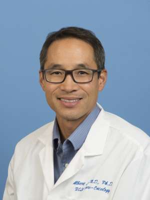 Albert Lai合作论文数Department of Neurology, David Geffen School of Medicine, University of California, Los Angeles34
 Joey Orpilla合作论文数33
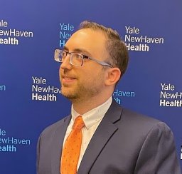 Joseph Antonios合作论文数Sch Med, Yale Univ32Jingwen Yao合作论文数School of Medicine, University of California, San Francisco30Janet Treger合作论文数29
 Noriko Salamon合作论文数Department of Radiology, UCLA UCLA29Catalina Raymond合作论文数28
 Phioanh Leia Nghiemphu合作论文数28Anthony C. Wang合作论文数Department of Neurosurgery, University of Michigan Health System24Horacio Soto合作论文数24
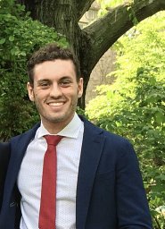 Francesco Sanvito合作论文数Neuroradiol Unit, IRCCS San Raffaele Sci Inst20
 Alex K. C. Lee合作论文数Dept Chem Biomol Engn, Hong Kong Univ Sci Technol20Kim Won合作论文数19
 Matthew Sun合作论文数Department of Neurological Surgery, University of California19
 Lu Sun合作论文数Harbin Univ Commerce, Harbin 150028, Heilongjiang, Peoples R China18
 Shaina Sedighim合作论文数Miller School of Medicine, University of Miami17
 Akifumi Hagiwara (萩原彰文)合作论文数Department of Radiology, School of Medicine, Juntendo University17
 Gang Li合作论文数Department of Biostatistics, Fielding School of Public Health, University of California, Los Angeles16
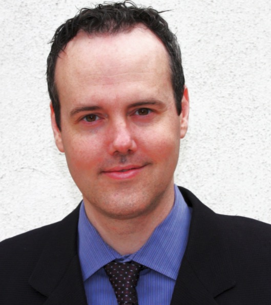 Whitney B. Pope合作论文数Department of Radiology, David Geffen School of Medicine, University of California16Tucker Alexander M合作论文数Department of Child Health, University of Bristol16
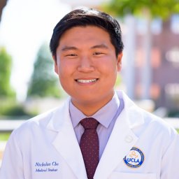 Nicholas S Cho合作论文数Med Scientist Training Program, David Geffen UCLA15Thomas Lai合作论文数14
 Kunal J. Patel合作论文数Departments of 1 Neurological Surgery.14
 Tom Davidson合作论文数13
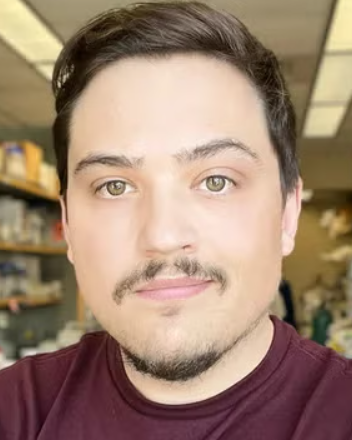 Terry J. Prins合作论文数Department of Neurology, David Geffen School of Medicine, UCLA13
 Aaron Mochizuki合作论文数Neurology & Neurological Sciences Department, Stanford University13Kunal S. Patel合作论文数12
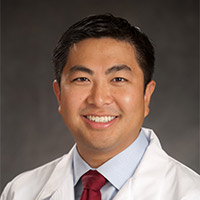 Isaac Yang合作论文数University of California12Melody Hsu合作论文数11Sun Lu O合作论文数11Li Tie合作论文数11
 Marco Giovannini合作论文数Ludwig Institute for Cancer Research and Department of Clinical Genetics, Karolinska Hospital;Molecular Genetics Laboratory, The Salk Institute;and Laboratory of Tumor Genetics, Institut Curie;The Salk Institute, Institut Curie11Namjo P Shin合作论文数10
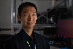 Willy Hugo合作论文数National University of Singapore10
 Jeremie Vitte合作论文数Department of Human Genetics, Hadassah University Hospital10Mildred Galvez合作论文数9
 Maya Harary合作论文数Department of Neurosurgery, University of California, Los Angeles9Moughon Diana合作论文数David Geffen School of Medicine, University of California9Sonoko Oshima合作论文数8Joshua Casaos合作论文数Department of Neurosurgery, Los Angeles (UCLA),8Chencai Wang合作论文数8Frances Chow合作论文数Department of Anesthesiology, Pharmacology & Therapeutics, Faculty of Medicine, The University of British Columbia8Odesa Sylvia K合作论文数8




