Do steady-state protein levels accurately predict mRNA levels? Based on the central dogma (DNA RNA protein) current protein levels are representative of mRNA present at an earlier time. However, most cellular mRNA protein comparative studies try to relate steady-state protein levels to current mRNA levels in cells. Protein steady-states are more correctly related to protein production, protein degradation and other complex cellular conditions. Using Structural Equation Modelling (SEM) we relate linear protein measurements to latent mRNA in E.coli. This method can be used to find the optimal protein measurements that explain underlying mRNA expression, and better understand the proteomic and transcriptomic relationship in E.coli gene expression.
The equilibrium unfolding transitions of Cro repressor variants, dimeric variant Cro F58W and monomer Cro K56[DGEVK]F58W, have been studied by urea and guanidine hydrochloride to probe the folding mechanism. The unfolding transitions of a dimeric variant are well described by a two state process involving native dimer and unfolded monomer with a free energy of unfolding, ΔG0,un0, of ∼10–11kcal/mol. The midpoint of transition curves is dependent on total protein concentration and ΔG0,un0 is independent of protein concentration, as expected for this model. Unfolding of Cro monomer is well described by the standard two state model. The stability of both forms of protein increases in the presence of salt but decreases with the decrease in pH. Because of the suggested importance of a N2⇔2F dimerization process in DNA binding, we have also studied the effect of sodium perchlorate, containing the chaotropic perchlorate anion, on the conformational transition of Cro dimer by CD, fluorescence and NMR (in addition to urea and guanidine hydrochloride) in an attempt both to characterize the thermodynamics of the process and to identify conditions that lead to an increase in the population of the folded monomers. Data suggest that sodium perchlorate stabilizes the protein at low concentration (<1.5M) and destabilizes the protein at higher perchlorate concentration with the formation of a “significantly folded” monomer. The tryptophan residue in the “significantly folded” monomer induced by perchlorate is more exposed to the solvent than in native dimer.
Dimers of Cro are required to recognize operator DNA and repress transcription, but dimerization is weak compared to DNA binding. Fluorophore-conjugated, single-cysteine variants of Cro have been used to investigate the equilibria and kinetics of dimer assembly. Equilibrium distributions of mixed dimers, monitored by fluorescence resonance energy transfer (FRET), confirm that labeled variants have equilibrium dimer dissociation constants in the micromolar concentration range. Subunit exchange experiments yield first order rate constants for dimer dissociation that range from 0.02 s(-1) to 0.04 s(-1). Association rate constants calculated from the ratios of dissociation equilibrium and rate constants range from 0.7x10(4) M(-1) s(-1) to 3x10(4) M(-1) s(-1), depending on the site of the fluorescent label. At nanomolar concentrations of subunits, assembly can be driven by addition of DNA. The bimolecular association rate constants measured under these conditions are not dramatically enhanced, ranging from 7x10(4) M(-1) s(-1) to 9x10(4) M(-1) s(-1). The association rate is second order in protein but independent of DNA concentration between 10 nM and 200 nM. The association of subunits under native conditions is more than four orders of magnitude slower than the fast assembly phase measured previously in refolding experiments, and is unaffected by peptidyl-prolyl isomerases. Stabilization of the folded structure of the protein by residue substitution in Cro F58W or reduced temperature increases the ratio of dimers to monomers and decreases the rate of subunit exchange. These data suggest that native monomers have compact structures with substantial barriers to unfolding and that unfolded or partially folded monomers are the preferred substrates for dimer assembly. Cro binding in vivo may be under kinetic rather than thermodynamic control. The slow assembly of Cro dimers demonstrated here provides a new perspective on the lysis/lysogeny switch of bacteriophage lambda.
The thermodynamic stabilities of three monomeric variants of the bacteriophage lambda Cro repressor that differ only in the sequence of two amino acids at the apex of an engineered beta-hairpin have been determined. The sequences of the turns are EVK-XX-EVK, where the two central residues are DG, GG, and GT, respectively. Standard-state unfolding free energies, determined from circular dichroism measurements as a function of urea concentration, range from 2.4 to 2.7 kcal/mole, while those determined from guanidine hydrochloride range from 2.8 to 3.3 kcal/mole for the three proteins. Thermal denaturation yields van't Hoff unfolding enthalpies of 36 to 40 kcal /mole at midpoint temperatures in the range of 53 to 58degreesC. Extrapolation of the thermal denaturation free energies with heat capacities of 400 to 600 cal/mole deg gives good agreement with the parameters determined in denaturant titrations. As predicted from statistical surveys of amino acid replacements in beta-hairpins, energetic barriers to transformation from a type I' turn (DG) to a type II' turn (GT) can be quite small.
Cro binds to operator sites in lambda DNA as a dimer. Dimerization of this small repressor protein is weak, however, and proline residues in the dimer interface suggest that folding and assembly of active repressors may be complex. Cro and selected variants have been studied by circular dichroism and fluorescence. Fluorescent probes include a unique tryptophan residue in the dimer interface and extrinsic resonance energy transfer probes that monitor dimerization. Both folding and unfolding are characterized by two distinct kinetic phases. Fast processes that are complete within the 5-10 ms dead time of stopped flow experiments account for the majority of the change in the CD signal and abrupt changes in both tryptophan fluorescence and energy transfer. The slow phases show all the hallmarks of proline isomerization. The rates of the slow phases are between 0.005 and 0.02 s(-1), are relatively independent of protein and denaturant concentration, display activation energies of 20 kcal/mol, and are accelerated by the peptidyl-prolyl isomerase SlyD. Although CD measurements indicate that more than 70% of the secondary structure is regained in the refolding burst phase, intermolecular fluorescence resonance energy transfer experiments indicate that less than 25% of these subunits are assembled into dimers. Full folding and dimerization requires isomerization of the non-native prolyl isomers over hundreds of seconds.
A robust selection system affording phage display of the DNA-binding helix-turn-helix protein Cro is presented. The aim of the work was to construct an experimental system allowing for the construction and isolation of Cro-derived protein with new DNA-binding properties. A derivative of the phage lambda Cro repressor, scCro8, in which the protein subunits had been covalently connected via a peptide linker was expressed in fusion with the gene 3 protein of Escherichia coli filamentous phage. The phage-displayed single-chain Cro was shown to retain the DNA binding properties of its wild-type Cro counterpart regarding DNA sequence specificity and binding affinity. A kinetic analysis revealed the rate constant of dissociation of the single-chain Cro-phage/DNA complex to be indistinguishable from that of the free single-chain Cro. Affinity selection using a biotinylated DNA with a target consensus operator sequence allowed for a 3000-fold enrichment of phages displaying single-chain Cro over control phages. The selection was based on entrapment of phage/DNA complexes formed in solution on streptavidin-coated paramagnetic beads. The expression system was subsequently used to isolate variant scCro8 proteins, mutated in their DNA-binding residues, that specifically recognized new, unnatural target DNA ligands.
It was previously shown that the Cro repressor from phage lambda, which is a dimer, can be converted into a stable monomer by a five-amino acid insertion. Phe58 is the key residue involved in this transition, switching from interactions which stabilize the dimer to those which stabilize the monomer. Structural studies, however, suggested that Phe58 did not penetrate into the core of the monomer as well as it did into the native dimer. This was strongly supported by the finding that certain core-repacking mutations, including in particular, Phe58-->Trp, increased the stability of the monomer. Unexpectedly, the same substitution also increased the stability of the native dimer. At the same time it decreased the affinity of the dimer for operator DNA. Here we describe the crystal structures of the Cro F58W mutant, both as the monomer and as the dimer. The F58W monomer crystallized in a form different from that of the original monomer. In contrast to that structure, which resembled the DNA-bound form of Cro, the F58W monomer is closer in structure to wild-type (i.e. non-bound) Cro. The F58W dimer also crystallizes in a form different from the native dimer but has a remarkably similar overall structure which tends to confirm the large changes in conformation of Cro on binding DNA. Introduction of Trp58 perturbs the position occupied by the side-chain of Arg38, a DNA-contact residue, providing a structural explanation for the reduction in DNA-binding affinity. The improved thermal stability is seen to be due to the enhanced solvent transfer free energy of Trp58 relative to Phe58, supplemented in the dimer structure, although not the monomer, by a reduction in volume of internal cavities.
The solution structure of a monomeric variant of the lambda Cro repressor has been determined by multidimensional NMR. Cro K56[DGEVK] differs from wild‐type Cro by the insertion of five amino acids at the center of the dimer interface. 1 H and 15 N resonances for 70 of the 71 residues have been assigned. Thirty‐two structures were calculated by hybrid distance geometry/simulated annealing methods using 463 NOE‐distance restraints, 26 hydrogen‐bond, and 39 dihedral‐angle restraints. The root‐mean‐square deviation (RMSD) from the average structure for atoms in residues 3‐60 is 1.03 ± 0.44 Å for the peptide backbone and 1.6 ± 0.73 Å for all nonhydrogen atoms. The overall structure conforms very well to the original design. Although the five inserted residues form a beta hairpin as expected, this engineered turn as well as other turns in the structure are not well defined by the NMR data. Dynamics studies of backbone amides reveal T1/T2 ratios of residues in the α2‐α3, β2‐β3, and engineered turn that are reflective of chemical exchange or internal motion. The solution structure and dynamics are discussed in light of the conformational variation that has been observed in other Cro structures, and the importance of flexibility in DNA recognition.
The structure has been determined at 3.0 Å resolution of a complex of engineered monomeric Cro repressor with a seven‐base pair DNA fragment. Although the sequence of the DNA corresponds to the consensus half‐operator that is recognized by each subunit of the wild‐type Cro dimer, the complex that is formed in the crystals by the isolated monomer appears to correspond to a sequence‐independent mode of association. The overall orientation of the protein relative to the DNA is markedly different from that observed for Cro dimer bound to a consensus operator. The recognition helix is rotated 48° further out of the major groove, while the turn region of the helix‐turn‐helix remains in contact with the DNA backbone. All of the direct base‐specific interactions seen in the wild‐type Cro‐operator complex are lost. Virtually all of the ionic interactions with the DNA backbone, however, are maintained, as is the subset of contacts between the DNA backbone and a channel on the protein surface. Overall, 25% less surface area is buried at the protein‐DNA interface than for half of the wild‐type Cro‐operator complex, and the contacts are more ionic in character due to a reduction of hydrogen bonding and van der Waals interactions. Based on this crystal structure, model building was used to develop a possible model for the sequence‐nonspecific interaction of the wild‐type Cro dimer with DNA. In the sequence‐specific complex, the DNA is bent, the protein dimer undergoes a large hinge‐bending motion relative to the uncomplexed form, and the complex is twofold symmetric. In contrast, in the proposed nonspecific complex the DNA is straight, the protein retains a conformation similar to the apo form, and the complex lacks twofold symmetry. The model is consistent with thermodynamic, chemical, and mutagenic studies, and suggests that hinge bending of the Cro dimer may be critical in permitting the transition from the binding of protein at generic sites on the DNA to binding at high affinity operator sites.
The overall affinity of the bacteriophage lambda Cro repressor for its operator DNA site is limited by dimer dissociation at submicromolar concentrations. Since Cro dimer-operator complexes form at nanomolar concentrations of Cro subunits where free dimers are rare, these dimers must bind with compensating high affinities. Previous studies of the covalent dimer Cro V55C suggest little change in DNA binding affinity even though the dimeric species is quantitatively populated; this is an apparent contradiction to the expectation of high intrinsic dimer-DNA affinity. In contrast to the disulfide linkage at the center of the dimer interface in Cro V55C, polypeptide linkers that join the two subunits allow single-chain Cro repressors to bind operator DNA with picomolar affinities. A series of five single-chain Cro repressors have been expressed from fused tandem cro genes. Each contains a peptide linker of 8-16 hydrophilic residues that connects the C-terminus of one subunit to the N-terminus of the next. All bind to operator DNA with at least 100-fold higher affinity than Cro V55C. Proteins containing the longest and shortest linkers have been purified and characterized in detail. Both exhibit similar CD spectra to wild-type Cro and enhanced thermal stability. Sedimentation equilibrium experiments show that single-chain Cro repressors do not associate at concentrations up to 30 microM. The rate of dissociation of Cro-DNA complexes is almost unchanged by covalent linkage. Biophysical characterization of Cro variants such as these, where DNA binding is uncoupled from subunit assembly, is necessary for a quantitative understanding of the structural and energetic determinants of DNA recognition in this simple model system.
 Akmm Mollah合作论文数6
 Tony R. Hazbun合作论文数Departments of Genome Sciences and Medicine, University of Washington6Ronald A. Albright合作论文数5
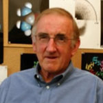 Brian Matthews合作论文数Department of Physics, College of Arts and Sciences, University of Oregon5
 Bob Sauer合作论文数Department of Biology, Massachusetts Institute of Technology4Mt Record合作论文数3
 M. Thomas Record Jr.合作论文数Department of Biochemistry, College of Agricultural and Life Sciences, University of Wisconsin-Madison3
 Gene L Bidwell合作论文数Department of Biochemistry, University of Mississippi Medical Center3Jana R合作论文数3W. John Satumba合作论文数3Ae Fong合作论文数2H Maity合作论文数2
 James U. Bowie合作论文数Exozymes;Department of Chemistry & Biochemistry , University of California, Los Angeles2Fields J D合作论文数2Rupert P B合作论文数2
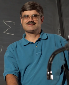 Maurice Eftink合作论文数Department of Chemistry and Biochemistry, College of Liberal Arts, The University of Mississippi2B Richey合作论文数2Ds Cayley合作论文数1Cf Anderson合作论文数1
 Pamela Mills合作论文数1RS Adelstein合作论文数1CM Manubay合作论文数1Gr Bellomy合作论文数1K L Knight合作论文数1S Cayley合作论文数1
 Jacqueline Harris合作论文数Sch Med, Johns Hopkins Univ1
 Wendell Lim合作论文数Department of Cellular & Molecular Pharmacology, University of California, San Francisco;Center for Systems & Synthetic Biology, University of California, San Francisco1Nilsson M T合作论文数1
 Jung-Hye Roe合作论文数School of Biological Sciences and Institute of Microbiology, Seoul National University1Haifeng Jia (贾海峰)合作论文数1Charles F. Anderson合作论文数1John F. Reidhaar-Olson合作论文数1Thomas Madej合作论文数1Christine W. Duarte合作论文数1James C. Hu合作论文数1
 Mikael Widersten合作论文数Biomedical Center, Uppsala University1
 Thomas C. Farrar合作论文数Department of Chemistry, University of Wisconsin-Madison1
 Richard Breyer合作论文数Department of Pharmacology, Vanderbilt University1
 Douglas J. Fishkind合作论文数Harvard Medical School, Department of Anatomy and Cellular Biology, Laboratory of Human Reproduction and Reproductive Biology, 45 Shattuck Street, Boston, Massachusetts 02130 USA1




