CRISPR-based high-throughput mutagenesis screens enable systematic mapping of mutations to phenotypes, yet deciphering mutation-phenotype links remains challenging. Here, we present ProTiler-Mut, a versatile computational framework that leverages tiling mutagenesis screens, which introduce variants across entire protein sequences, to analyze mutation effects at the levels of residues, substructures, and protein-protein interactions (PPIs). Applying ProTiler-Mut to multi-condition base-editing (BE) screens targeting DNA damage response proteins and T cell regulators, we define a separation-of-function (SoF) category beyond the conventional loss-of-function (LoF) and gain-of-function (GoF) classes, where SoF mutations show the strongest enrichment for ClinVar-annotated pathogenic variants. ProTiler-Mut also identifies candidate substructures that enable functional inference of unscreened pathogenic mutations and prioritizes candidate phenotype-associated PPIs potentially disrupted by functional variants. Using ProTiler-Mut, in cells with elevated programmed cell death 1 (PD-1) expression, we identify pathogenic GoF mutations that constitute a substructure that may disrupt mitogen-activated protein kinase (MAPK)1-RSK1 interactions and lead to MAPK activation. Finally, we show that ProTiler-Mut is applicable across different mutagenesis screening platforms. A record of this paper’s transparent peer review process is included in the supplemental information.
N 6-Methyladenosine (m6A) is a prevalent RNA modification that regulates multiple aspects of RNA metabolism, including RNA localization, stability, decay, and translation. m6A deposition is catalyzed by distinct methyltransferase complexes, including the METTL3/14 complex and METTL16, which recognize different RNA sequence motifs. The biological effects of m6A are mediated by effector proteins that selectively recognize m6A-modified RNA. To identify previously uncharacterized m6A-binding proteins, we developed an RNA-binding protein domain array to systematically screen for candidate m6A effectors. Using this approach, we identified the spliceosomal protein SF3B4 as a potential m6A reader. RNA pulldown assays using m6A-modified RNA probes demonstrated selective enrichment of endogenous SF3B4, supporting its ability to recognize m6A-modified RNA. To define the RNA targets of SF3B4, we performed SF3B4 RIP-seq alongside m6A RIP-seq, followed by RIP-qPCR validation of overlapping targets. Motif analysis revealed that SF3B4 preferentially associates with the conserved GRAGRA (R = A/G) motif, consistent with the RNA sequence recognized by the METTL16 methyltransferase. Notably, transcripts of the BCR and MET oncogenes were identified as shared targets of SF3B4 and METTL16. Together, these findings identify SF3B4 as a previously unrecognized m6A effector and suggest that it participates in RNA metabolic processes downstream of METTL16-mediated m6A modification.
Abstract Spindlin1 (SPIN1) is an epigenetic reader involved in oncology, whose third Tudor domain remains largely underexplored. Here, we applied a minimalist pruning strategy to the 26-mer peptide DOCpep3 to identify the core pharmacophore for SPIN1 Tudor 3 domain binding. This yielded truncated linear peptide analogues (e.g., 1, 3, and 4) displaying low nanomolar affinities, significantly outperforming the parent peptide. Orthogonal biophysical validation (MST and SPR) confirmed competitive target engagement. Furthermore, circular dichroism spectroscopy revealed that these pruned ligands induce distinct structural rearrangements in SPIN1, establishing high-affinity chemical probes for drug discovery.
Spatiotemporal regulation of Dicer is essential for small RNA biogenesis and fertility, yet how its helicase domain is controlled remains unclear. Using Caenorhabditis elegans, we identify a regulatory role for the arginine-rich GRARR motif within helicase domain motif VI of DCR-1. Mutating conserved arginines in this sequence disrupts maternal 26 G endo-siRNA production, impairs oocyte meiosis I and II, and reduces fertility. Biochemically, an asymmetrically dimethylated DCR-1 GRA[R495*]R peptide enhances interaction with ERI-5, a tandem-Tudor protein in the ERIC complex, while loss of DCR-1(R495) diminishes this interaction in vivo. Genetically, eri-5 deletion phenocopies the dcr-1 R495K mutant, supporting a functional partnership in 26 G siRNA biogenesis. Notably, these defects parallel those seen in DCR-1 phosphorylation mutants in the catalytic domain. AlphaFold modeling suggests that arginine methylation in the helicase domain and serine phosphorylation in catalytic domain may operate in a coordinated manner to modulate DCR-1 conformation, effector recruitment, and proper execution of the oocyte meiotic program.
Protein arginine methyltransferase 5 (PRMT5) is a synthetic lethal target in methylthioadenosine phosphorylase-deleted (MTAP-null) cancers. Second-generation methylthioadenosine (MTA)-cooperative PRMT5 inhibitors preferentially target MTAP-null cells while largely sparing MTAP-wildtype (MTAP-WT) cells, thereby improving tumor selectivity over first-generation PRMT5 inhibitors. Despite encouraging efficacy and safety signals in early clinical studies, the modest objective response rates (ORRs) observed with these inhibitors suggest that intrinsic or acquired resistance may limit their clinical benefit. Here, we investigated acquired resistance to the MTA-cooperative PRMT5 inhibitor BMS-986504/MRTX1719 in MTAP-null non-small cell lung cancer (NSCLC) cells and sought to identify therapeutic vulnerabilities that emerge upon resistance. Using multiple in vitro-derived resistant models, we found that acquired resistance was accompanied by cross-resistance to mechanistically distinct PRMT5 inhibitors. Notably, this phenotype was not fully explained by altered PRMT5 activity or changes in MTA levels. High-throughput drug screening of paired sensitive and resistant cells revealed increased sensitivity to MEK inhibitors following acquisition of MRTX1719 resistance in KRAS-wildtype NSCLC cells. Consistently, resistant cells exhibited rewired MAPK-related transcriptional programs. Together, these findings identify MEK inhibition as a reproducible collateral vulnerability associated with acquired MRTX1719 resistance in MTAP-null NSCLC models and support further evaluation of MEK inhibition as a potential treatment-switching strategy following resistance.
The inaugural FASEB conference Protein Arginine Methylation: Mechanism to Therapeutics was held in Tsukuba, Japan (January 5-8, 2026) and brought together investigators studying protein arginine methyltransferases (PRMTs) and its methylarginine product. Post-translational arginine methylation, found as monomethylarginine (MMA/Rme1), asymmetric dimethylarginine (ADMA/Rme2a), and symmetric dimethylarginine (SDMA/Rme2s), is increasingly recognized as a central regulator of RNA metabolism, chromatin function, and cellular stress responses. Presentations highlighted emerging directions for the field, including mechanisms linking PRMT activity to RNA processing and gene expression, roles in genome stability, identification of new methylarginine reader proteins, and connections between PRMT function and cellular metabolism. Work on immune and antiviral pathways and neuroscience further expanded the biological scope of PRMT activity. At the same time, significant advances in chemical probes, degraders, and selective inhibitors are enabling more precise interrogation of PRMT biology and accelerating efforts to translate these discoveries into therapeutic strategies.
High-throughput mutagenesis screens are powerful tools for mapping mutations to phenotypes. However, deciphering the molecular mechanisms that link mutations to phenotypic outcomes remains a significant challenge. Here, we present ProTiler-Mut, a versatile computational framework that harnesses tiling mutagenesis screens, which introduce variants across entire protein sequences, to facilitate investigation of mutation-to-phenotype associations at multiple levels, including individual residues, protein substructures, and protein-protein interactions (PPIs). As demonstrated through our analyses of base editing (BE) screens targeting DNA Damage Response (DDR) proteins and T cell regulators, ProTiler-Mut provides novel insights into the mutation-phenotype linkages, including: i) refined classification of mutation that reveals separation-of-function (SOF) category beyond the conventional binary classification of loss-of-function (LOF) and gain-of-function (GOF); ii) definition of phenotype-associated hotspot substructures that enable the inference of the function of unscreened pathogenic mutations; and iii) identification of phenotype-associated PPIs disrupted by functional mutations. Through ProTiler-Mut analyses, we identified a substructure harboring pathogenic GOF mutations that disrupt interactions between the kinases MAPK1 and RSK1, leading to MAPK1 activation and elevated expression of the immune checkpoint receptor PD-1. Furthermore, we demonstrate the applicability of ProTiler-Mut to various mutagenesis screening platforms, highlighting its broad utility and generalizability. ### Competing Interest Statement The authors have declared no competing interest. NIH, , R35GM137927, R35GM153387, R01CA197774 John S. Dunn Foundation, https://ror.org/03yhprz62,
Tubulin is crucial in several cellular processes, including intracellular organization, organelle transport, motility, and chromosome segregation. Intracellular tubulin concentration is tightly regulated by an autoregulation mechanism, in which excess free tubulin promotes tubulin mRNA degradation. However, the details of how changes in free tubulin levels initiate this autoregulation remain unclear. In this study, we identified coactivator-associated arginine methyltransferase 1 (CARM1)-phosphatidylinositol 3-kinase class 2α (PI3KC2α) axis as a novel regulator of tubulin autoregulation. CARM1 stabilizes PI3KC2α by methylating its R175 residue. Once PI3KC2α is not methylated, it becomes unstable, leading to decreased cellular levels. Loss of PI3KC2α results in the release of tetratricopeptide repeat domain 5 (TTC5), which initiates tubulin autoregulation. Thus, PI3KC2α, along with its CARM1-mediated arginine methylation, regulates the initiation of tubulin autoregulation. Additionally, disruption of the CARM1-PI3KC2α axis decreases intracellular tubulin levels, leading to a synergistic increase in the cytotoxicity of microtubule-targeting agents (MTAs). Taken together, our study demonstrates that the CARM1-PI3KC2α axis is a key regulator of TTC5-mediated tubulin autoregulation and that disrupting this axis enhances the anti-cancer activity of MTAs.
Histone post-translational modifications (PTMs) are crucial to eukaryotic genome regulation, with a range of reported functions and mechanisms of action. Though often studied individually, it has long been recognized that the modifications function by combinatorial synergy or antagonism. Interplay may involve PTMs on the same histone, within the same nucleosome (containing a histone octamer), or between nucleosomes in higher-order chromatin. Given this, the field must distinguish ever greater complexity, and the context in which it is studied, with brevity and precision. The proteoform was introduced to define individual forms of a protein by sequence and PTMs, followed by the nucleoform to describe the particular gathering of histones within an individual nucleosome. There is now a need to define specific forms of these entities in prose while providing space for experimental nuance. To this end, we introduce a nomenclature that can express discrete PTMs, proteoforms, nucleoforms, or situations where defined PTMs exist in an uncertain context. Though specifically designed for the chromatin field, adaptions of the framework could be used to describe-and thus dissect-how proteoforms are configured in functionally distinct complexes across biology.
SARS-CoV-2 is the betacoronavirus causing the COVID-19 pandemic. Although the SARS-CoV-2 genome and transcriptome were reported previously, the function of individual viral proteins is largely unknown. Utilizing biochemical and molecular biology methods, we identified that four SARS-CoV-2 RNA-binding proteins (RBPs) regulate the host RNA metabolism by direct interaction with mature miRNA let-7b revealed by Nuclear Magnetic Resonance spectroscopy (NMR). SARS-CoV-2 RBP Nsp9 primarily binds mature miRNA let-7b, a direct ligand of the Toll-like Receptor 7 (TLR7), one of the potential SARS-CoV-2 therapeutics. Nsp9 suppresses host gene expression possibly by promoting let-7b-mediated silencing of a cellular RNA polymerase, POLR2D. In addition, Nsp9 inhibits extracellular release of let-7b and subsequent antiviral activity via TLR7. These results demonstrate that SARS-CoV-2 hijacks the host RNA metabolism to suppress antiviral responses and to shut down cellular transcription. Our findings of how a natural ligand of TLR7, miRNA let-7b, is suppressed by SARS-CoV-2 RBPs will advance our understanding of COVID-19 and SARS-CoV-2 therapeutics.
Microtubules, composed of αβ-tubulin dimers, undergo dynamic polymerization and are fundamental to cell structure and function. In the current study, we discovered that phosphatidylinositol 3-kinase class 2α (PI3KC2α) acts as a novel regulatory factor in microtubule dynamics. Specifically, asymmetric dimethylation of PI3KC2α at the R175 residue (R175me2a) by coactivator-associated arginine methyltransferase 1 (CARM1) enhances its interaction with α-tubulin, stabilizing microtubule assembly. Furthermore, lysine Demethylase 4 A (KDM4A) serves as an arginine demethylase for PI3KC2α R175me2a. During mitosis, protein kinase C (PKC)-mediated phosphorylation of KDM4A results in its dissociation from PI3KC2α, preventing demethylation and increasing R175me2a levels. This facilitates spindle formation and highlights the critical role of reversible arginine methylation in regulating mitotic spindle dynamics. Cumulatively, these findings reveal the coordinated interplay between CARM1 and KDM4A in modulating microtubule behavior through PI3KC2α R175 methylation, offering new insights into the regulatory mechanisms of mitotic progression.
Several empirical and theoretical studies suggest the presence of multiple enhancers per gene that collectively regulate gene expression, and that common sequence variation impacting on the activities of these enhancers is a major source of inter-individual gene expression variability. However, for the vast majority of genes, enhancers and the underlying regulatory variation remains unknown. Even for the genes with well-characterized enhancers, the nature of the combined effects from multiple enhancers and their variants, when known, on gene expression regulation remains unexplored. Here, we have evaluated the combined effects from five SCN5A enhancers and their regulatory variants that are known to collectively correlate with SCN5A cardiac expression and underlie QT interval association in the general population. Using small deletions centered at the regulatory variants in episomal reporter assays in a mouse cardiomyocyte cell line, we demonstrate that the variants and their flanking sequences play critical role in individual enhancer activities, likely being a transcription factor (TF) binding site. By oligonucleotide-based pulldown assays on predicted TFs, we identify the TFs likely driving allele-specific enhancer activities. Using all 32 possible allelic synthetic constructs in reporter assays, representing the five bi-allelic enhancers, we demonstrate combined additive effects on overall enhancer activities. Using transient enhancer assays in zebrafish embryos we demonstrate that four elements act as enhancers in vivo. Together, these studies uncover the TFs driving the enhancer activities of QT interval associated SCN5A regulatory variants, reveal the additive effects from allelic combinations of these regulatory variants, and prove their potential to act as enhancers in vivo.
Ion channel-controlled cell volume regulation is of fundamental significance to the physiological function of sperm. In addition to volume regulation, LRRC8A-dependent volume-regulated anion channel (VRAC) activity is involved in cell cycle progression, insulin signaling, and cisplatin resistance. Nevertheless, the contribution of LRRC8A and its dependent VRAC activity in the germ cell lineage remain unknown. By utilizing a spontaneous Lrrc8a mouse mutation (c.1325delTG, p.F443*) and genetically engineered mouse models, we demonstrate that LRRC8A-dependent VRAC activity is essential for male germ cell development and fertility. Lrrc8a-null male germ cells undergo progressive degeneration independent of the apoptotic pathway during postnatal testicular development. Lrrc8a-deficient mouse sperm exhibit multiple morphological abnormalities of the flagella (MMAF), a feature commonly observed in the sperm of infertile human patients. Importantly, we identified a human patient with a rare LRRC8A hypomorphic mutation (c.1634G>A, p.Arg545His) possibly linked to Sertoli cell-only syndrome (SCOS), a male sterility disorder characterized by the loss of germ cells. Thus, LRRC8A is a critical factor required for germ cell development and volume regulation in the mouse, and it might serve as a novel diagnostic and therapeutic target for SCOS patients.
As the most common and malignant primary brain tumor in adults, glioblastoma (GBM) remains highly refractory to current therapeutic efforts, and patient outcome has not markedly improved in the past decades. Significant intratumor heterogeneity drives aggressive invasion, immune evasion, and tumor recurrence. Novel therapeutic paradigms informed by deeper mechanistic insights of gliomagenesis are urgently needed. Quaking (Qki, encoded by Qk) is frequently altered in GBM patients, with the Qk gene mutated or deleted in 35% percent of GBM cases and the Qki protein absent in 50-60%. Our lab has established Qki as an important tumor suppressor in GBM, where Qk deletion in a genetic mouse model induces the formation of tumors with high penetrance that recapitulate human GBM. Mechanistically, Qki acts as a transcriptional coactivator for the expression of lipid metabolism genes, in which loss of Qki dysregulates lipid homeostasis and compromises the integrity and dynamics of various membrane structures. While several downstream processes governed by Qki have been delineated, upstream regulators of Qki are largely unknown. By examining cancer patient genomic data, we observe recurrent mutations to specific Qki arginine residues that implicate the dysregulation of arginine methylation. This post-translational modification is catalyzed by protein arginine methyltransferases, which play key roles in transcriptional regulation and co-activator function. Through in vitro methylation assays, we demonstrate that Qki is selectively methylated by the coactivator-associated arginine methyltransferase 1 (CARM1), also known as PRMT4. This methylation occurs specifically in the C-terminal Quaking regulatory domain (QRD), where cancer mutations have been reported. We observe that Qki directly interacts with known CARM1 substrates including RNA polymerase II and MED12 of the Mediator complex and is co-detected with these binding partners at the promoters of lipid metabolism genes. Through mutagenesis studies, we identified arginine 242 (R242) and arginine 256 (R256) as specific CARM1-mediated methylation sites on Qki. Mutations to these sites in glioma stem cells compromise Qki’s activity as a transcriptional coactivator. Moreover, brain-specific deletion of CARM1 in a novel genetic mouse model results in neurological phenotypes reminiscent of those observed previously with Qki deletion. Altogether, we provide evidence that CARM1-mediated arginine methylation represents a novel regulatory mechanism for Qki, an essential tumor suppressor in GBM. Kaylene Lu, Tanner Wright, Seula Shin, Mark T. Bedford, Jian Hu. Coactivator-associated arginine methyltransferase 1 regulates the transcriptional coactivator function of glioblastoma tumor suppressor Quaking [abstract]. In: Proceedings of the American Association for Cancer Research Annual Meeting 2025; Part 1 (Regular Abstracts); 2025 Apr 25-30; Chicago, IL. Philadelphia (PA): AACR; Cancer Res 2025;85(8_Suppl_1):Abstract nr 1404.
Poly(ADP-ribosyl)ation (PARylation) is a post-translational modification mediated by ADP-ribosyltransferases, known as PARPs, which attach ADP-ribose units onto proteins, forming negatively charged multimeric chains. This modification relaxes chromatin at DNA damage sites, facilitating repair machinery access. Additionally, PAR polymers serve as docking platforms for effector proteins, termed PAR "readers", commonly involved in DNA repair. The recruitment of these proteins is mediated through conserved protein domains, including RNA recognition motifs (RRMs). Using an array of hundreds of recombinant RNA-binding domains, we systematically examined RRM interactions with PAR chains of varying lengths. Despite their chemical similarity to RNA, only a small subset of RRMs binds PAR. We identified the RRMs of poly(A)-binding protein (PABPN1) and nucleolin (NCL) as readers of short- and long-chain PAR, respectively. PABPN1 binds short chains via a single RRM unit, while NCL engages long chains using three of its four RRMs. Both proteins are recruited to DNA damage sites marked by PARP activity in a laser micro-irradiation assay, and their RRMs exhibit competitive binding to PAR and RNA. These findings highlight the capacity of specific RRMs to recognize structurally similar ribonucleotide and ADP-ribose polymers, expanding our understanding of RRM versatility and the functional interplay between PARylation and RNA binding.
Abstract Aberrant protein arginine methylation has been observed in multiple cancer types, making it an attractive drug target. Proteins can undergo asymmetric arginine methylation by type I protein arginine methyltransferases (PRMTs), predominately by PRMT1 and to a lesser extent PRMT4, or symmetric arginine methylation by type II PRMTs, predominately PRMT5. Here, we performed targeted proteomics following inhibition of PRMT1, PRMT4, and PRMT5 across cancer cell lines. We found that inhibition of both type I and type II PRMTs suppressed levels of total and phosphorylated ATR protein in cancer cell lines, and down-regulated expression of the ATR gene. Loss of ATR from PRMT inhibition resulted in defective DNA replication stress response activation in following exogenous replication stress. Since PARP inhibitors are known to induce replication stress, we next combined PRMT inhibition with PARP inhibition and found inhibition of PRMT1 or PRMT5 greatly exacerbated PARP inhibitor induced DNA damage. Based on this observation, we assessed the combination of PARP and PRMT inhibition in a panel of cell lines. While inhibition of both type I and type II PRMTs were synergistic with PARP inhibition in both cells with intact and deficient homologous recombination, type I PRMT inhibition resulted in higher toxicity in non-malignant cells. Therefore, we validated the synergy of combined PARP/PRMT5 inhibition in primary patient-derived organoids. Finally, we demonstrate that the combination of PARP and PRMT5 inhibition improves overall survival in both BRCA-mutant and wild-type patient-derived xenograft models without any detectable hematological toxicities typically associated with PARPi combination therapies. Taken together, these results demonstrate that PRMT5 inhibition may be a well-tolerated approach to improve tumor sensitivity to PARP inhibition. Citation Format: Deepa Bisht, Yang Li, Lacey E. Dobrolecki, Christina Sallas, Xudong Zhang, Travis D. Kerr, Yalong Wang, Sharad Awasthi, Babita Kaundal, Siqi Wu, Weiyi Peng, Marc L. Mendillo, Yiling Lu, Collene R. Jeter, Guang Peng, Jinsong Liu, Shannon N. Westin, Anil K. Sood, Michael T. Lewis, Jishnu Das, S. Stephen Yi, Mark T. Bedford, Daniel J. McGrail, Nidhi Sahni. PRMT blockade induces defective DNA replication stress response via ATR suppression and synergizes with PARP inhibition [abstract]. In: Proceedings of the American Association for Cancer Research Annual Meeting 2024; Part 1 (Regular Abstracts); 2024 Apr 5-10; San Diego, CA. Philadelphia (PA): AACR; Cancer Res 2024;84(6_Suppl):Abstract nr 3371.
Phosphorylation of proteins on tyrosine (Tyr) residues evolved in metazoan organisms as a mechanism of coordinating tissue growth1. Multicellular eukaryotes typically have more than 50 distinct protein Tyr kinases that catalyse the phosphorylation of thousands of Tyr residues throughout the proteome1-3. How a given Tyr kinase can phosphorylate a specific subset of proteins at unique Tyr sites is only partially understood4-7. Here we used combinatorial peptide arrays to profile the substrate sequence specificity of all human Tyr kinases. Globally, the Tyr kinases demonstrate considerable diversity in optimal patterns of residues surrounding the site of phosphorylation, revealing the functional organization of the human Tyr kinome by substrate motif preference. Using this information, Tyr kinases that are most compatible with phosphorylating any Tyr site can be identified. Analysis of mass spectrometry phosphoproteomic datasets using this compendium of kinase specificities accurately identifies specific Tyr kinases that are dysregulated in cells after stimulation with growth factors, treatment with anti-cancer drugs or expression of oncogenic variants. Furthermore, the topology of known Tyr signalling networks naturally emerged from a comparison of the sequence specificities of the Tyr kinases and the SH2 phosphotyrosine (pTyr)-binding domains. Finally we show that the intrinsic substrate specificity of Tyr kinases has remained fundamentally unchanged from worms to humans, suggesting that the fidelity between Tyr kinases and their protein substrate sequences has been maintained across hundreds of millions of years of evolution.
 Cari A. Sagum合作论文数32
 Alexsandra Espejo合作论文数32
 Donghang Cheng合作论文数22
 Gianluca Sbardella合作论文数Dipartimento di Scienze Farmaceutiche, Università degli Studi di Salerno16
 Xiaodong Cheng合作论文数Department of Epigenetics and Molecular Carcinogenesis, Division of Basic Sciences, The University of Texas MD Anderson Cancer Center14Lu Yue合作论文数12
 Yanzhong Yang合作论文数College of Medicine, Mayo Clinic12
 Leilei Shi合作论文数Sun Yat Sen Univ11Yalong Wang合作论文数University of Calgary(University of Calgary),Calgary,Canada11
 Steven G. Clarke合作论文数Department of Chemistry & Biochemistry, College of Physical Sciences, University of California, Los Angeles;Molecular Biology Institute, University of California, Los Angeles11
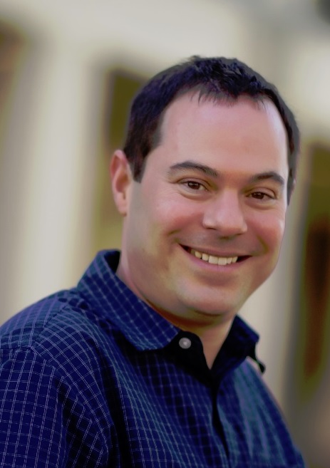 Brian D. Strahl合作论文数Department of Biochemistry and Biophysics, School of Medicine, The University of North Carolina at Chapel Hil;Center for Structural Biology, School of Medicine, The University of North Carolina at Chapel Hil11
 Marius Sudol合作论文数Department of Physiology, Institute for Molecular and Cell Biology, Yong Loo Lin School of Medicine, National University of Singapore11
 Xiaobing Shi合作论文数Department of Epigenetics, Van Andel Institute11
 Guozhen Gao合作论文数College of Life Sciences, Wuhan University10Taiping Chen合作论文数10
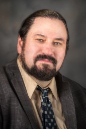 John R. Horton合作论文数Department of Biochemistry, Emory University School of Medicine10
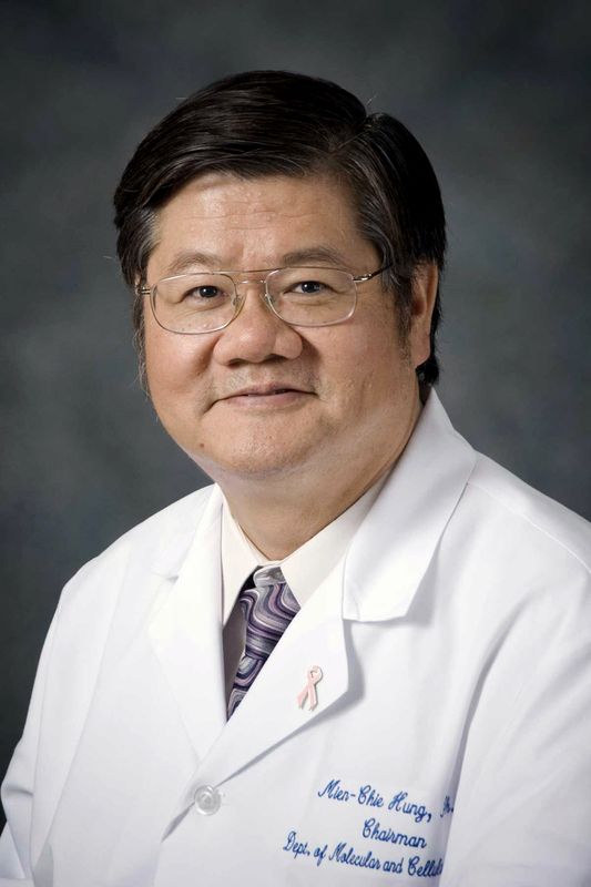 Mien-Chie Hung (洪明奇)合作论文数China Medical University;China Medical University Hospital9
 Vemulapalli Vidyasiri合作论文数Department of Epigenetics and Molecular Carcinogenesis, The University of Texas M. D. Anderson Cancer Center9
 Gayatri Sitaram合作论文数Department of Molecular Carcinogenesis, The University of Texas MD Anderson Cancer Center9
 Jianqiang Bao (鲍坚强)合作论文数Division of Life Sciences and Medicine, University of Science and Technology of China;The First Affiliated Hospital of University of Science and Technology of China9
 Han Xu合作论文数Harvard School of Dental Medicine, Harvard University9Sabrina Castellano合作论文数Dipartimento di Scienze Farmaceutiche, Università degli Studi di Salerno9
 Alessandra Di Lorenzo合作论文数8
 Stephane Richard合作论文数Lady Davis Institute, Jewish General Hospital, McGill University8
 Stephen V. Frye合作论文数Lineberger Comprehensive Cancer Center, School of Medicine, The University of North Carolina at Chapel Hill;Center for Integrative Chemical Biology and Drug Discovery, University of North Carolina at Chapel Hill;Division of Chemical Biology and Medicinal Chemistry, University of North Carolina at Chapel Hill;Meryx Inc8
 Weiyi Peng合作论文数Department of Melanoma Medical Oncology, The University of Texas MD Anderson Cancer Center8
 Jianjun Shen合作论文数Department of Epigenetics and Molecular Carcinogenesis, Division of Basic Science Research, The University of Texas MD Anderson Cancer Center8
 Wei Li合作论文数Department of Biological Chemistry, School of Medicine, University of California, Irvine;Division of Computational Biomedicine, School of Medicine, University of California, Irvine;Dan L. Duncan Cancer Center, University of California, Irvine8
 Philip Leder合作论文数Department of Genetics, Harvard Medical School8Astrid Spannhoff合作论文数8Rehnuma Shifat合作论文数7
 He Wei合作论文数Univ Texas MD Anderson Canc Ctr, Dept Epigenet & Mol Carcinogenesis, Smithville, TX 78957 USA7
 Ronald N. Harty合作论文数School of Veterinary Medicine, University of Pennsylvania7
 Frederick F. Lang合作论文数Department of Neurosurgery, Division of Surgery, The University of Texas MD Anderson Cancer Center;UT Health Graduate School of Biomedical Sciences, The University of Texas MD Anderson Cancer Center7Xing Zhang合作论文数7Jaeho Lee合作论文数7
 Cheryl Arrowsmith合作论文数Department of Medical Biophysics, Temerty Faculty of Medicine, University of Toronto;Princess Margaret Cancer Centre7
 Lindsey Ingerman James合作论文数Division of Chemical Biology and Medicinal Chemistry, Eshelman School of Pharmacy, The University of North Carolina at Chapel Hil7Erik P. Sulman合作论文数Division of Oncology, Children's Hospital of Philadelphia7
 Juan Fueyo合作论文数Department of Neuro-Oncology, Division of Cancer Medicine, The University of Texas MD Anderson Cancer Center7
 Candelaria Gomez-Manzano合作论文数Division of Cancer, Department of Neuro-Oncology, The University of Texas MD Anderson Cancer Center;ivision of Cancer Medicine, Department of Neuro-Oncology - Research, DThe University of Texas MD Anderson Cancer Center7Carrie Capps合作论文数6Raoul V Kuiper合作论文数Laboratory for Health Protection Research, National Institute for Public Health and the Environment (RIVM), Bilthoven, The Netherlands6
 Dmitri Kireev合作论文数University of North Carolina, University of North Carolina at Chapel Hill;Tel: (919) 843-5486.;University of North Carolina, University of North Carolina at Chapel Hill6
 David Johnson合作论文数6
 Scott Rothbart合作论文数Ctr Epigenet, Van Andel Res Inst6
 Monica Viviano合作论文数6
 Jianji Chen合作论文数Dalian University of Technology, Dalian Univ. of Technol.6Harry Van Steeg合作论文数6




