稀な腫瘍である混合型大細胞神経内分泌癌と紡錘細胞癌を含む異時性多発肺癌の1切除例を経験したので報告する.症例は60歳代,男性で,健診の胸部レントゲンで異常陰影を指摘され当院を受診した.CTで左上葉S5に充実性結節,S1+2, S3にスリガラス陰影を認め,胸腔鏡下左上葉切除術を施行した.病理検査でS5の結節は腺癌成分を含む混合型大細胞神経内分泌癌(T1bN0M0)と診断された.その他の結節は腺癌であった.術後補助化学療法を施行したが,術後1年で右中葉に孤立性の充実性結節が出現した.混合型大細胞神経内分泌癌の転移を疑い,化学療法を行ったが短期間で増大した.発見より8ヵ月後に右中葉切除術を施行した結果,紡錘細胞癌(T2aN0M0)と診断され,異時性多発癌と考えられた.現在再手術後1年経過し無再発生存中である.
乳癌胸骨転移再発に対し,胸腹壁再建に用いられるデュアルメッシュ®にて胸骨再建を施行し,良好な経過を得ている症例を経験したので報告する.症例は56歳の女性.2001年に右乳癌に対して胸筋温存乳房切除術施行後,内分泌療法を施行していたところ,2005年に右鎖骨上,縦隔のリンパ節および胸骨に転移が出現した.その後,内分泌療法を変更し,化学療法・放射線療法を追加したが,胸骨のみに腫瘍が残存した.同病変は増大傾向を示し,疼痛を伴うようになったため,2011年に胸骨体部切除術を施行した.再建にはデュアルメッシュ®を用いた.組織診断は乳癌の転移であり,切離断端は陰性であった.呼吸による胸郭変動はなく,疼痛も軽快した.現在のところ再発の兆候はない.
症例は84歳,男性.前立腺癌術後の経過観察中に胸部CTにて後縦隔に腫瘍性病変を指摘された.確定診断および治療の目的に手術が施行された.完全胸腔鏡下に病変を完全切除した結果,MALTリンパ腫と診断された.切除部位に術後補助放射線治療を行い現在無再発生存中である.胸膜原発のMALTリンパ腫は極めて稀であり,今後のさらなる症例集積が重要と考えられた.
cells for cardiothoracic surgery research and treatment?
Objective: We sought to evaluate the utility of integrated breath-hold single-photon emission tomography and computed tomography imaging compared with that of simple calculation with the lung segment-counting technique for predicting residual pulmonary function in patients undergoing surgical intervention for lung cancer.Methods: A prospective series of 22 patients undergoing anatomic lung resection for cancer were enrolled in this study. Postoperative residual forced expiratory volume in 1 second was predicted by measuring the radioactivity counts of the affected lobes or segments to be resected within the entire lungs by placement of regions of interest on single-photon emission tomography and computed tomography images. Residual forced expiratory volume in 1 second was also estimated by using the segment-counting technique.Results: Both predicted values agreed well with postoperative forced expiratory volume in 1 second. Although the residual forced expiratory volume in 1 second predicted by means of single-photon emission tomography and computed tomography correlated well with that predicted by using segment counting, the values were significantly underestimated by the segment-counting technique in 4 outliers with severe emphysema. There were 2 patients with borderline pulmonary functional reserve whose residual forced expiratory volume in 1 second values were predicted more accurately by means of single-photon emission tomography and computed tomography than by using segment counting.Conclusion: Integrated breath-hold single-photon emission tomography and computed tomography images allow the accurate prediction of postoperative pulmonary function but without statistical superiority over the simple segment-counting technique. Further study of the usefulness of single-photon emission tomography and computed tomography in patients with severe emphysema and borderline lung function should prove valuable because the segment-counting technique underestimates pulmonary functional reserve in these patients.
OBJECTIVE:Video-assisted thoracic surgery followed by fast-track rehabilitation has been claimed to accelerate physiological recovery after lung lobectomy for cancer; however, we are still uncertain when the exercise and oxygenation capacity recover and how to determine the rehabilitation time required by each patient. The aim of this study was to evaluate the rehabilitation time after this type of surgery and determine the best predictors of rehabilitation time. METHODS:We measured exercise and oxygenation capacity daily during the perioperative period on a prospective series of 40 patients who had scheduled to undergo video-assisted lung lobectomy for cancer. Postoperative rehabilitation was confirmed when patients had regained more than 80% of their baseline exercise capacity and more than 98% of their baseline oxygenation capacity without the use of routine tubes for oxygen supplementation, fluid transfusion, bladder catheterization, chest drainage, and epidural catheterization. The hypoxemia index, which we found to have correlated with early-postoperative oxygenation capacity, was calculated preoperatively using baseline arterial oxygen saturations and the severity of emphysema on computed tomography. RESULTS:The median rehabilitation time was 3 days. Stepwise Cox regression analysis revealed that the postoperative predicted forced expiratory volume in 1s (relative ratio 1.043, p < 0.01) and the hypoxemia index (relative ratio 1.343, p = 0.02) were the best independent determinants of the postoperative rehabilitation time. CONCLUSIONS:By conducting daily physiological assessments, we identified the rehabilitation time and its determinants in patients who underwent video-assisted lung lobectomy for cancer. Our results are valuable for planning patient-specific fast-track surgery in the hospital setting.
STUDY OBJECTIVES:To determine the ability of quantitative CT, with special reference to area of emphysema, to predict early postoperative oxygenation capacity and outcome after lung lobectomy for cancer.METHODS:Sixty-two consecutive patients scheduled to undergo lung lobectomy for cancer were enrolled in this study. The area of emphysema (< - 910 Hounsfield units) was measured on a three-dimensional CT lung model. Arterial oxygen saturation (Sao(2)) was calculated from Pao(2) measured 1 day before and 1 day after surgery with patients at rest breathing room air. A patient was considered to have recovered at the completion of a standardized management regimen.RESULTS:Postoperative Sao(2) (postSao(2)) was predicted by the baseline value and the area of emphysema with the use of a regression equation. Ten of the 62 patients (16%) had postoperative cardiopulmonary complications (CPCs). The median time to postoperative recovery was 3 days (range, 1 to 17 days). Predicted postSao(2) and predicted postoperative FEV(1) were shown to be significant independent predictors of postoperative CPCs as well as postoperative recovery time.CONCLUSION:Determining the area of emphysema by quantitative CT is useful in predicting early postoperative oxygenation capacity. Predicted oxygenation capacity and predicted ventilatory capacity independently affect perioperative outcomes. Therefore, using quantitative CT in combination with spirometry may improve risk prediction in patients undergoing lung lobectomy for cancer. However, the role of quantitative CT in grading nonemphysematous lung diseases, such as interstitial lung diseases, must be investigated.
症例は34歳,女性.平成15年8月ごろより左下腹部の腫瘤を自覚していた.平成16年2月近医を受診し腹部CTにて左後腹膜腫瘍を指摘され,当科紹介となった.初診時,左下腹部に弾性軟な5×3cm大の腫瘤を触知した.腹部CTでは左後腹膜腔に5×3cmの造影効果の乏しいlow densityな腫瘤を認め, S状結腸が内側に圧排されていた.後腹膜嚢腫の診断のもと,腫瘍摘出術を施行した.腫瘤は薄い嚢胞壁を有し,周囲への浸潤は認めなかった.内腔には淡黄色透明の漿液が充満していた.内容液のCA19-9およびCEAはそれぞれ1,087, 100U/mlおよび53.5ng/mlと高値を示した.細胞診はClassIで,嚢胞壁に悪性所見を認めなかった.免疫組織化学染色を行ったところCA19-9のみ陽性であった.術後経過は良好で第7病日に軽快退院となった.
我々は乳癌術後12年目に原発性肺癌手術を契機に発見された乳癌胸膜転移の1例を経験したので報告する. 症例は72歳, 女性で, 主訴は胸部レントゲン写真上の異常陰影である. 2002年の肺癌検診にて胸部レントゲン写真上の異常陰影を指摘された. 当院放射線科にて精査の後, 右下葉の原発性肺癌と診断され, 手術目的にて当科紹介入院となった. 術前胸部CT検査にて同一肺葉の胸膜下に多発の微小腫瘤を認めていたが良性腫瘍と診断されていた. 手術は右下葉切除術+リンパ節郭清が施行された. 術中胸腔内洗浄細胞診では癌細胞塊を認め, 右下葉の病理学的検索により原発性肺癌 (pT1N0M0) と乳癌の胸膜転移と診断された. 術後経過は良好で軽快退院した. 術後1年3ヵ月の現在ホルモン療法を行っているが明らかな再発は認められない.
Background: Application of the sentinel node concept to lung cancer is still controversial. Patients with peripheral small lung cancers would gain the most benefit from this concept, if it were valid. We sought to determine whether it is possible to choose between limited lymph node sampling and systematic lymphadenectomy from the distribution of sentinel lymph nodes in patients with node-negative disease on the basis of imaging.Methods: Sixty-five consecutive patients with M NO MO non-small cell lung cancer were enrolled. A radioisotope tracer (4 mCi of technetium-99m tin colloid, 2.0 mL) was injected in the vicinity of the tumor before surgical intervention with computed tomographic guidance. The radioactivity of each resected lymph node was measured separately with a hand-held gamma probe after complete tumor resection. Sentinel nodes were identified, and the accuracy of sentinel node mapping was examined. Whether the location of the sentinel node depended on the site of the primary tumor was also examined.Results: Of the 65 patients, 3 were excluded because of the final pathologic results. Successful radionuclide migration occurred in 39 (62.9%) of the 62 patients. There was 1 (2.6%) false-negative result among 39 patients with a sentinel node, and therefore the sensitivity was 90%, and the specificity was 100%. The most common sentinel lymph nodes were at level 12 (46.7%), followed by level 11 (18.3%), the mediastinum (16.7%), and level 10 (11.7%).Conclusion: The sentinel node concept is valid in patients with cT1 NO MO lung cancer. The lobar lymph nodes were identified as sentinel nodes more frequently than other lymph nodes. We need to make further efforts to increase the sentinel node identification rate. However, we believe that if sentinel nodes are identified, sentinel node mapping can allow the accurate intraoperative diagnosis of pathologic NO status in patients with cT1 NO MO lung cancer.
Ventricular aneurysms, shock, and late followup in patients with heart failure f P. M. McCarthy 326 Does size matter?What is your infarct rate after coronary artery bypass grafting?᭹
症例は73歳, 男性.既往に1996年3月に胃癌に対し胃全摘術 (D2郭清) が施行されている.この時の病理診断は中~低分化型腺癌, ss, n2 (+), stage IIIaであった.1999年9月に経過観察のため撮影された胸部CTにて右S8に直径15mmと6mmの結節影を指摘された.15mmの結節には小棘形成と胸膜陥入を, 6mmの結節には胸膜嵌入を認めた.B8bの気管支擦過細胞診でclassVの診断を得た.以上よりT4N0M0の原発性肺癌と診断され右下葉切除術と縦隔リンパ節郭清が施行された.術後病理では, 15mmの結節が胃癌原発巣に酷似した低分化型腺癌であるため胃癌の肺転移, 6mmの結節は原発性肺癌 (気管支肺胞上皮癌) と診断された.また肺門部リンパ節に胃癌からの転移を認めた.胃癌術後患者に多発肺腫瘤を認めた場合, 原発性と転移性の両者を念頭におき手術術式を選択すべきである.
 Kazuhiro Ueda合作论文数7
 Yoshikazu Kaneda合作论文数7Kimikazu Hamano合作论文数3Mitsutaka Jinbo合作论文数3makoto tamai合作论文数2Sano Fumiho合作论文数2Tao-Sheng Li合作论文数2Kazuyoshi Suga合作论文数2Masatoshi Shigeta合作论文数2Masataro Hayashi合作论文数2Takahisa Matsuoka合作论文数2Hisashi Sakano合作论文数2Y Minamiya合作论文数1P Larini合作论文数1Marc Ruel合作论文数1Koichi Saeki合作论文数1Sota Yoshimine合作论文数1Katherine Cornelius合作论文数1
 Helen M Holtby合作论文数1
 Marcello Carlo Ambrogi合作论文数1Naohiro Yamamoto合作论文数1Toshiki Tanaka合作论文数1Toshiki TANAKA合作论文数1Walter H. Merrill合作论文数1H Sakano合作论文数1yuji fujita合作论文数1S Saccani合作论文数1Andrea Agostinelli合作论文数1Alberto Pochettino合作论文数1Pinuccia Faviana合作论文数Department of Surgery- Medical- Molecular- and Critical Area Pathology, Pathological Anatomy III Univ1
 Brian McCrindle合作论文数The Hospital for Sick Children, Department of Paediatrics, University of Toronto;CHSS Data Center1
 Jehangir J Appoo合作论文数Libin Cardiovascular Institute of Alberta, University of Calgary1Vigilance Deon W合作论文数1Kurichi Jibby E合作论文数1AlFaleh Hussam F合作论文数1Gustav Steinhoff合作论文数1Sugi K合作论文数1
 Joseph Woo合作论文数Department of Cardiothoracic Surgery, Stanford University School of Medicine1Kwan L. Chan合作论文数Faculty of Medicine, University of Ottawa1Nakamura Tamami合作论文数1Sawabata N合作论文数1Krajisnik Sanja合作论文数1
 Joseph S. Coselli合作论文数Department of Surgery, Baylor College of Medicine1
 William G Williams合作论文数Division of Cardiovascular Surgery, SickKids1Kensuke Esato合作论文数Yamaguchi University, 1st Department of Surgery1
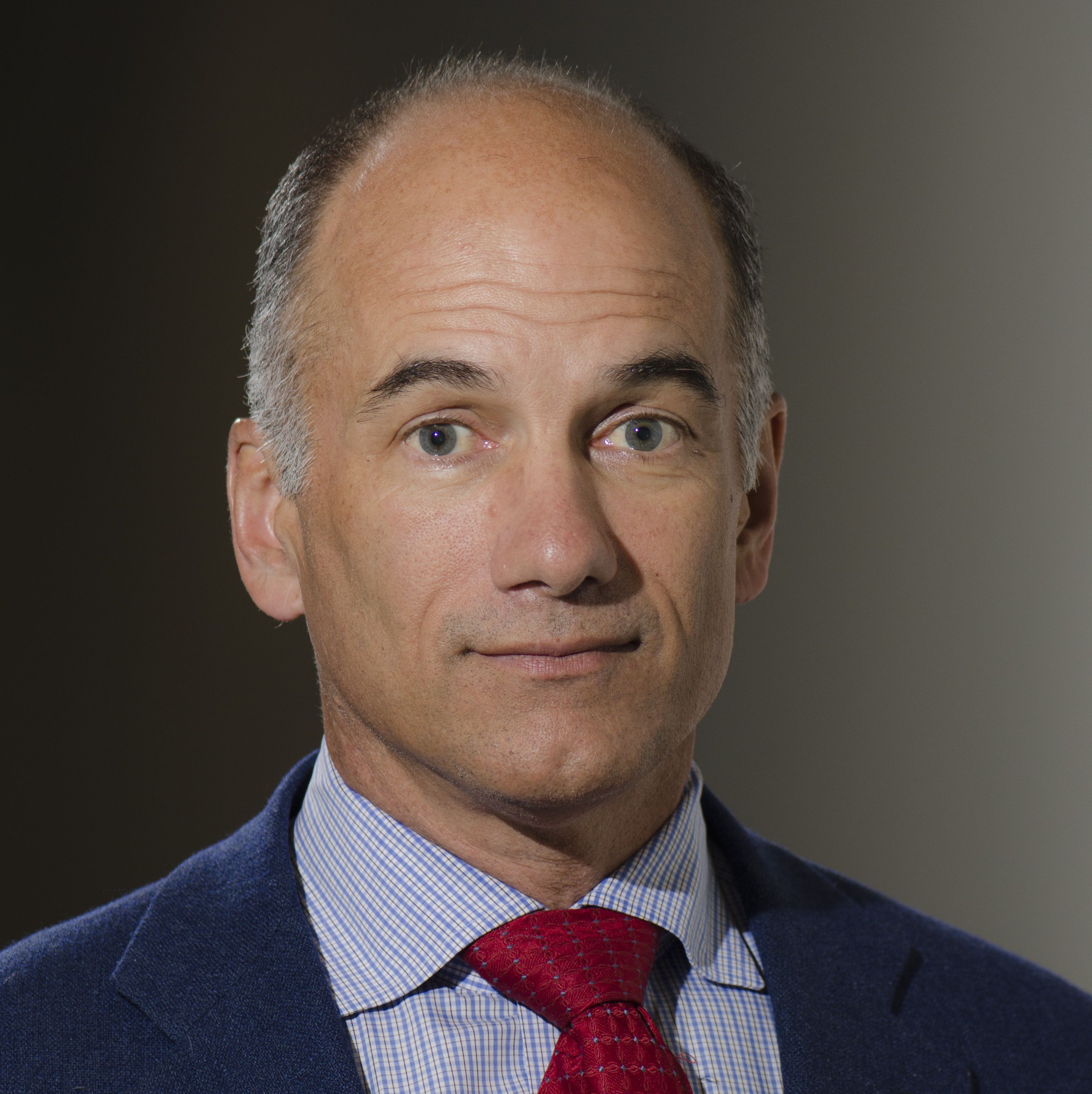 Christopher Caldarone合作论文数Department of Surgery, Baylor College of Medicine1
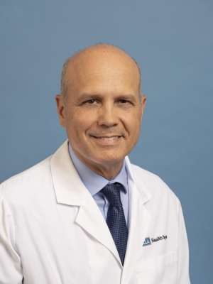 Glen S. Van Arsdell合作论文数Children's Heart Center, University of California Los Angeles1Nobuyuki Tanaka合作论文数1




