The high-temperature requirement A1 (HTRA1), a serine protease, has been demonstrated to play a pivotal role in the extracellular matrix (ECM) and has been reported to be associated with the pathogenesis of age-related macular degeneration (AMD). To delineate its role in the retina, the phenotype of homozygous Htra1-KO (Htra1-/-) mice was characterized to examine the effect of Htra1 loss on the retina and retinal pigment epithelium (RPE) with age. The ablation of Htra1 led to a significant reduction in rod and cone photoreceptor function, primary cone abnormalities followed by rods, and atrophy in the RPE compared with WT mice. Ultrastructural analysis of Htra1-/- mice revealed RPE and Bruch's membrane (BM) abnormalities, including the presence of sub-RPE deposits at 5 months (m) that progressed with age accompanied by increased severity of pathology. Htra1-/- mice also displayed alterations in key markers for inflammation, autophagy, and lipid metabolism in the retina. These results highlight the crucial role of HTRA1 in the retina and RPE. Furthermore, this study allows for the Htra1-/- mouse model to be utilized for deciphering mechanisms that lead to sub-RPE deposit phenotypes including AMD.
Traditional cancer treatments use nonspecific drugs and monoclonal antibodies to target tumor cells. Chimeric antigen receptor (CAR)-T cell therapy, however, leverages the immune system's T-cells to recognize and attack tumor cells. T-cells are isolated from patients and modified to target tumor-associated antigens. CAR-T therapy has achieved FDA approval for treating blood cancers like B-cell acute lymphoblastic leukemia, large B-cell lymphoma, and multiple myeloma by targeting CD-19 and B-cell maturation antigens. Bi-specific chimeric antigen receptors may contribute to mitigating tumor antigen escape, but their efficacy could be limited in cases where certain tumor cells do not express the targeted antigens. Despite success in blood cancers, CAR-T technology faces challenges in solid tumors, including lack of reliable tumor-associated antigens, hypoxic cores, immunosuppressive tumor environments, enhanced reactive oxygen species, and decreased T-cell infiltration. To overcome these challenges, current research aims to identify reliable tumor-associated antigens and develop cost-effective, tumor microenvironment-specific CAR-T cells. This review covers the evolution of CAR-T therapy against various tumors, including hematological and solid tumors, highlights challenges faced by CAR-T cell therapy, and suggests strategies to overcome these obstacles, such as utilizing single-cell RNA sequencing and artificial intelligence to optimize clinical-grade CAR-T cells.
ABSTRACTInflammation is an innate immune response triggered by harmful stimuli, such as pathogens, tissue injury, or toxins. The purpose is to eliminate the source of infection and initiate the healing process. However, an excessive acute inflammatory response can lead to severe and life-threatening complications, as seen during recent pandemics. In the context of viral infections, the activation of the TLR7/8 signaling pathway has been implicated in excessive cytokine secretion. In this study, we aimed to replicate the perturbed inflammatory environment resulting from the activation of the TLR7/8 specific agonists, imiquimod, and resiquimod.We utilized macrophage-like cells, as macrophages are the first responders during infections and secrete key pro-inflammatory cytokines (TNF-α, IL-6, and IL-1β) to recruit immune cells to the site of infection. Herbal medicines have been traditionally used for centuries to enhance respiratory immune function. In the present study, we employed a prophylactic approach, where macrophage-like THP1 cells, differentiated with PMA, were pre-treated with select herbal extracts/formulations prior to TLR7/8 activation in the presence of agonists.Several medicinal plants and formulations known for their therapeutic potential in respiratory ailments were investigated, includingWithania somnifera, Tinospora cordifolia, Glycyrrhiza glabra,and AYUSH-64, an herbal formulation. The gene expression and corresponding secreted levels of various inflammatory mediators were measured using RT-PCR and ELISA methods, respectively, in treated and untreated differentiated THP1 cells induced with TLR7/8 agonists. Comparatively, the gene expression of inflammatory markers was significantly higher in resiquimod-induced cells than in imiquimod-treated cells. Notably,Withania somniferademonstrated pronounced prophylactic efficacy compared to other herbs/formulations, as evidenced by reduction in expression of majority of investigated inflammatory marker genes.
The androgen receptor (AR) is often activated in prostate cancer patients undergoing androgen-ablative therapy because of the activation of cellular pathways that stimulate the AR despite low androgen levels. In many of these tumors, the cAMP-dependent protein kinase A (PKA) pathway is activated. Previous studies have shown that PKA can synergize with low levels of androgen to enhance androgen signaling and consequent cell proliferation, leading to castration-resistant prostate cancer. However, the mechanism by which PKA causes AR stimulation in the presence of low/no androgen is not established yet. Here, using immunofluorescence immunoblotting assays, co-immunoprecipitation, siRNA-mediated gene silencing, and reporter gene assays, we demonstrate that PKA activation is necessary for the phosphorylation of heat shock protein (HSP90) that binds to unliganded AR in the cytoplasm, restricting its entry into the nucleus. We also found that PKA-mediated phosphorylation of the Thr89 residue in HSP90 releases AR from HSP90, enabling AR binding to HSP27 and its migration into the nucleus. Substitution of the Thr89 in HSP90 prevented its phosphorylation by PKA and significantly reduced AR transactivation and cellular proliferation. We further observed that the transcription of AR target genes, such as prostate-specific antigen (PSA), is also lowered in the HSP90 Thr89 variant. These results suggest that using a small-molecule inhibitor against the HSP90 Thr89 residue in conjunction with existing androgen-ablative therapy may be more effective than androgen-ablative therapy alone in the treatment of prostate cancer patients.
The androgen receptor (AR) is activated in patients with castration resistant prostate cancer (CRPC) despite low circulating levels of androgen, suggesting that intracellular signaling pathways and non-androgenic factors may contribute to AR activation. Many G-protein coupled receptors (GPCR) and their ligands are also activated in these cells indicating that they may play a role in development of Prostate Cancer (PCa) and CRPC. Although a cross talk has been suggested between the two pathways, yet, the identity of GPCRs which may play a role in androgen signaling, is not established yet. By using blast analysis of 826 GPCRs, we identified a GPCR, GPCR 205, which exhibited maximum similarity with the ligand binding domain of the AR. We demonstrate that adhesion GPCR 205, also known as GPR56, can be activated by androgens to stimulate the Rho signaling pathway, a pathway that plays an important role in prostate tumor cell metastasis. Testosterone stimulation of GPR56 also activates the cAMP/ Protein kinase A (PKA) pathway, that is necessary for AR signaling. Knocking down the expression of GPR56 using siRNA, disrupts nuclear translocation of AR and transcription of prototypic AR target genes such as PSA. GPR56 expression is higher in all twenty-five prostate tumor patient's samples tested and cells expressing GPR56 exhibit increased proliferation. These findings provide new insights about androgen signaling and identify GPR56 as a possible therapeutic target in advanced prostate cancer patients.
 Donita Garland合作论文数10
 Radha Ayyagari合作论文数10
 Biswas Pooja合作论文数9Anne Marie Berry合作论文数6
 Shikha Pachauri合作论文数Homi Bhabha National Institute4Kelsey Dang合作论文数3Ryan Lancione合作论文数3Danielle Lazaro合作论文数3
 Anil Chekuri合作论文数<institution>Center for Genomic Medicine, Massachusetts General Hospital Research Institute</institution>, Boston, MA3Allen Wang合作论文数Center for Epigenomics & Department of Cellular & Molecular Medicine, University of California, San Diego, San Diego, United States.3Danae Woodard合作论文数2
 Gunjan Dagar合作论文数2
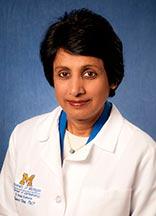 Naheed Wali Khan合作论文数Department of Ophthalmology and Visual Sciences, Kellogg Eye Center, Medical School, University of Michigan2Preissl Sebastian合作论文数2
 Peter X. Shaw合作论文数Dept Ophthalmol, Univ Calif San Diego2
 Monica M. Jablonski合作论文数Retinal Degeneration Research Center, University of Texas Health Sciences Center2Sudhir Rawal合作论文数1
 Ashna Gupta合作论文数1Julie Pratibha Singh合作论文数1Anne Berry合作论文数1Berzhan Kurmanov合作论文数1
 Ravi Chauhan合作论文数1
 Ammira Akil合作论文数Department of Human Genetics, Sidra Medicine1Sabah Nisar合作论文数Department of Physiology and Biophysics, Weill Cornell Medical College1
 Ravi Datta Sharma合作论文数Amity Institute of Integrative Sciences and Health, Amity University Haryana1
 T.J. Hollingsworth合作论文数1Sheema Hashem合作论文数Molecular and Metabolic Imaging Laboratory, Sidra Medicine1
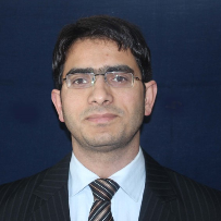 Muzafar A. Macha合作论文数Department of Biochemistry and Molecular Biology, College of Medicine, University Nebraska Medical Center1
 Ajaz Bhat合作论文数Functional and Molecular Imaging Laboratory, Cancer Research Department, Sidra Medical and Research Center1
 Masoodi Tariq Ahmad合作论文数Department of Biotechnology, K L University1
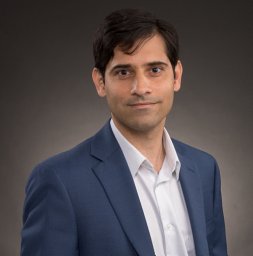 Bagga Puneet合作论文数Centre for Cellular and Molecular Biology (CCMB),, Council of Scientific and Industrial Research (CSIR),1
 Sudhir Kumar合作论文数Department of Microbiology & Immunology, School of Medicine, Emory University1
 Mohammad Haris合作论文数Perelman School of Medicine, University of Pennsylvania1
 Mayank Singh合作论文数Computational Linguistics and Complex Social Networks Group, Department of Computer Science and Engineering, Indian Institute of Technology, Gandhinagar1
 Shahab Uddin合作论文数Translational Research Institute, Hamad Medical Corporation;Laboratory of Animal Research Center, Qatar University1Rakesh K. Kumar合作论文数1Chio Oka合作论文数Division of Gene Function in Animals, Nara Institute of Science and Technology1Sameer Mirza合作论文数Department of Biochemistry, All India Institute of Medical Sciences1
 Madhu Dikshit合作论文数Pharmacology Division, Central Drug Research Institute;Carleton University1
 Takeshi Iwata合作论文数1
 Rakesh K Tyagi合作论文数Special Centre for Molecular Medicine, Jawaharlal Nehru University1
 Gargi Bagchi合作论文数Department of Biology, Boston University1
 Maysaloun Merhi合作论文数UMR 1089-Xénobiotiques, Institut INRA–ENVT1




