Advanced cellular immune therapeutics have transformative potential for an expanding range of diseases, including cancer, autoimmunity, and various immune-related conditions. Despite these advances, significant barriers need to be addressed to achieve improved efficacy, access, safety, and robust manufacturing. In this insight article, we present research activities in Ireland that focus on a) understanding and directing cellular heterogeneity for improved cellular therapeutics through control of metabolism during the biomanufacturing process and b) designing, biomanufacturing, and translating personalized advanced cellular immune therapeutics by creating an All-Ireland Research Center that leverages academic, manufacturing, regulatory, and clinical expertise to deliver a sustainable all-island ecosystem of innovation in cellular therapeutics.
Bone tissue engineering (BTE) emerged as a practical approach to tackle prosthetic industry limitations. We merge aspects from developmental biology, engineering and medicine with the aim to produce fully functional bone tissue. Mesenchymal stem cells have the capability of self-renewal and specific lineage differentiation. Herein lies their potential for BTE. Among MSCs, human dental pulp stem cells have a higher proliferation rate, shorter doubling times, lower cellular senescence, and enhanced osteogenesis than hBM-SCs under specific conditions. In addition, these cells are readily accessible and can be extracted through a subtle extraction procedure. Thus, they garner fewer moral concerns than most MSCs available and embody a promising cell source for BTE therapies able to replace hBM-MSCs. Interestingly, their study has been limited. Conversely, there is a need for their further study to harness their true value in BTE, with special emphasis in the design of bioprocesses able to produce viable, homogenous bone constructs in a clinical scale. Here, we study the osteogenic differentiation of hDPSCs encapsulated in alginate hydrogels under suspended culture in a novel perfusion bioreactor. The system is compared with traditional 3D static and fed-batch culture methodologies. The novel system performed better, producing higher alkaline phosphatase activity, and more homogeneous, dense and functional bone constructs. Additionally, cell constructs produced by the in-house-designed system were richer in mature osteoblast-like and mineralizing osteocyte-like cells. In conclusion, this study reports the development of a novel bioprocess able to produce hDPSC-based bone-like constructs, providing new insights into hDPSCs’ therapeutic potential and a system able to be transferred from the laboratory bench into medical facilities.
Proteins can perform ideal therapeutic functions. However, their large size and significant surface hydrophilicity and charge prohibit them from reaching intracellular targets. These chemical features also render them poorly encapsulated by nanoparticles used for intracellular delivery. In this work, a novel combination of protein vesicles and hydrophobic ion pairing (HIP) was used to load protein cargo and achieve cytosolic delivery to overcome the limitations of previous protein vesicle properties. Protein vesicles are thermally self-assembling nanoparticles made from elastin-like polypeptide (ELP) fused to an arginine-rich leucine zipper and a globular protein fused to a glutamate-rich leucine zipper. To impart stimuli-responsive disassembly, physiological stability, and small size, the ELP sequence was modified to include histidine and tyrosine residues. HIP was used to load and release protein cargo requiring endosomal escape for cytosolic function. HIP vesicles enabled delivery of cytochrome c, a cytosolically active protein, and a significant reduction in viability in both a traditional two-dimensional (2D) human cancer cell line culture and a biomimetic three-dimensional (3D) organoid model of acute myeloid leukemia. By examining the uptake of positively and negatively charged fluorescent protein cargos loaded by HIP, this work revealed the necessity of HIP for cytosolic cargo delivery and how HIP loading influences protein vesicle self-assembly and disassembly using microscopy, small-angle X-ray scattering, and nanoparticle tracking analysis. HIP protein vesicles have the potential to broaden the use of intracellular proteins as therapeutics for various diseases and extend protein vesicles to deliver other biomacromolecules, as the strategy developed here resulted in the first cytosolic protein cargo delivery using protein vesicles.
Background. Chimeric antigen receptor (CAR) T cell therapy, a "living drug" immunotherapy, harnesses the power of T-cells from a patient (autologous) or healthy donor (allogeneic) to target and kill cancer cells and has shown unprecedented outcomes in patients with relapsed and refractory malignancies. Treatment with CAR-T cells requires the application of unique skillsets in recognised specialist centres for successful outcomes and requires management by the multidisciplinary team incorporating the specialist pharmacist. Method. A multimodal research strategy was employed for this literature review whereby PubMed, Google Scholar, Embase, Stella Library Search, EMA website, and EBMT website were sources of information. The search was limited from 2020 onwards with key terms referring to CAR-T cell therapy. Results and Discussion. There are six CAR-T cell products currently approved by the European Medicines Agency (EMA) and Food and Drug Administration (FDA) which target haematological malignancies with abundant clinical trials underway exploring new and improved CAR designs and antigen targets. As CAR-T cell therapy is an advanced therapy medicinal product (ATMP), there is need for an extensive regulatory framework underpinning its safety and efficacy. The clinical pharmacist plays an integral role in the provision of safe and effective CAR-T cell therapy including governance, operational and clinical aspects of treatment. Pharmacists may also be involved through provision of "Qualified Person" (QP) expertise in clinical trials and for release within hospitals under certain circumstances. There is a need for harmonised and accessible guidance on the clinical delivery of ATMPs such as CAR-T cells, with fully delineated responsibilities of pharmacists involving the oversight and supervision of CAR-T cell treatment. Conclusion. There is an unmet need to provide suitable and applicable literature for clinical pharmacists who are involved in the delivery of CAR-T cells. We have provided an overview of T-cell biology and an explanation of CAR-T cell design and the biomanufacturing process. We reviewed the complex and multifaceted treatment cycle requiring considerable logistics, and described the involvement of the clinical pharmacist in each part of this cycle from patient selection to postinfusion care. Finally, we look to the challenges and future opportunities that will require the involvement of the clinical pharmacist.
Stem cells have been widely used to produce artificial bone grafts. Nonetheless, the variability in the degree of stem cell differentiation is an inherent drawback of artificial graft development and requires robust evaluation tools that can certify the quality of stem cell-based products and avoid source-tissue-related and patient-specific variability in outcomes. Omics analyses have been utilised for the evaluation of stem cell attributes in all stages of stem cell biomanufacturing. Herein, metabolomics in combination with machine learning was utilised for the benchmarking of osteogenic differentiation quality in 2D and 3D cultures. Metabolomics analysis was performed with the use of gas chromatography–mass spectrometry (GC-MS). A set of 11 metabolites was used to train an XGboost model which achieved excellent performance in distinguishing between differentiated and undifferentiated umbilical cord blood mesenchymal stem cells (UCB MSCs). The model was benchmarked against samples not present in the training set, being able to efficiently capture osteogenesis in 3D UCB MSC cultures with an area under the curve (AUC) of 82.6%. On the contrary, the model did not capture any differentiation in Wharton’s Jelly MSC samples, which are well-known underperformers in osteogenic differentiation (AUC of 56.2%). Mineralisation was significantly correlated with the levels of fumarate, glycerol, and myo-inositol, the four metabolites found most important for model performance (R2 = 0.89, R2 = 0.94, and R2 = 0.96, and p = 0.016, p = 0.0059, and p = 0.0022, respectively). In conclusion, our results indicate that metabolomics in combination with machine learning can be used for the development of reliable potency assays for the evaluation of Advanced Therapy Medicinal Products.
Image-based spatial omics methods such as fluorescence in situ hybridization (FISH) generate molecular profiles of single cells at single-molecule resolution. Current spatial transcriptomics methods focus on the distribution of single genes. However, the spatial proximity of RNA transcripts can play an important role in cellular function. We demonstrate a spatially resolved gene neighborhood network (spaGNN) pipeline for the analysis of subcellular gene proximity relationships. In spaGNN, machine-learning-based clustering of subcellular spatial transcriptomics data yields subcellular density classes of multiplexed transcript features. The nearest-neighbor analysis produces heterogeneous gene proximity maps in distinct subcellular regions. We illustrate the cell-type-distinguishing capability of spaGNN using multiplexed error-robust FISH data of fibroblast and U2-OS cells and sequential FISH data of mesenchymal stem cells (MSCs), revealing tissue-source-specific MSC transcriptomics and spatial distribution characteristics. Overall, the spaGNN approach expands the spatial features that can be used for cell-type classification tasks.
Hematoimmunopoiesis takes place in the adult human bone marrow (BM), which is composed of heterogeneous niches with complex architecture that enables tight regulation of homeostatic and stress responses. There is a paucity of representative culture systems that recapitulate the heterogeneous three-dimensional (3D) human BM microenvironment and that can endogenously produce soluble factors and extracellular matrix that deliver culture fidelity for the study of both normal and abnormal hematopoiesis. Native BM lymphoid populations are also poorly represented in current in vitro and in vivo models, creating challenges for the study and treatment of BM immunopathology. BM organoid models leverage normal 3D organ structure to recreate functional niche microenvironments. Our focus herein is to review the current state of the art in the use of 3D BM organoids, focusing on their capacities to recreate critical quality attributes of the in vivo BM microenvironment for the study of human normal and abnormal hematopoiesis.
Purpose:Two-dimensional (2D)-based cell culture systems, limited by their inherent heterogeneity and scalability, are a bottleneck in the production of high-quality cells for downstream biomedical applications. Finding the optimal conditions for large-scale stem cell culture while maintaining good cellular status is challenging. The aim of this study was to assess the effects of three-dimensional (3D) culture on the viability, proliferation, self-renewal, and differentiation of human induced pluripotent stem cells (IPSCs).Patients and Methods:Various culture conditions were evaluated to determine the optimal conditions to maintain the viability and proliferation of human IPSCs in a 3D environment: static versus dynamic culture, type of adhesion protein added to alginate (Matrigel™ versus gelatin), and the addition of Y-27632t on long-term 3D culture. The proliferation ability of the cells was evaluated via the MTS proliferation assay; the expression levels of the pluripotency markers Nanog and Oct3/4, PAX6 as an ectoderm marker, and laminin-5 and fibronectin as markers of extracellular matrix synthesis were assessed; and HIF1α and HIF2α levels were measured using quantitative reverse transcription polymerase chain reaction.Results:Using a high-aspect-ratio vessel bioreactor with a gentle, low-sheer, and low-turbulence environment with sufficient oxygenation and effective mass transfer of nutrients and waste, we verified its ability to promote cell proliferation and self-renewal. The findings showed that human IPSCs have the ability to maintain pluripotency in a feeder-free system and by inhibiting ROCK signaling and using hypoxia to improve single-cell viability in 3D culture. Furthermore, these cells demonstrated increased self-renewal and proliferation when inoculated as single cells in 3D alginate beads by adding RI during the culture period.Conclusion:Dynamic 3D culture is desirable for the large-scale expansion of undifferentiated human IPSCs.
Hallmarks of acute myeloid leukemia (AML) include (1) formation of a self-supporting and self-regulating tumor microenvironment (TME) in the bone marrow (BM) rendering pathological niches that orchestrate retention, proliferation and survival of AML blasts, and (2) a dynamic metabolism that promotes therapy resistance and disease progression. Efforts to understand AML biology and enhance current therapies are hampered by the paucity of state-of-the-art human models that can recreate the three-dimensional (3D) BM TME and recapitulate the heterogeneity, complexity, dynamics, and metabolic shifts observed in human disease. Previously, we described the development of the first long-term self-propagating serum- and cytokine-free human BM organoid generated from an AML patient-derived xenograft (PDX) that captured metabolic shifts during AML progression. Here, we characterize the dynamic changes observed in the self-constituted TME with respect to AML proliferation, metabolism, and upon reconstitution of secondary organoids. BM was collected from NSG mice engrafted with human primary AML cells (PDX-AML model) or naïve control mice and seeded into polyurethane scaffolds in serum- and cytokine-free medium at 4e6 cells per scaffold, optimized previously. Scaffolds were recharged with autologous PDX-AML BM on day (D) 13 and cultured until D70. Organoids were assessed for (1) inter- and intra-scaffold AML kinetics, (2) the TME, (3) metabolic composition and (4) propagation into secondary organoids. AML cell kinetics from organoid culture supernatants reflected those of scaffold-extracted populations (n=3). CD33 +CD44 + AML cells expanded and/or were maintained from 7% (D0) to >10% (D70), being >15% at most culture timepoints. Confocal microscopy revealed reconstitution of typical AML-TME niche interactions within organoids, including expression of fibronectin, VCAM-1 and N-Cadherin, and formation of proliferative niches composed of AML cells with heterogeneous Osteopontin- and Osterix-expressing support cells. Human AML-supportive cytokines were consistently observed in organoid supernatants (N=3), with SDF-1, IL-1α, IL-8, GM-CSF, FGF-2 and Osteopontin present throughout the culture; some cytokines had biphasic kinetics with factors critical for TME formation present early (e.g. Osteopontin, GM-CSF), and those required for AML proliferation dominant later (e.g. FLT3L, IL-8), coinciding also with metabolic shifts during culture. Despite the absence of IL-6, single molecule RNA fluorescence in situ hybridization confirmed IL-6 expression by niche-resident AML blasts suggesting autocrine signaling networks beyond those identified in supernatants. Cells from D70 organoids were able to re-establish secondary organoids (n=2) which also exhibited long-term (D70) AML maintenance (>11% blasts), and regenerated the AML TME. Previously, we identified 3 culture stages defined by metabolic shifts: 1) pre/early recharge (D4-D19), (2) intermediate (D25-D40) and, (3) late (from D55) where organoids exhibited highest proliferation. To further characterize these shifts, intracellular metabolites were extracted from organoids (n=5) at D10, D25, D40, D55 and D70, analyzed with gas-chromatography mass spectrometry (GC-MS) and evaluated through unsupervised hierarchical clustering and multivariate analysis. Significance Analysis of Microarrays (SAM) analysis demonstrated dysregulation of alanine, serine, isoleucine, glycolate, hexanoic acid and glycolysis when comparing stage 1 with stage 2, and in isoleucine, leucine, serine, succinate and tryptophan metabolism when comparing stage 2 with stage 3. When proliferative and non-proliferative timepoints were compared, amino acid and glycolysis-driven metabolic switches were identified, typical of AML metabolism. We have characterized a long-term 3D human AML BM biomimicry in serum- and cytokine-free conditions with a self-organizing TME that recapitulates many features of AML BM, including robust human AML cell maintenance within niches, cytokine and metabolic kinetics to support different phases of progression, and the ability to propagate into secondary organoids. This dynamic, physiologically-relevant ex vivo platform can enable studies in human AML pathogenesis/progression and is poised to test novel therapeutics directed towards AML cells, the TME and metabolic targets.
Healthy hearts use more fatty acids (FA) than glucose for ATP synthesis but diabetic cardiomyopathy (DbCM) occurs with higher FA dependency. It remains controversial whether glucotoxicity or lipotoxicity or both account for DbCM. We recently discovered that insulin signaling inhibition and eventual FOXO1 activation stimulate cardiac KLF5 expression, which drives lipotoxicity and causes cardiac dysfunction. In the present study, we investigated the relative contribution of glucose in the activation of cardiac KLF5 and DbCM. We induced Type-1 diabetes (T1D) in C57BL/6 mice via intraperitoneal injections of streptozotocin (STZ). In contrast to late-stage diabetes (12 weeks post-STZ), cardiac KLF5 mRNA and protein levels were not increased in the early T1D stage (4 weeks post-STZ) although mice have mild cardiac dysfunction already. Seahorse analysis in adult cardiomyocytes isolated from mice with early T1D showed higher glucose and lower FA dependency compared to non-diabetic mice and mice in late T1D. To confirm whether hyperglycemia causes cardiac dysfunction, we treated diabetic mice with Dapagliflozin (DAPA, SGLT2 inhibitor) or STF-31, a GLUT1 inhibitor. These treatments restored normal dependency on fatty acids and prevented cardiac dysfunction. GC-MS analysis showed that the reversal of fuel dependency from glucose to fatty acids in late T1D is accompanied by increased glucose content opposite to the early T1D. Accordingly cardiac KLF5 is increased in late T1D, accompanied by severe cardiac dysfunction. The expression changes of KLF5 are mirrored by transcriptional activity of FOXO1 -shown by expression of FOXO1 targets- in early and late T1D. The changes in transcriptional activity are accompanied by differential FOXO1 acetylation, which is controlled by Sirtuin-1 and modulates its DNA affinity. Analysis of mouse cardiac tissue in early T1D and a human cardiomyocyte cell line (AC16) that was treated with high glucose showed higher Sirtuin-1 expression and stronger protein-protein interaction with FOXO1. To this end, mice that were subjected to treatment with either DAPA or STF31 for 12 Wks had improved cardiac function, lower cardiac KLF5 expression and decreased expression of cardiac KLF5 gene targets. Interestingly, GLUT1 mRNA levels were increased in late T1D compared to early T1D. Cardiomyocyte-specific KLF5 overexpression or adenovirus-mediated KLF5 overexpression in AC16 cells stimulated GLUT1 expression. Collectively, in early T1D, hearts rely more on glucose utilization in mitochondria. In late T1D, SIRT1-FOXO1-KLF5 axis causes lipotoxicity and subsequent induction of GLUT1 expression that contributes to glucotoxicity. Inhibition of GLUT1-dependent glucose uptake alleviates diabetic cardiomyopathy via inhibition of both early glucose dependency and late KLF5 activation.
Background: As stem cells are considered a promising cell source for tissue engineering, many culture strategies have been extensively studied to generate in vitro stem cell-based tissue constructs. However, most approaches using conventional tissue culture plates are limited by the lack of biological relevance in stem cell microenvironments required for neotissue formation. In this study, a novel perfusion rotating wall vessel (RWV) bioreactor was developed for mass-production of stem cell-based 3D tissue constructs. Methods: An automated RWV bioreactor was fabricated, which is capable of controlling continuous medium perfusion, highly efficient gas exchange with surrounding air, as well as low-intensity pulsed ultrasound (LIPUS) stimulation. Embryonic stem cells encapsulated in alginate/gelatin hydrogel were cultured in the osteogenic medium by using our bioreactor system. Cellular viability, growth kinetics, and osteogenesis/mineralization were thoroughly evaluated, and culture media were profiled at real time. The in vivo efficacy was examined by a rabbit cranial defect model. Results: Our bioreactor successfully maintained the optimal culture environments for stem cell proliferation, osteogenic differentiation, and mineralized tissue formation during the culture period. The mineralized tissue constructs produced by our bioreactor demonstrated higher void filling efficacy in the large bone defects compared to the group implanted with hydrogel beads only. In addition, the LIPUS modules mounted on our bioreactor successfully reached higher mineralization of the tissue constructs compared to the groups without LIPUS stimulation. Conclusion: This study suggests an effective biomanufacturing strategy for mass-production of implantable mineralized tissue constructs from stem cells that could be applicable to future clinical practice.
Healthy hearts rely more on fatty acid (FA) rather than glucose utilization. It remains unclear whether diabetic cardiomyopathy (DbCM) is accounted for by glucotoxicity or lipotoxicity. Previously, we discovered that either insulin deficiency or insulin resistance causes FOXO1-KLF5 activation in human and murine hearts, which drives cardiac lipotoxicity and oxidative stress. Now, we investigate how glucose activates cardiac KLF5 and causes DbCM. We hypothesized that higher cardiac glucose content in diabetes potentiates FOXO1-KLF5 activation and causes glucolipotoxicity. We mimicked Type-1 diabetes (T1D) in C57BL/6 mice via 5 daily intraperitoneal injections of streptozotocin (STZ). In contrast to late T1D (12 Wks post-STZ), cardiac KLF5 expression levels are not increased in the early T1D (4 Wks post-STZ). However, mice developed cardiac dysfunction in early T1D. Seahorse analysis in adult cardiomyocytes isolated from mice with early T1D showed suppression of FA dependence in the expense of higher glucose dependence compared to non-diabetic mice. To confirm whether hyperglycemia or higher cardiac glucose content accounts for cardiac dysfunction in early T1D, we applied anti-hyperglycemia treatment (Dapagliflozin, DAPA, SGLT2 inhibitor) or GLUT1 inhibition (STF-31). Either of the two treatments restored cardiac FA dependence and prevented both glucose preference and cardiac dysfunction in early T1D. GC-MS analysis in hearts of mice with late T1D, which have shifted back to increased FA dependence, showed increased cardiac glucose content -presumably unused glucose- and subsequent increase of cardiac KLF5, as previously shown, which exacerbates cardiac dysfunction. New data revealed that GLUT1 levels were increased in late T1D compared to early T1D, as well as that this is driven by KLF5 activation. The lack of activation of KLF5 expression in early T1D, which reverses in late T1D, is mirrored by FOXO1 transcriptional activity as shown by expression of FOXO1 targets and lower FOXO1 acetylation in late T1D, which is controlled by Sirtuin-1. Analysis of hearts from mice with late T1D and a human cardiomyocyte cell line (AC16) that was treated with high glucose indicated higher Sirtuin-1 expression and Sirtuin-1 binding on FOXO1. Accordingly, prevention of glucose transport to hearts of diabetic mice via treatment with either DAPA or STF-31 for 12 Wks lowered cardiac expression of KLF5 and its targets and improved cardiac function. Conclusively, DbCM begins in early T1D with lower mitochondrial FA utilization that is compensated by higher glucose utilization and is exacerbated in late T1D with activation of SIRT1-FOXO1-KLF5 axis that causes combined lipotoxicity and glucotoxicity. Inhibition of GLUT1 alleviates DbCM via prevention of both early glucose dependence and late KLF5 activation.
A significant limitation to improving treatment for Acute Myeloid Leukemia (AML) is the lack of representative culture systems that (1) recapitulate the three-dimensional (3D) human AML bone marrow (BM) Tumor Microenvironment (TME) where AML cells are protected during treatment, and (2) account for metabolic shifts that lead to drug resistance and relapse. Current in vitro two-dimensional plate cultures and 3D systems lack TME heterogeneity and complexity resulting in oversimplification of disease biology and utilize either (1) AML cell lines, (2) artificially manufactured TME with allogeneic feeder layers, or (3) exogenously added cytokines and serum, with inherent clonal selection and bias. AML patient-derived xenograft (PDX) mouse models remain inadequate for the study of AML due to 50% engraftment failure and stromal damage incurred during conditioning. Herein, we have developed the first human personalized long-term dynamic cytokine- and serum-free in vitro BM AML ex vivo organoid using PDX-derived cells, distinguishing our system from existing technologies. BM was harvested from busulfan-conditioned NSG mice engrafted with an AML PDX when human CD33+ AML blood cells reached >30%; NSG controls were harvested concurrently. We evaluated 3D organoid cultures for: (1) optimal seeding density, (2) TME seeding conditions (AML or naïve) and (3) metabolic shifts. PDX cells were seeded onto 3D polyurethane scaffolds in serum- and cytokine-free medium at different cell densities: 2e6, 4e6 and 6e6 cells per scaffold (n=3) and recharged with autologous PDX cells on day (D)13-14 (Leukemic seeding + Leukemic recharge; L+L). L+L organoids were compared with non-recharged controls. Organoids seeded with 4e6 and 6e6 cells/scaffold proliferated from D34 and exhibited higher dynamic growth kinetics over time when compared with controls (p<0.05). In contrast, 2e6 cultures proliferated after D56 similar to the non-recharged control (p>0.05), suggesting inadequate organoid seeding density. When comparing cell sources, naïve NSG BM cells seeded at 4e6 cells/scaffold (n=3) and recharged with PDX cells (Naïve seeding + Leukemic recharge; N+L) displayed similar kinetics to L+L, suggesting that PDX cells could harness a naïve microenvironment to proliferate, as typified in vivo. Organoids successfully supported proliferation of CD33+CD44+ AML cells, which had similar phenotype to the input population by flow cytometry and morphology, even at D70. AML cells increased from 6% (D0) to >50% at D70 and a murine CD45+ population was observed throughout culture (11% in L+L; 4% in N+L at D70) indicating ongoing interactions between normal and AML cells as described in vivo. Using confocal microscopy, 3D niches spontaneously formed with cell-specific production of IL-6 and IL-8 (assessed by single molecule RNA fluorescence in situ hybridization; RNA FISH) creating a platform for the study of niche biology. Extracellular metabolites from AML-PDX organoid supernatants (n=3) were evaluated with a Bioprofiler and analyzed by principal component analysis with hierarchical clustering. Three culture stages were identified: 1) pre/early recharge phase characterized by low glucose consumption and low lactate production, (2) D20 to D41 phase exhibiting low glutamine uptake and, (3) from D44 onwards where the organoid exhibited leukemic metabolism characterized by high glucose and glutamine consumption with high lactate, ammonia and glutamate production. The last stage of growth was concurrent with the highest cell counts, suggesting that organoids proliferated more after switching to leukemic metabolism, as observed in vivo. Though N+L organoids exhibited similar metabolic stages, the second stage started later (D26), suggesting that a latent leukemic metabolic switch occurred due to early exposure to a naïve microenvironment. We have created a long-term 3D personalized human BM AML-PDX biomimicry in serum- and cytokine-free conditions that not only out-lives the murine PDX donor, but also is an ideal system to understand BM TME and AML niche biology through the evaluation of constituent cells, cytokines and metabolic needs and shifts in a dynamic ex vivo culture. The organoids support self-organizing AML niches and mirror leukemic metabolism while preserving parental cell phenotype, representing a physiologically-relevant heterogeneous and dynamic platform to test novel therapeutics.
Neuroblastoma (NB) is the most common extracranial tumor in children resulting in substantial morbidity and mortality. A deeper understanding of the NB tumor microenvironment (TME) remains an area of active research but there is a lack of reliable and biomimetic experimental models. This study utilizes a 3D bioprinting approach, in combination with NB spheroids, to create an in vitro vascular model of NB for exploring the tumor function within an endothelialized microenvironment. A gelatin methacryloyl (gelMA) bioink is used to create multi-channel cubic tumor analogues with high printing fidelity and mechanical tunability. Human-derived NB spheroids and human umbilical vein endothelial cells (HUVECs) are incorporated into the biomanufactured gelMA and cocultured under static versus dynamic conditions, demonstrating high levels of survival and growth. Quantification of NB-EC integration and tumor cell migration suggested an increased aggressive behavior of NB when cultured in bioprinted endothelialized models, when cocultured with HUVECs, and also as a result of dynamic culture. This model also allowed for the assessment of metabolic, cytokine, and gene expression profiles of NB spheroids under varying TME conditions. These results establish a high throughput research enabling platform to study the TME-mediated cellular-molecular mechanisms of tumor growth, aggression, and response to therapy.
Advanced ScienceVolume 9, Issue 23 2270142 Inside Front CoverOpen Access A 3D Bioprinted in vitro Model of Neuroblastoma Recapitulates Dynamic Tumor-Endothelial Cell Interactions Contributing to Solid Tumor Aggressive Behavior (Adv. Sci. 23/2022) Liqun Ning, Liqun NingSearch for more papers by this authorJenny Shim, Jenny ShimSearch for more papers by this authorMartin L. Tomov, Martin L. TomovSearch for more papers by this authorRui Liu, Rui LiuSearch for more papers by this authorRiya Mehta, Riya MehtaSearch for more papers by this authorAndrew Mingee, Andrew MingeeSearch for more papers by this authorBoeun Hwang, Boeun HwangSearch for more papers by this authorLinqi Jin, Linqi JinSearch for more papers by this authorAthanasios Mantalaris, Athanasios MantalarisSearch for more papers by this authorChunhui Xu, Chunhui XuSearch for more papers by this authorMorteza Mahmoudi, Morteza MahmoudiSearch for more papers by this authorKelly C. Goldsmith, Kelly C. GoldsmithSearch for more papers by this authorVahid Serpooshan, Vahid SerpooshanSearch for more papers by this author Liqun Ning, Liqun NingSearch for more papers by this authorJenny Shim, Jenny ShimSearch for more papers by this authorMartin L. Tomov, Martin L. TomovSearch for more papers by this authorRui Liu, Rui LiuSearch for more papers by this authorRiya Mehta, Riya MehtaSearch for more papers by this authorAndrew Mingee, Andrew MingeeSearch for more papers by this authorBoeun Hwang, Boeun HwangSearch for more papers by this authorLinqi Jin, Linqi JinSearch for more papers by this authorAthanasios Mantalaris, Athanasios MantalarisSearch for more papers by this authorChunhui Xu, Chunhui XuSearch for more papers by this authorMorteza Mahmoudi, Morteza MahmoudiSearch for more papers by this authorKelly C. Goldsmith, Kelly C. GoldsmithSearch for more papers by this authorVahid Serpooshan, Vahid SerpooshanSearch for more papers by this author First published: 15 August 2022 https://doi.org/10.1002/advs.202270142AboutPDF ToolsRequest permissionExport citationAdd to favoritesTrack citation ShareShare Give accessShare full text accessShare full-text accessPlease review our Terms and Conditions of Use and check box below to share full-text version of article.I have read and accept the Wiley Online Library Terms and Conditions of UseShareable LinkUse the link below to share a full-text version of this article with your friends and colleagues. Learn more.Copy URL Share a linkShare onEmailFacebookTwitterLinkedInRedditWechat Graphical Abstract 3D Bioprinted in vitro Model of Neuroblastoma Neuroblastoma (NB) accounts for 50% of all cancers in infants with substantial mortality. Due to the lack of robust experimental models, the role of NB microenvironment in tumor progression is poorly understood. Through integrating 3D bioprinting and NB spheroid technologies, in article number 2200244, Kelly C. Goldsmith, Vahid Serpooshan, and co-workers create an in vitro vascular model of NB and examine the tumor function within an endothelialized microenvironment. Volume9, Issue23August 15, 20222270142 RelatedInformation
Background Bone tissue engineering emerged as a practical approach to tackle the prosthetic industry limitations. Merging aspects from developmental biology, engineering and medicine with the aim to produce fully-functional bone tissue. Mesenchymal stem cells (MSCs) harbor the capability of self-renewal and specific lineage differentiation. Herein lies their potential for bone tissue engineering. Among MSCs, human dental pulp stem cells (hDPSCs) lodge higher proliferation rate, shorter doubling times, lower cellular senescence, and enhanced osteogenesis than hBM-SCs. In addition, these cells have ease in access and a subtle extraction procedure. Thus, harbouring fewer moral concerns than most MSCs available and embodying a promising cell source for BTE therapies able to replace hBM-MSCs. Interestingly, their study has been limited. Conversely, there is a need for their further study to harness their BTE true value, with special emphasis in the design of bioprocesses able to produce viable, homogenous bone constructs in a clinical scale. Methods Here, we study the in vitro osteogenic differentiation of hDPSCs encapsulated in alginate hydrogels under suspended culture in a novel and scalable perfusion bioreactor, establishing culture conditions; and compare it with three-dimensional (3D) static and fed-batch culture. Results hDPSC-based bone-like constructs produced in the novel system performed above the compared culture strategies, displaying higher alkaline phosphatase activity, more homogeneous, denser and functional bone constructs. In addition, cell constructs produced by the in-house designed system were richer in mature osteoblasts. Conclusion This study reports the development of a novel bioprocess able to produce hDPSC-alginate-based bone-like constructs to be used as bone fillers, while providing new insights into hDPSCs therapeutic potential and a system able to be transferred from the laboratory bench into medical facilities.
 Nicki Panoskaltsis合作论文数102
 Efstratios N. (Stratos) Pistikopoulos合作论文数Artie McFerrin Department of Chemical Engineering, Texas A&M University;Texas A&M Energy Institute64
 Michael Georgiadis合作论文数Laboratory of Process System Engineering, Department of Chemical Engineering, Chemical Engineering, School of Engineering, Aristotle University of Thessaloniki;Tsinghua University36
 Eleftherios Tsiridis合作论文数The Academic Unit of Orthopaedic Surgery, A Floor, Clarendon Wing, Leeds General Infirmary, Great George Street, Leeds LS1 3EX, UK34
 Alexander Bismarck合作论文数Institute of Materials Chemistry & Research, University of Vienna31
 Manolis Heliotis合作论文数23Ruth Misener合作论文数Department of Computing, Faculty of Engineering, Imperial College London22
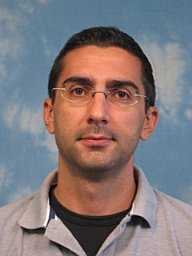 Alexandros Kiparissides合作论文数Centre for Process Systems Engineering, Imperial College London22
 Mark C Allenby合作论文数Queensland University of Technology17Eirini Velliou合作论文数17
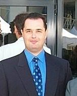 Margaritis Kostoglou合作论文数Laboratory of Chemical and Environmental Technology, School of Chemistry, Faculty of Sciences, Aristotle University of Thessaloniki16
 Mayasari Lim合作论文数Biological Systems Engineering Laboratory, Imperial College London16
 Michail Klontzas合作论文数University Hospital of Heraklion, University of Crete15
 Maria Fuentes-Gari合作论文数15Julia M. Polak合作论文数15
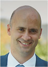 Michalis Koutinas合作论文数Department of Chemical Engineering and Chemical Technology, Imperial College London15Eleni Pefani合作论文数14
 Susana Brito dos Santos合作论文数12
 Simon Graham合作论文数11
 Maria Papathanasiou合作论文数Department of Chemical Engineering, Faculty of Engineering, Imperial College London11
 Jh David Wu合作论文数11
 Zakareya Gamie合作论文数9
 Cathy Ye合作论文数Department of Engineering Science, Linacre College, University of Oxford;Oxford Centre for Tissue Engineering and Bioprocessing, University of Oxford;Institute of Biomedical Engineering, University of Oxford;Oxford Suzhou Centre for Advanced Research;Ivy Farm Technologies9david garciamunzer合作论文数8
 Vahid Serpooshan合作论文数Stanford Cardiovasc Inst, Stanford Univ8Hwang Yu-Shik合作论文数8
 Andrew Livingston合作论文数School of Engineering and Materials Science, Queen Mary University of London8
 Tomov Martin L合作论文数Colleges of Nanoscale Science and Engineering, SUNY Polytechnic Institute8
 Spyros Vernardis合作论文数Institute of Chemical Engineering Sciences, Foundation for Research and Technology-Hellas8
 Teresa Mortera-Blanco合作论文数8
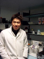 Jae Min Cha合作论文数College of Medicine, Yonsei University8
 Maria Rende合作论文数8Jhd Wu合作论文数7
 Kontoravdi Cleo合作论文数Department of Chemical Engineering, Imperial College London7Chen Yi-Guang合作论文数The Jackson Laboratory7
 Robert J. Macfarlane合作论文数Department of Materials Science and Engineering, MIT7
 Hugo M. Macedo合作论文数Biological Systems Engineering Laboratory, Imperial College London7
 Yunyi Kang合作论文数7
 Iliana Fauzi合作论文数6Sai Raviteja Bhamidipati合作论文数Dept Mech Engn 66, Texas A&M Univ6
 Bowen Jing合作论文数Georgia Institute of Technology, Emory University6
 Andreas Leonidou合作论文数Worcestershire Acute Hospitals, Alexandra Hospital6
 Xiaoyun Xu合作论文数Department of Chemical Engineering, Imperial College London6
 Bauser-Heaton Holly合作论文数Lucile Packard Children's Hospital, Stanford University6
 Liqun Ning合作论文数Northwestern Polytechnical University, Northwestern Polytechnical Univ6
 Gizem Buldum合作论文数6
 Brooks Lindsey合作论文数Wallace H. Coulter Department of Biomedical Engineering, College of Engineering, Georgia Institute of Technology6
 Richard Oberdieck合作论文数6
 Xicai Yue合作论文数Department of Bioengineering, Imperial College London6




