Supplementary Figure 7 contains dot plots showing KLK2 and STEAP1 IHC H-scores for AR+/NE-metastatic sites in the UW-RA cohort.
Supplementary Figure 12 contains additional RNA-seq analyses in AR+/NE-tumors from the UW-RA cohort and SU2C-IDT cohort.
Tier 1 druggable genome targets and their associated limma values from KLK2-high vs KLK2-low and STEAP1-high vs STEAP1-low differential expression analyses.
Differentially expressed genes from STEAP1-high versus STEAP1-low analysis in UW-TAN and ECDT cohorts.
Distribution of intra-tumoral and intra-patient hypergeometric heterogeneity indices for KLK2, STEAP1, and PSMA based on IHC H-scores across samples in the UW-RA cohort.
Supplementary Figure 9 contains donut plots showing the distribution of tumor expressing only one marker, both markers or neither marker for each KLK2-STEAP1, STEAP1-PSMA, and KLK2-PSMA pair across molecular phenotypes and across H-score thresholds.
Previous studies have highlighted that some T cell subsets in tissues can provide signals to support tissue cell homeostasis and differentiation. If and how T cell-tissue cell signaling is altered in healthy compared to inflamed tissues is poorly understood. Here, we address if communication between human T cells and tissue cells changes from steady state to an acutely inflamed state in the human placenta. We used single cell analysis strategies to examine invasive cytotrophoblasts (iCTBs) and immune cells isolated from third trimester healthy and acutely inflamed human placentas. We performed cell communication analysis to predict cell-cell communication networks, and found evidence that iCTBs provided signals to support the recruitment of T cells, as well as the formation of tissue-resident memory CD8 T cells (Trm). In exchange, Trm provide signals to support iCTB homeostasis. During acute inflammation, iCTBs and macrophages underwent profound transcriptional changes, while most T cell subsets only underwent limited transcriptional changes. This was not due to T cell exhaustion or tolerance, as T cells were functionally intact. Cell communication analysis and validation at the protein level provide evidence that T cells can maintain their homeostatic support to iCTBs during acute inflammation.
Supplementary Figure 10 shows UMAP projections of single-cell RNA-seq data from five additional AR+/NE-metastatic prostate cancer samples.
Distribution of expression and co-expression between KLK2, STEAP1, and PSMA based on IHC H-scores across samples in the UW-RA cohort.
Gene set enrichment results from KLK2-high versus KLK2-low analysis in UW-TAN and ECDT.
Distribution of KLK2 and STEAP1 single cell mRNA expression in cells from ARPC samples across MSK scRNA dataset.
Abstract Kallikrein 2 (KLK2) and six-transmembrane epithelial antigen of the prostate 1 (STEAP1) are two cell surface targets with relevance for prostate cancer therapy. The objective of this study was to characterize the expression landscape of KLK2 and STEAP1 in metastatic castration-resistant prostate cancer (mCRPC) and to define associated transcriptomic, genomic, and epigenomic features. We analyzed a total of 1,095 patient samples from three mCRPC cohorts, including in situ studies of rapid autopsy cases and patient-derived xenograft models. We found that KLK2 and STEAP1 expression is strongly enriched in androgen receptor (AR)–positive tumors and largely absent in neuroendocrine and double-negative phenotypes. Within AR+ tumors, pairwise comparisons revealed coexpression and high combined positivity rates for STEAP1, KLK2, and prostate-specific membrane antigen, suggesting that cotargeting any two of these antigens increases overall tumor coverage. Analysis of samples from a rapid autopsy cohort, which enabled assessment of intra- and intertumoral diversity, showed comparable degrees of expression heterogeneity for KLK2 and STEAP1. Antigen expression correlated positively with AR genomic alterations and serum prostate-specific antigen levels and negatively with RB1 and PTEN loss. Transcriptomic and epigenome analyses demonstrated distinct mechanisms governing antigen expression: KLK2 showed a strict AR dependence with coordinated AR/FOXA1/HOXB13 binding and enhancer activation, whereas STEAP1 was only partially AR-dependent and additionally regulated by locus-specific DNA methylation changes. Furthermore, KLK2 and STEAP1 expression states were associated with distinct transcriptional programs and immune microenvironmental features. Implications: These findings establish KLK2 and STEAP1 as key prostate adenocarcinoma-lineage antigens and provide critical insights to inform the rational design and clinical development of cell surface antigen–directed therapies in prostate cancer.
Results from predictive model integrating somatic alterations and serum PSA to estimate the probability that a sample is positive for the given antigen.
Supplementary Figure 11 shows additional KLK2 and STEAP1 IHC H-score boxplots stratified by alteration status.
Abstract Triple-negative breast cancer (TNBC) exhibits the worst overall survival compared to other breast cancer subtypes and has the most limited treatment options. Molecularly, most TNBCs are classified as basal-like and are associated with aggressive metastasis and drug resistance.A major hurdle in the development of effective therapeutics is tumor cell state and phenotypic plasticity that confer fitness advantages for growth, therapeutic resistance, and invasion. In this study, we sought to identify transcription factors that regulate basal-like plasticity for collective invasion. Using mouse mammary tumor organoids and time course single-cell RNA sequencing, we show that transcription factor KLF4 is a potent suppressor of basal-like invasion plasticity.Unexpectedly, we observed that KLF4 overexpression potently induced a molecular program recapitulating the classical differentiation of basal epidermal cells processively toward a biologically dead cornified outer skin layer. Overexpression of KLF4 induced hallmark markers of epidermal differentiation such as keratin 10, desmosomal cadherins, loricrin, and multicellular features of maturing epidermis, including cell piling, keratinization, and increased desmosomes, all putative anti-invasive features. Induction of epidermal differentiation, specifically in basal but not mesenchymal or luminal breast tumor cells, resulted in terminal differentiation in vitro and irreversible loss of colony-forming potential, indicative of tumor subtype-specific vulnerability. In vivo, KLF4 potently suppressed metastatic outgrowth of human basal TNBC in an intracardiac metastasis assay. In human breast tumors, we observed 50% of tumors expressed KLF4 RNA yet lacked KLF4 protein. Mechanistically, we show that basal-like TNBCs actively degrade KLF4protein to limit epidermal differentiation, which was overcome by proteasome inhibitors. In sum, these data uncover epidermal differentiation as a latent plasticity switch of highly metastatic basal-like breast cancer cells, making them irreversibly more cohesive, and point to KLF4protein stabilization as a therapeutic strategy. Citation Format: Justin Hui, Andrea E. Doak, Nicole Rhoads, Ruijin Yang, Jimin Park, Cory L. Simpson, Manu Setty, Kevin J. Cheung. Induction of epidermal like differentiation irreversibly limits the invasive and metastatic potential of basal like breast cancer cells [abstract]. In: Proceedings of the American Association for Cancer Research Annual Meeting 2026; Part 1 (Regular Abstracts); 2026 Apr 17-22; San Diego, CA. Philadelphia (PA): AACR; Cancer Res 2026;86(7 Suppl):Abstract nr 4765.
Supplementary Figure 2 shows representative IHC micrographs in benign and control samples.
Supplementary Figure 5 shows representative STEAP1 IHC micrographs of LuCaP PDX tumors across phenotypes.
 Ilsa Coleman合作论文数Fred Hutchinson Cancer Research Center28
 Erolcan Sayar合作论文数27
 Brian Hanratty合作论文数27
 Michael C Haffner合作论文数Johns Hopkins Univ, Sidney Kimmel Comprehens Canc Ctr, Baltimore, MD USA27
 Lawrence D. True合作论文数Department of Laboratory Medicine & Pathology, School of Medicine, University of Washington;University of Washington Health Sciences Center27
 Heather H. Cheng合作论文数Divisions of Human Biology,;Medicine, and;Divisions of Human Biology,;Medicine, and27
 Daniel W. Lin合作论文数Department of Urology, University of Washington27Schweizer Michael T合作论文数The University of Chicago Hospitals27
 John Lee合作论文数North Carolina State University27Peter S. Nelson合作论文数27
 Colm Morrissey合作论文数Department of Urology, University of Washington27
 Roman Gulati合作论文数Fred Hutchinson Cancer Research Center27
 Eva Corey合作论文数Department of Urology, University of Washington27
 Gavin Ha合作论文数27William S. Chen合作论文数26Ruben Raychaudhuri合作论文数26Montgomery Bruce合作论文数26
 Jun Luo合作论文数Department of Urology, School of Medicine, Johns Hopkins University26
 Dana Pe'er合作论文数Phyiology, Biophysics & Systems Biology, Weill Cornell Graduate School of Medical Sciences, Cornell University22
 Christina Leslie合作论文数Memorial Sloan Kettering Cancer Center12
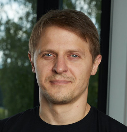 Linas Mažutis合作论文数Life Sciences Center, Vilnius University10Roshan Sharma合作论文数Memorial Sloan Kettering Cancer Center, Sloan Kettering Institute8
 Eric Holland合作论文数Division of Human Biology, Fred Hutchinson Cancer Research Center;Seattle Translational Tumor Research, Fred Hutchinson Cancer Research Center;Department of Neurological Surgery, University of Washington5Vaidotas Kiseliovas合作论文数4Dominik J. Otto合作论文数4Lisa Sikkema合作论文数These authors made an equal contribution to this work.4Ignas Masilionis合作论文数Memorial Sloan Kettering Cancer Center, Sloan Kettering Institute4
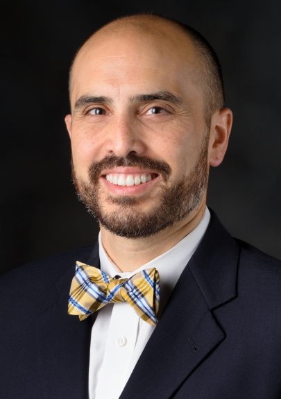 Jason T Huse合作论文数Department of Pathology and Translational Molecular Pathology, University of Texas MD Anderson Cancer Center;Brain Tumor Tissue Bank, University of Texas MD Anderson Cancer Center4
 Khan Aly A合作论文数4
 Zhao Jimmy L合作论文数Division of Biology and Biological Engineering, California Institute of Technology4
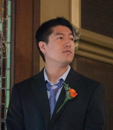 Chan Joseph M合作论文数Center for Computational Biology and Bioinformatics, Columbia University College of Physicians and Surgeons4Sonja Nowotschin合作论文数4
 Charles Sawyers合作论文数Memorial Sloan Kettering Cancer Center;Howard Hughes Medical Institute4Jillian R. Love合作论文数3Amina Lazrak合作论文数3
 Peter Vandenberghe合作论文数3
 Ronan Chaligne合作论文数3Ying-Yi Kuo合作论文数3Sitara Persad合作论文数Memorial Sloan Kettering Cancer Center3K B Nichols合作论文数3Virginia Teijeiro合作论文数Dev Biol Program, Sloan Kettering Inst3Christof Schneider合作论文数Univ Ulm, Dept Internal Med 3, Ulm, Germany3Zi-Ning Choo合作论文数3Lauren Fairchild合作论文数Mem Sloan Kettering Canc Ctr, Computat Biol Program, 1275 York Ave, New York, NY 10021 USA3
 Katia Basso合作论文数Vagelos College of Physicians and Surgeons, Columbia University3Mavrakis Konstantinos J合作论文数Cancer Biology &Genetics Program, Memorial Sloan-Kettering Cancer Center3
 Hana Andrlová合作论文数Department of Hematology and Oncology, University Medical Center3Olson Oakley C合作论文数Cancer Biology and Genetics Program, Memorial Sloan-Kettering Cancer Center3




