Ion channels play a central role in the regulation of nearly every cellular process. Dating back to the classic 1952 Hodgkin-Huxley model of the generation of the action potential, ion channels have always been thought of as independent agents. A myriad of recent experimental findings exploiting advances in electrophysiology, structural biology, and imaging techniques, however, have posed a serious challenge to this long-held axiom, as several classes of ion channels appear to open and close in a coordinated, cooperative manner. Ion channel cooperativity ranges from variable-sized oligomeric cooperative gating in voltage-gated, dihydropyridine-sensitive CaV1.2 and CaV1.3 channels to obligatory dimeric assembly and gating of voltage-gated NaV1.5 channels. Potassium channels, transient receptor potential channels, hyperpolarization cyclic nucleotide-activated channels, ryanodine receptors (RyRs), and inositol trisphosphate receptors (IP3Rs) have also been shown to gate cooperatively. The implications of cooperative gating of these ion channels range from fine-tuning excitation-contraction coupling in muscle cells to regulating cardiac function and vascular tone, to modulation of action potential and conduction velocity in neurons and cardiac cells, and to control of pacemaking activity in the heart. In this review, we discuss the mechanisms leading to cooperative gating of ion channels, their physiological consequences, and how alterations in cooperative gating of ion channels may induce a range of clinically significant pathologies.
Oscillations in membrane potential induced by synaptic inputs and intrinsic ion channel activity play a role in regulating neuronal excitability, but the precise mechanisms underlying their contributions remain largely unknown. Here we used electrophysiological and modeling approaches to investigate the effects of Gaussian white noise injected currents on the membrane properties and discharge characteristics of hypoglossal (HG) motoneurons in P16-21 day old rats. We found that the noise-induced membrane potential oscillations facilitated spike initiation by hyperpolarizing the cells' voltage threshold by 3.1 ± 1.0 mV and reducing the recruitment current for the tonic discharges by 0.26 ± 0.1 nA, on average (n = 59). Further analysis revealed that the noise reduced both recruitment and decruitment currents by 0.26 ± 0.13 and 0.33 ± 0.1 nA, respectively, and prolonged the repetitive firing. The noise also increased the slopes of frequency-current (F-I) relationships by 1.1 ± 0.2 Hz/nA. To investigate the potential mechanisms underlying these findings, we constructed a series of HG motoneuron models based on their electrophysiological properties. The models consisted of five compartments endowed with transient sodium (NaT), delayed-rectify potassium [K(DR)], persistent sodium (NaP), calcium-activated potassium [K(AHP)], L-type calcium (CaL) and H-current channels. In general, all our experimental results could be well fitted by the models, however, a modification of standard Hodgkin-Huxley kinetics was required to reproduce the changes in the F-I relationships and the prolonged discharge firing. This modification, corresponding to the noise generated by the stochastic flicker of voltage-gated ion channels (channel flicker, CF), was an adjustable sinusoidal function added to kinetics of the channels that increased their sensitivity to subthreshold membrane potential oscillations. Models with CF added to NaP and CaL channels mimicked the noise-induced alterations of membrane properties, whereas models with CF added to NaT and K(DR) were particularly effective in reproducing the noise-induced changes for repetitive firing observed in the real motoneurons. Further analysis indicated that the modified channel kinetics enhanced NaP- and CaL-mediated inward currents thus increasing the excitability and output of HG motoneurons, whereas they produced relatively small changes in NaT and K(DR), thus balancing these two currents and triggering variability of repetitive firing. This study provided insight into the types of membrane channel mechanisms that might underlie oscillation-induced alterations of neuronal excitability and motor output in rat HG motoneurons.
All movements are generated by the activation of motoneurons, and hence their input-output properties define the final step in processing of all motor commands. A major challenge to understanding this transformation has been the striking nonlinear behavior of motoneurons conferred by the activation of persistent inward currents (PICs) mediated by their voltage-gated Na+ and Ca2+ channels. In this review, we focus on the contribution that these PICs make to motoneuronal discharge and how the nonlinearities they engender impede the construction of a comprehensive model of motor control.
In the mSOD1 model of ALS, the excitability of motoneurons is poorly controlled, oscillating between hyperexcitable and hypoexcitable states during disease progression. The hyperexcitability is mediated by excessive activity of voltage-gated Na+ and Ca2+ channels that is initially counteracted by aberrant increases in cell size and conductance. The balance between these opposing actions collapses, however, at the time that the denervation of muscle fibers begins at about P50, resulting in a state of hypo-excitability and cell death. We propose that this process of neurodegeneration ensues from homeostatic dysregulation of excitability and have tested this hypothesis by perturbing a signal transduction pathway that plays a major role in controlling biogenesis and cell size. Our 『homeostatic dysregulation hypothesis' predicted that neonatal mSOD1 motoneurons would be much more sensitive to such perturbations than wild type controls and our results strongly support this hypothesis. Our results have important implications for therapeutic approaches to ALS.
Membrane ion channels are expressed into clusters in excitable cells. At present, however, the mechanisms leading to the formation and maintenance of this complex spatial organization are unknown. In this study, we used super resolution microscopy to determine the time course of TRPV4 and CaV1.2 channel cluster formation. Our data indicates that TRPV4 and CaV1.2 channel cluster area and density increase rapidly after the initiation of channel expression, but both eventually reach a plateau. At every time point, the probability distribution functions of cluster size were described by a single exponential. We generated a stochastic model to simulate CaV1.2 and TRPV4 cluster formation. The model has parameters that reflect channel cluster nucleation, growth, and removal probabilities. Probabilities were determined based on the cluster density and the mean cluster size in experiments. Key features of our stochastic model include a recycling mechanism of channel nucleation, growth, and removal. Furthermore, the model assumes a relatively rapid channel turnover rate, which we found critical to reproduce the experimentally-determined steady-state channel organization. Importantly, our model incorporates a self-regulating mechanism for CaV1.2 and TRPV4 channel cluster formation that assumes a feedback process between channel activity and cluster formation. Finally, our model can reproduce the distributions of TRPV4, CaV1.2, CaV1.3 and BK channels in cardiac and smooth muscle cells as well as in neurons. To conclude, our in vitro and in silico data support the view that TRPV4, CaV1.2, CaV1.3, BK, and TRPV4 channels are randomly inserted in the membranes of muscle cells and neurons and that clusters grow in the membrane of these cells until they reach a steady-state likely determined by a feedback mechanism.
Ion channels are often found arranged into dense clusters in the plasma membranes of excitable cells, but the mechanisms underlying the formation and maintenance of these functional aggregates are unknown. Here, we tested the hypothesis that channel clustering is the consequence of a stochastic self-assembly process and propose a model by which channel clusters are formed and regulated in size. Our hypothesis is based on statistical analyses of the size distributions of the channel clusters we measured in neurons, ventricular myocytes, arterial smooth muscle, and heterologous cells, which in all cases were described by exponential functions, indicative of a Poisson process (i.e., clusters form in a continuous, independent, and memory-less fashion). We were able to reproduce the observed cluster distributions of five different types of channels in the membrane of excitable and tsA-201 cells in simulations using a computer model in which channels are "delivered" to the membrane at randomly assigned locations. The model's three parameters represent channel cluster nucleation, growth, and removal probabilities, the values of which were estimated based on our experimental measurements. We also determined the time course of cluster formation and membrane dwell time for Ca(v)1.2 and TRPV4 channels expressed in tsA-201 cells to constrain our model. In addition, we elaborated a more complex version of our model that incorporated a self-regulating feedback mechanism to shape channel cluster formation. The strong inference we make from our results is that Ca(v)1.2, Ca(v)1.3, BK, and TRPV4 proteins are all randomly inserted into the plasma membranes of excitable cells and that they form homogeneous clusters that increase in size until they reach a steady state. Further, it appears likely that cluster size for a diverse set of membrane-bound proteins and a wide range of cell types is regulated by a common feedback mechanism.
Various membrane ion channels have been shown to cluster in excitable cells. However, the mechanism of spatiotemporal organization of cluster complexes is unknown. We used super resolution microscopy to study the time course of membrane expression of CaV1.2 and TRPV4 channels in transfected tsA‐201 cells. Channel clusters vary in size but can be fit exponentially at all time points. Similarly, mean cluster size and cluster density increase rapidly, but eventually reach a plateau around 24 hours post transfection. Using our findings, we devised a model for stochastic cluster assembly based on probabilities of three factors: cluster nucleation, growth and removal. This model reproduced distributions from previous studies of various channels in different tissues and suggests that a feedback process is responsible for determining a setpoint of cluster density and size.This abstract is from the Experimental Biology 2019 Meeting. There is no full text article associated with this abstract published in The FASEB Journal.
CaV1.3 channels regulate excitability in many neurons. As is the case for all voltage-gated channels, it is widely assumed that individual CaV1.3 channels behave independently with respect to voltage-activation, open probability, and facilitation. Here, we report the results of super-resolution imaging, optogenetic, and electrophysiological measurements that refute this long-held view. We found that the short channel isoform (CaV1.3S), but not the long (CaV1.3L), associates in functional clusters of two or more channels that open cooperatively, facilitating Ca2+ influx. CaV1.3S channels are coupled via a C-terminus-to-C-terminus interaction that requires binding of the incoming Ca2+ to calmodulin (CaM) and subsequent binding of CaM to the pre-IQ domain of the channels. Physically-coupled channels facilitate Ca2+ currents as a consequence of their higher open probabilities, leading to increased firing rates in rat hippocampal neurons. We propose that cooperative gating of CaV1.3S channels represents a mechanism for the regulation of Ca2+ signaling and electrical activity.
In the heart, reliable activation of Ca2+ release from the sarcoplasmic reticulum during the plateau of the ventricular action potential requires synchronous opening of multiple CaV1.2 channels. Yet the mechanisms that coordinate this simultaneous opening during every heartbeat are unclear. Here, we demonstrate that CaV1.2 channels form clusters that undergo dynamic, reciprocal, allosteric interactions. This ‘functional coupling’ facilitates Ca2+ influx by increasing activation of adjoined channels and occurs through C-terminal-to-C-terminal interactions. These interactions are initiated by binding of incoming Ca2+ to calmodulin (CaM) and proceed through Ca2+/CaM binding to the CaV1.2 pre-IQ domain. Coupling fades as [Ca2+]i decreases, but persists longer than the current that evoked it, providing evidence for ‘molecular memory’. Our findings suggest a model for CaV1.2 channel gating and Ca2+-influx amplification that unifies diverse observations about Ca2+ signaling in the heart, and challenges the long-held view that voltage-gated channels open and close independently.
The linear-nonlinear cascade model (LN model) has proven very useful in representing a neural system's encoding properties, but has proven less successful in reproducing the firing patterns of individual neurons whose behavior is strongly dependent on prior firing history. While the cell's behavior can still usefully be considered as feature detection acting on a fluctuating input, some of the coding capacity of the cell is taken up by the increased firing rate due to a constant "driving" direct current (DC) stimulus. Furthermore, both the DC input and the post-spike refractory period generate regular firing, reducing the spike-timing entropy available for encoding time-varying fluctuations. In this paper, we address these issues, focusing on the example of motoneurons in which an afterhyperpolarization (AHP) current plays a dominant role regularizing firing behavior. We explore the accuracy and generalizability of several alternative models for single neurons under changes in DC and variance of the stimulus input. We use a motoneuron simulation to compare coding models in neurons with and without the AHP current. Finally, we quantify the tradeoff between instantaneously encoding information about fluctuations and about the DC.
CaV1.3 channels play a critical role in pace-making of sinoatrial node cells and in the repetitive firing of neurons. We investigated the function and spatial organization of CaV1.3 channels in tsA-201 cells and in human iPSC-derived cardiomyocytes (hiPSC-CM) using patch-clamp, TIRF and super resolution GSD microscopy. The activation of CaV1.3 channels in these cells produced local Ca2+ signals called "CaV1.3 sparklets". Sparklet activity varied regionally with some regions of the surface membrane showing higher activity than others. The amplitude of the elementary CaV1.3 sparklet was about 40 nM. Sparklets with discrete amplitudes of 80, 120 and 160 nM were frequently observed, suggesting the simultaneous opening of 1-4 CaV1.3 channels. We used GSD super resolution imaging, with a spatial resolution of approximately 30 nm, to determine the organization of CaV1.3 channels. As shown in the super resolution map in Fig. 1, we found that CaV1.3 channels were expressed in clusters, of variable sizes and geometry, distributed at irregular intervals throughout the cell membrane. Our data suggest that CaV1.3 channels organize in clusters and that the simultaneous activation of channels within these clusters can produce high-amplitude Ca2+ signals. (NIH Grants HL085870, HL085686, NS077863)
Although the loss of motoneurons is an undisputed feature of amyotrophic lateral sclerosis (ALS) in man and in its animal models (SOD1 mutant mice), how the disease affects the size and excitability of motoneurons prior to their degeneration is not well understood. This study was designed to test the hypothesis that motoneurons in mutant SOD1(G93A) mice exhibit an enlargement of soma size (i.e., cross-sectional area) and an increase in Cav1.3 channel expression at postnatal day 30, well before the manifestation of physiological symptoms that typically occur at p90 (Chiu et al. 1995). We made measurements of spinal and hypoglossal motoneurons vulnerable to degeneration, as well as motoneurons in the oculomotor nucleus that are resistant to degeneration. Overall, we found that the somata of motoneurons in male SOD1(G93A) mutants were larger than those in wild-type transgenic males. When females were included in the two groups, significance was lost. Expression levels of the Cav1.3 channels were not differentiated by genotype, sex, or any interaction of the two. These results raise the intriguing possibility of an interaction between male sex steroid hormones and the SOD1 mutation in the etiopathogenesis of ALS.
 Nobutaka Hirokawa (廣川信隆)合作论文数Department of Cell Biology and Anatomy, Graduate School of Medicine, The University of Tokyo68
 uwe windhorst合作论文数66
 Randall K Powers合作论文数Department of Physiology;School of Medicine;University of Washington;School of Medicine, University of Washington37
 Charles J Heckman合作论文数Department of Neuroscience, Feinberg School of Medicine, Northwestern University12Dg Stuart合作论文数9
 Dixon Rose Ellen合作论文数Department of Physiology and Cell Biology, University of Nevada School of Medicine9
 Luis Fernando Santana合作论文数Department of Physiology and Membrane Biology, School of Medicine, University of California, Davis;Department of Biochemistry and Molecular Medicine, School of Medicine, University of California, Davis9
 Claudia M Moreno合作论文数Instituto de Fisiología Celular, Universidad Nacional Autónoma de México8Sawczuk A合作论文数7
 Manuel F Navedo合作论文数Department of Physiology and Biophysics, University of Washington6Can Yuan合作论文数5
 Robert M. Reinking合作论文数4
 Tajada Sendoa合作论文数Departamento de Bioquímica y Biología Molecular y Fisiología e Instituto de Biología y Genética Molecular (IBGM),, Universidad de Valladolid y4
 Daisuke Sato合作论文数3
 Collin Matsumoto合作论文数Sch Med, Univ Calif Davis3
 Paul Lucassen合作论文数Swammerdam Institute for Life Sciences, Faculty of Science, University of Amsterdam3
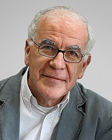 Dick F. Swaab合作论文数University of Amsterdam3
 Farrel Richard Robinson合作论文数3S.M Lucas合作论文数3George P. Moore合作论文数School of Engineering, University of Southern California3
 Colleen E. Clancy合作论文数Department of Physiology and Membrane Biology, School of Medicine, University of California, Davis3P Bawa合作论文数2Ximena Opitz-Araya合作论文数2
 Gonzalo Hernandez-Hernandez合作论文数Univ Calif Davis2E Henneman合作论文数2Paul Ruenzel合作论文数2Botterman B R合作论文数2Moritz Anna T合作论文数2McDonagh J C合作论文数2Gregory Newkirk合作论文数2Donald B. Percival合作论文数Department of Statistics, University of Washington;Applied Physics Laboratory, University of Washington2
 Adrienne L. Fairhall合作论文数Dept Physiol & Biophys, Univ Washington2
 T Richard Nichols (T Nichols)合作论文数School of Biological Sciences, College of Sciences, Georgia Institute of Technology2xin long guang chuan (信隆 廣川)合作论文数1Samantha C O'Dwyer合作论文数1
 Edward K Stauffer合作论文数1
 Renkai Ge合作论文数1JR Rosenberg合作论文数1Qiang Zhang合作论文数1Kemal Sitki Türker合作论文数1Hamm Thomas M合作论文数1
 William Rymer合作论文数Department of Biomedical Engineering, McCormick School of Engineering, Northwestern University;Shirley Ryan AbilityLab1
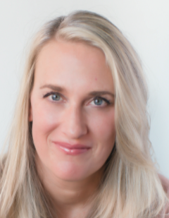 Vicki M Tysseling合作论文数Feinberg School of Medicine, Northwestern University1
 Robert Ruff合作论文数Department of Neurosciences, Case Western Reserve University1Cameron W E合作论文数1
 Rebecca Mease合作论文数Inst Neurosci, Tech Univ Munich1Lindsay A D合作论文数1
 James S Trimmer合作论文数Department of Physiology and Membrane Biology, School of Medicine, University of California, Davis Health1Fisher Erika合作论文数1




