Florencite is a hydrous light rare-earth elements (LREE) aluminium phosphate [REEAl3(PO4)(2)(OH)(6)], that amongst the REE-rich minerals is quite common. The main end-members are Ce-, La- and Nd-rich terms that were found in several genetic environments. Despite the large occurrence worldwide, to the authors' knowledge, florencite has attracted very few studies, particularly concerning the characterization of its Raman spectrum. We present a detailed study of the Raman spectrum of florencite, combining experimental measurements and theoretical calculations. Experimental Raman spectra (in the 100-1300 cm(-1) spectral range) are measured on four florencite samples characterized by different chemical composition, that is, different REE abundance. The results highlight a remarkable coincidence between different Raman spectra measured on each sample, despite the significantly different chemical compositions in terms of their REE content. The same similarities were also observed in the computed spectra at the ab initio level; moreover, the calculations allowed the attributions of the different Raman signals to specific vibrational modes.
The new mineral bonacinaite (IMA2018-056), Sc(AsO4) center dot 2H(2)O, was found on the dumps of the Varenche Mine (Saint-Barth & eacute;lemy, Nus, Aosta Valley, Italy), an old manganese mine, where it occurs as a low-temperature hydrothermal mineral associated mainly with quartz, granular braunite, undefined manganese oxides, arseniopleite, manganberzeliite and thortveitite. Bonacinaite forms colourless (with faint to distinct violet tints), pseudohexagonal, thick tabular crystals, up to 0.25 mm in size, sometimes with annular internal zones showing violet tinges, or as small, faintly violet lath-shaped crystals. The crystals are transparent and brittle, with vitreous lustre. The calculated density of an almost pure bonacinaite crystal is 2.82 g cm(-3). Optically, bonacinaite is biaxial negative, alpha=1.598(4), beta=1.618(3), and gamma=1.638(3) (measured with a Na light source, 589 nm); 2V (measured) is large, and 2V (calculated) = -88.9 degrees. The empirical formula, based on six O atoms per formula unit is (Sc0.90Mn0.083+Fe0.013+Pb0.01)(Sigma 1.00)[(As0.95P0.06)(Sigma 1.01)O-4] center dot 2H(2)O. Bonacinaite has monoclinic symmetry, with space group P2(1)/n and unit-cell parameters (single-crystal data powder diffraction data) , , & Aring;, , & Aring;(3) and Z=4. The crystal structure was refined from single-crystal intensity data obtained from a distinctly Al- and P-bearing crystal to R-1(F) = 3.7 % for 1178 reflections. Bonacinaite is isotypic with the other members of the metavariscite group: kolbeckite, metavariscite and phosphosiderite.
Piccoliite, ideally NaCaMn23+(AsO4)(2)O(OH), is a new mineral discovered in the Fe-Mn ore hosted in metaquartzites of the Montaldo di Mondoi mine, Corsaglia Valley, Cuneo Province, Piedmont, Italy. It occurs as small and rare black crystals and aggregates hosted by a matrix of quartz, associated with calcite and berzeliite/manganberzeliite. It has been also found in the Valletta mine near Canosio, Maira Valley, Cuneo Province, Piedmont, Italy, where it occurs embedded in quartz associated with grandaite, hematite, tilasite/adelite and rarely thorianite. The mineral is opaque (thin splinters may be very dark red), with brown streak and has a resinous to vitreous lustre. It is brittle with irregular fracture. No cleavage has been observed. The measured Mohs hardness is similar to 5-5.5. Piccoliite is non fluorescent. The calculated density is 4.08 g.cm(-3). Chemical spot analyses by electron microprobe analysis using wavelength dispersive spectroscopy resulted in the empirical formula (based on 10 anions per formula unit) (Na0.64Ca0.35)(Sigma 0.99)(Ca0.75Na0.24)(Sigma 0.99)(Mn1.083+Fe0.593+Mg0.20Ca0.10)(Sigma 1.97)(As2.03V0.03Si0.01)(Sigma 2.07)O-9(OH) and (Na0.53Ca0.47)(Sigma 1.00)(Ca0.76Na0.23Sr0.01)(Sigma 1.00)(Mn0.633+Fe0.493+Mg0.48Mn0.344+Ca0.06)(Sigma 2.00)(As1.97P0.01Si0.01)(Sigma 1.99)O-9(OH) for the Montaldo di Mondovi and Valletta samples, respectively. The mineral is orthorhombic, Pbcm, with single-crystal unit-cell parameters a = 8.8761(9), b = 7.5190(8), c = 11.689(1) angstrom and V = 780.1(1) angstrom(3) (Montaldo di Mondovi sample) and a = 8.8889(2), b = 7.5269(1), c = 11.6795(2) angstrom, V = 781.43(2) angstrom(3) (Valletta sample) with Z = 4. The seven strongest powder X-ray diffraction lines for the sample from Montaldo di Mondovi are [d angstrom (I-rel; hkl)]: 4.85 (57; 102), 3.470 (59; 120, 113), 3.167 (100; 022), 2.742 (30; 310, 213), 2.683 (53; 311, 023), 2.580 (50; 222, 114) and 2.325 (19; 320, 214, 223). The crystal structure (R-1 = 0.0250 for 1554 unique reflections for the Montaldo di Mondovi sample and 0.0260 for 3242 unique reflections for the Valletta sample) has MnO5(OH) octahedra forming edge-shared dimers; these dimers are connected through corner-sharing, forming two-up-two-down [M-[6](2)((TO4)-T-[4])(4)phi(2)] chains [M = Mn; T = As; phi = O(OH)] running along [001]. These chains are bonded in the a and b directions by sharing corners with AsO4 tetrahedra, giving rise to a framework of tetrahedra and octahedra hosting seven-coordinated Ca2+ and Na+ cations. The crystal structure of piccoliite is closely related to that of pilawite-(Y) as well as to carminite-group minerals that also show the same type of chains but with different linkage. The mineral is named after the mineral collectors Gian Paolo Piccoli and Gian Carlo Piccoli (father and son) (1926-1996 and b. 1953, respectively), the latter having discovered the type material at the Montaldo di Mondovi mine.
The new mineral species graulichite-(La), ideally LaFe33+(AsO4)2(OH)6, has been discovered in the Patte d'Oie mine, Bou Skour mining district, Morocco. It occurs as yellow rhombohedral crystals, up to 0.1 mm in size, with a resinous luster, associated with malachite, agardite-(La), conichalcite, and a still undetermined REE carbonate. Crystals are chemically zoned and two homogeneous domains were identified, corresponding to the empirical chemical formulae (calculated on the basis of 6 cations per formula unit, assuming the occurrence of 14 O atoms) (La0.34Ce0.20Ca0.11Sr0.07Pb0.05K0.04)Σ0.81(Fe2.163+Al0.84Cu0.20)Σ3.20(As1.23P0.39S0.37)Σ1.99O14H6.13 (domain #1) and (La0.38Ce0.22Sr0.10Ca0.09Pb0.05K0.06)Σ0.90(Fe2.603+Al0.49Cu0.20)Σ3.29(As0.91P0.50S0.40)Σ1.81O14H6.53 (domain #2). Single-crystal unit-cell parameters are a=7.252(13), c=16.77(3) Å, V=764(3) Å3, space group R-3m. The eight strongest reflections in the observed X-ray powder diffraction pattern are (d in Å, visually estimated intensity): 5.86, medium; 3.045, strong; 2.511, medium-weak; 2.239, medium; 1.960, medium-weak; 1.813, medium-weak; 1.689, medium-weak; 1.478, medium. Graulichite-(La) belongs to the dussertite group within the alunite supergroup. It is the La analogue of graulichite-(Ce) and the Fe3+ analogue of arsenoflorencite-(La).
Discreditation of the monoclinic tourmaline mineral species luinaite-(OH), ideally (Na,▯)(Fe2+,Mg)3Al6(BO3)3Si6O18(OH)4 was approved by the IMA-CNMNC (proposal 21-L) and is described. We analyzed two luinaite-(OH) samples: one from the type locality Cleveland tin mine, Luina, Waratah, Tasmania, Australia, and the other from Blue Mountain Saddle (Bald Hornet Claim), North Bend, King County, Washington, DC, USA. Biaxial (−) crystals representative of the studied samples were spectroscopically (Mössbauer, polarized Fourier transform infrared, optical absorption spectroscopy), chemically (nuclear microprobe analysis and electron microprobe analysis), and structurally characterized (single-crystal X-ray diffraction). Results show the occurrence of a triclinic structure for the studied luinaite-(OH) samples, which differs only in terms of a slight structural distortion from typical trigonal tourmaline structure (the topology of the structure is retained). As a result, following the IMA-CNMNC and tourmaline nomenclature rules, the triclinic luinaite-(OH) from the type locality (Australia) can be considered as the triclinic dimorph of schorl, as its chemical composition corresponds to schorl, and thus it should be referred as schorl-1A. Similarly, the triclinic sample from the USA can be considered as the triclinic dimorph of oxy-dravite, as its chemical composition corresponds to oxy-dravite, and then is referred to as oxy-dravite-1A.
AbstractLombardoite, ideally Ba2Mn3+(AsO4)2(OH), and aldomarinoite, ideally Sr2Mn3+(AsO4)2(OH), are two new minerals of the arsenbrackebuschite group in the brackebuschite supergroup, discovered in Fe–Mn ore in metaquartzites of the abandoned mine of Valletta, Canosio, Val Maira, Cuneo Province, Piedmont, Italy. They occur as red–brown and orange brown, respectively, as subhedral crystals (< 0.5 mm) in thin masses, associated with quartz, aegirine, baryte, calcite, hematite, muscovite and Mn minerals such as cryptomelane, braunite and manganberzeliite. Both minerals are translucent, have yellow–orange streak and vitreous lustre. Both are brittle. Estimated Mohs hardness is 6–6½ for lombardoite (by analogy to canosioite), and 4½–5 for aldomarinoite (by analogy to tokyoite). Calculated densities are 5.124 g/cm3 for lombardoite and 4.679 g/cm3 for aldomarinoite. Both minerals are biaxial (+). Lombardoite shows 2Vz(meas.) = 78(4)° and is pleochroic with X = yellowish brown, Y = brown and Z = reddish brown (Z > Y > X). Aldomarinoite has 2Vz(meas.) = 67.1(1)°, and is pleochroic with X = brown, Y = brownish orange and Z = yellowish brown (Z > Y > X). Point analyses by electron microprobe using wavelength dispersive spectroscopy resulted in the empirical formula (based on 9 O anions): (Ba1.96Sr0.17Pb0.04Na0.02Ca0.02)Σ2.21(Mn3+0.62Fe3+0.13Al0.06Mg0.11)Σ0.92[(As0.87V0.12P0.01)Σ1.00O4]2(OH) for lombardoite, and (Sr1.93Ca0.21Ba0.04Pb0.01)Σ2.19(Mn3+0.48Al0.35Fe3+0.21Mg0.01)Σ1.05[(As0.92V0.03)Σ0.95O4]2(OH) for aldomarinoite. The absence of H2O was confirmed by Raman spectroscopy and infrared spectroscopy. Both minerals are monoclinic, P21/m, with unit-cell parameters a = 7.8636(1) Å, b = 6.13418(1) Å, c = 9.1197(1) Å, β = 112.660(2)° and V = 405.94(1) Å3, for lombardoite and a = 7.5577(4) Å, b = 5.9978(3) Å, c = 8.7387(4) Å, β = 111.938(6)° and V = 367.43(3) Å3, for aldomarinoite. The eight strongest powder X-ray diffraction lines are [d, Å (Irel) (hkl)]: 6.985 (39) (10$\bar{1}$), 3.727 (33) (111), 3.314 (100) (21$\bar{1}$), 3.073 (24) (020), 3.036 (33) (21$\bar{2}$, 10$\bar{3}$), 2.810 (87) (12$\bar{1}$, 112), 2.125 (20) (301, 11$\bar{4}$) and 1.748 (24) (321) for lombardoite and 3.191 (89) (21$\bar{1}$), 2.997 (45) (020), 2.914 (47) (21$\bar{2}$, 10$\bar{3}$), 2.715 (100) (112), 2.087 (39) (12$\bar{3}$, 1.833 (32) (31$\bar{4}$), 1.689 (36) (321), 1.664 (21) (132) for aldomarinoite. The minerals are isostructural with brackebuschite: infinite chains of edge sharing octahedra running parallel to the b axis and decorated with AsO4 groups are connected along the a and c axes through Ba and Sr atoms in lombardoite and aldomarinoite, respectively. The minerals are named after Bruno Lombardo (1944–2014), geologist and petrologist at C.N.R. (National Research Council of Italy), and Aldo Marino (b. 1942) the mineral collector and founding member of the AMI – Italian Micromineralogical Association.
The new mineral species graulichite-(La), ideally LaFe33+(AsO4)(2)(OH)(6), has been discovered in the Patte d'Oie mine, Bou Skour mining district, Morocco. It occurs as yellow rhombohedral crystals, up to 0.1 mm in size, with a resinous luster, associated with malachite, agardite(La), conichalcite, and a still undetermined REE carbonate. Crystals are chemically zoned and two homogeneous domains were identified, corresponding to the empirical chemical formulae (calculated on the basis of 6 cations per formula unit, assuming the occurrence of 14 O atoms) (La0.34Ce0.20Ca0.11Sr0.07Pb0.05K0.04)(Sigma 0.81)(Fe2.163+Al0.84Cu0.20)(Sigma 3.20)(As1.23P0.39S0.37)(Sigma 1.99)O14H6.13 (domain #1) and (La0.38Ce0.22Sr0.10Ca0.09Pb0.05K0.06)(Sigma 0.90)(Fe2.603+Al0.49Cu0.20)(Sigma 3.29)(AS(0.91)P(0.50)S(0.40))(Sigma 1.81) O14H6.53 (domain #2). Single-crystal unit-cell parameters are a = 7.252(13), c = 16.77(3) angstrom, V = 764(3) angstrom(3), space group R-3m. The eight strongest reflections in the observed X-ray powder diffraction pattern are (d in angstrom, visually estimated intensity): 5.86, medium; 3.045, strong; 2.511, medium-weak; 2.239, medium; 1.960, medium-weak; 1.813, medium-weak; 1.689, medium-weak; 1.478, medium. Graulichite-(La) belongs to the dussertite group within the alunite supergroup. It is the La analogue of graulichite-(Ce) and the Fe3+ analogue of arsenoflorencite-(La).
A crystal fragment of schorl from Langesundsfjord (Norway), showing a zonation with a biaxial optic behavior in the rim, was studied by electron microprobe analysis, single-crystal X-ray diffraction, Mossbauer, infrared and optical absorption spectroscopy and optical measurements. Measured 2V(x) is 15.6 degrees. We concluded that biaxial character of the sample is not due to internal stress because it cannot be removed by heating and cooling. Diffraction data were refined with a standard R3m space group model, with a = 16.0013(2) angstrom, c = 7.2263(1) angstrom, and with a non-conventional triclinic R1 space-group model keeping the same hexagonal triple cell (a = 16.0093(5) angstrom, b = 16.0042(5) angstrom, c = 7.2328(2) angstrom, alpha = 90.008(3)degrees, beta = 89.856(3)degrees, gamma = 119.90(9)degrees), yielded Rall = 1.75% (3136 unique reflections) vs. R-all = 2.53% (17342 unique reflections), respectively. The crystal-chemical analysis resulted in the chemical formula (X)(Na0.98K0.01 square(0.01))(Sigma 1.00)(Y)(Fe(1.53)(2+)Al(0.68)Mg(0.35)Ti(0.20)Fe(0.20)(3+)Mn(0.0)2V(0.01)Zn(0.01))(Sigma 3.00)(Z) (Al5.10Fe0.502+Mg0.40)(Sigma 6.00)(Si6O18)(BO3)(3)(OH)(3)[(OH)(0.39)F-0.O-22(0.39)](Sigma 1.00), which agrees well in terms of calculated site-scattering (X 10.9 epfu, Y 63.7 epfu, Z 83.7 epfu) and refined site-scattering (X 11.4 epfu, Y 63.4 epfu, Z 83.6 epfu). About 0.19 apfu Fe2+ is at the Z sites in the R1 model that showed that one out of six independent Z sites (Zd) has higher refined site scattering [15.5 eps vs. mean 13.7(2) eps for the other five sites] and larger mean bond length [1.969 angstrom vs. 1.927(6) angstrom for the other five sites] and larger octahedral angle variance [53 degrees vs. 42(3)degrees]. All these features support local order of Fe2+ at the Zd site. Optical absorption spectra also show evidence of Fe2+ at the Z sites. The elongation of the Zd-octahedron is along a direction that forms an angle of ca. 73 degrees with a unit-cell edge and is coincident with the direction of the gamma-refraction index. All these data support the triclinic character of the structure of the optically biaxial part of the tourmaline sample from Langesundsfjord and provide evidence that even in the presence of excellent statistical agreement factors from excellent X-ray diffraction data, the lowering of symmetry due to cation ordering may have been overlooked in many other tourmaline samples in the absence of a check of the optical behaviour. According to the nomenclature rules, the studied triclinic schorl, should be named schorl-1A.
The new mineral species graulichite-(La), ideally LaFe33+(AsO4)2(OH)6, has been discovered in the Patte d'Oie mine, Bou Skour mining district, Morocco. It occurs as yellow rhombohedral crystals, up to 0.1 mm in size, with a resinous luster, associated with malachite, agardite-(La), conichalcite, and a still undetermined REE carbonate. Crystals are chemically zoned and two homogeneous domains were identified, corresponding to the empirical chemical formulae (calculated on the basis of 6 cations per formula unit, assuming the occurrence of 14 O atoms) (La0.34Ce0.20Ca0.11Sr0.07Pb0.05K0.04)Σ0.81(Fe2.163+Al0.84Cu0.20)Σ3.20(As1.23P0.39S0.37)Σ1.99O14H6.13 (domain #1) and (La0.38Ce0.22Sr0.10Ca0.09Pb0.05K0.06)Σ0.90(Fe2.603+Al0.49Cu0.20)Σ3.29(As0.91P0.50S0.40)Σ1.81O14H6.53 (domain #2). Single-crystal unit-cell parameters are a=7.252(13), c=16.77(3) Å, V=764(3) Å3, space group R-3m. The eight strongest reflections in the observed X-ray powder diffraction pattern are (d in Å, visually estimated intensity): 5.86, medium; 3.045, strong; 2.511, medium-weak; 2.239, medium; 1.960, medium-weak; 1.813, medium-weak; 1.689, medium-weak; 1.478, medium. Graulichite-(La) belongs to the dussertite group within the alunite supergroup. It is the La analogue of graulichite-(Ce) and the Fe3+ analogue of arsenoflorencite-(La).
AbstractPiccoliite, ideally NaCaMn3+2(AsO4)2O(OH), is a new mineral discovered in the Fe–Mn ore hosted in metaquartzites of the Montaldo di Mondovì mine, Corsaglia Valley, Cuneo Province, Piedmont, Italy. It occurs as small and rare black crystals and aggregates hosted by a matrix of quartz, associated with calcite and berzeliite/manganberzeliite. It has been also found in the Valletta mine near Canosio, Maira Valley, Cuneo Province, Piedmont, Italy, where it occurs embedded in quartz associated with grandaite, hematite, tilasite/adelite and rarely thorianite. The mineral is opaque (thin splinters may be very dark red), with brown streak and has a resinous to vitreous lustre. It is brittle with irregular fracture. No cleavage has been observed. The measured Mohs hardness is ~5–5.5. Piccoliite is non fluorescent. The calculated density is 4.08 g⋅cm–3. Chemical spot analyses by electron microprobe analysis using wavelength dispersive spectroscopy resulted in the empirical formula (based on 10 anions per formula unit) (Na0.64Ca0.35)Σ0.99(Ca0.75Na0.24)Σ0.99(Mn3+1.08Fe3+0.59Mg0.20Ca0.10)Σ1.97(As2.03V0.03Si0.01)Σ2.07O9(OH) and (Na0.53Ca0.47)Σ1.00(Ca0.76Na0.23Sr0.01)Σ1.00(Mn3+0.63Fe3+0.49Mg0.48Mn4+0.34Ca0.06)Σ2.00(As1.97P0.01Si0.01)Σ1.99O9(OH) for the Montaldo di Mondovì and Valletta samples, respectively. The mineral is orthorhombic, Pbcm, with single-crystal unit-cell parameters a = 8.8761(9), b = 7.5190(8), c = 11.689(1) Å and V = 780.1(1) Å3 (Montaldo di Mondovì sample) and a = 8.8889(2), b = 7.5269(1), c = 11.6795(2) Å, V = 781.43(2) Å3 (Valletta sample) with Z = 4. The seven strongest powder X-ray diffraction lines for the sample from Montaldo di Mondovì are [d Å (Irel; hkl)]: 4.85 (57; 102), 3.470 (59; 120, 113), 3.167 (100; 022), 2.742 (30; 310, 213), 2.683 (53; 311, 023), 2.580 (50; 222, 114) and 2.325 (19; 320, 214, 223). The crystal structure (R1 = 0.0250 for 1554 unique reflections for the Montaldo di Mondovì sample and 0.0260 for 3242 unique reflections for the Valletta sample) has MnO5(OH) octahedra forming edge-shared dimers; these dimers are connected through corner-sharing, forming two-up-two-down [[6]M2([4]TO4)4φ2] chains [M = Mn; T = As; φ = O(OH)] running along [001]. These chains are bonded in the a and b directions by sharing corners with AsO4 tetrahedra, giving rise to a framework of tetrahedra and octahedra hosting seven-coordinated Ca2+ and Na+ cations. The crystal structure of piccoliite is closely related to that of pilawite-(Y) as well as to carminite-group minerals that also show the same type of chains but with different linkage. The mineral is named after the mineral collectors Gian Paolo Piccoli and Gian Carlo Piccoli (father and son) (1926–1996 and b. 1953, respectively), the latter having discovered the type material at the Montaldo di Mondovì mine.
Abstract Armellinoite-(Ce), ideally Ca4Ce4+(AsO4)4⋅H2O, is a new mineral discovered in Fe–Mn ore in metaquartzites of the Montaldo mine, Corsaglia Valley, Cuneo Province, Piedmont, Italy. It occurs as very small and rare, pale yellow to brown–yellow pseudo-octahedral translucent crystals hosted by a matrix of quartz, hematite, cryptomelane/hollandite, tilasite, muscovite, braunite and montmorillonite. The mineral is translucent, with white streak and has a resinous to vitreous lustre. It is brittle with irregular fracture and fair cleavage parallel to {110} and {100}. Estimated Mohs hardness is ~3–3.5. Calculated density is 4.29 g⋅cm–3. Armellinote-(Ce) is uniaxial (–), ω = 1.795(5), ɛ = 1.765(5) (white light), non-pleochroic and non-fluorescent. Chemical point analyses by WDS-EPMA yielded the empirical formula (based on 17 O+F anions): A(Ca3.89Th0.08Sr0.02La0.03)Σ4.02B(Ce4+0.76Nd0.13Y0.08Gd0.03Sm0.02Pr0.01Dy0.01Ho0.01)Σ1.05[(As4.00P0.01)Σ4.01O4]4⋅(H2O0.85F0.15)Σ2.00. The presence of H2O was confirmed by Raman spectroscopy. The mineral is tetragonal, I41/a, with single-crystal unit-cell parameters a = 10.749(2), c = 12.030(2) Å and V = 1390.0(6) Å3, with Z = 4. The eight strongest X-ray powder diffraction lines are [d Å (Irel; hkl)]: 7.983 (36; 101), 4.443 (23; 2̄11), 2.957 (100; 3̄12), 2.398 (14; 420), 1.875 (22; 424, 325), 1.728 (19; 3̄16), 1.612 (13; 613) and 1.475 (26; 712, 552). The crystal structure (R1 = 0.0284 for 1275 unique reflections) has isolated TO4 (T = As5+) tetrahedra that link Ca2+- or Ce4+-centred polyhedra via common oxygen ligands to form 2D blocks or double-layered (DL) structural units parallel to (001). Armellinoite-(Ce) is isostructural with pottsite, ideally (Pb3Bi)Bi(VO4)4⋅H2O, and closely related to a larger number of anhydrous synthetic compounds. The mineral is named after the mineral collector Gianluca Armellino (b. 1962), who collected the discovery sample.
Demagistrisite, ideally BaCa2Mn3+4(Si3O10)(Si2O7)(OH)4·3H2O, is a new mineral found at the Cerchiara mine (eastern Liguria, La Spezia province, Italy). The ore consists of rhythmic interlaying of braunite-bearing metasediments (5–15 cm thick) and hematite-rich cherts. Demagistrisite occurs in association with cerchiaraite-(Mn), namansilite, noelbensonite, orientite, richterite, ruizite, and saponite in matrix consisting of braunite, calcite, cryptomelane, orthoclase, and quartz. Demagistrisite crystals occur as tightly intergrown blades or as millimeter-sized prisms and needles with square cross-section, typically with irregular terminations, and rarely terminated by a low-angle pyramid. The mineral is orange brown to red brown, streak is beige, and luster is vitreous, translucent to transparent. Fracture is irregular. In thin section, it is orange brown. The mineral is optically biaxial (–) with α 1.805(5), β 1.825(5), γ 1.8305(5) (white light); 2Vmeas 58(5)°, 2Vcalc 54.7°; optical orientation X = c, Y = b, Z = a. Dispersion is very strong, r > v. Pleochroism is strong with X orange yellow, Y red brown, Z red brown; X << Z < Y. It is unreactive in concentrated HCl at room temperature. Thirteen chemical analyses by WDS-EMPA gave the following empirical formula (based on 24 O apfu): (Ba0.69Ca1.25Mn2+0.70Sr0.21Na0.12Mg0.02)Σ2.99(Mn3+3.97Al0.03)Σ4(Si3O10)(Si2O7)(OH)3.87·3.13H2O. The mineral is orthorhombic, space group Amm2, with unit-cell parameters a 16.312(8), b 6.176(4), c 9.075(6) Å, V 914.2(10) Å3, and Z = 2. The seven strongest X-ray powder diffraction lines are [d Å (I%; hkl)]: 16.21 (49; 100), 4.86 (44; 111), 4.34 (56; 102,211), 2.871 (54; 220), 2.731 (100; 511,013), 2.671 (74; 320,113,502), and 2.426 (51; 222,313,611). The crystal structure (R1 = 0.0572 for 1485 reflections with I > 2σI) is based on straight edge-sharing chains of Mn3+-centered octahedra extending along [010], which are bridged by disilicate (Si2O7) and trisilicate (Si3O10) groups, yielding a framework. Cavities within this framework contain two large cation sites. The structure of demagistrisite can be considered transitional between the structures of orientite and noelbensonite. Demagistrisite is named in honor of Leandro de Magistris (1906–1990).
Abstract A gem-quality purplish-red tourmaline sample of alleged liddicoatitic composition from the Anjanabonoina pegmatite, Madagascar, has been fully characterised using a multi-analytical approach to define its crystal-chemical identity. Single-crystal X-ray diffraction, chemical and spectroscopic analysis resulted in the formula: X(Na0.41□0.35Ca0.24)Σ1.00Y(Al1.81Li1.00Fe3+0.04Mn3+0.02Mn2+0.12Ti0.004)Σ3.00ZAl6 [T(Si5.60B0.40)Σ6.00O18] (BO3)3 (OH)3 W[(OH)0.50F0.13O0.37]Σ1.00 which corresponds to the tourmaline species elbaite having the typical space group R3m and relatively small unit-cell dimensions, a = 15.7935(4) Å, c = 7.0860(2) Å and V = 7.0860(2) Å3. Optical absorption spectroscopy showed that the purplish-red colour is caused by minor amounts of Mn3+ (Mn2O3 = 0.20 wt.%). Thermal treatment in air up to 750°C strongly intensified the colour of the sample due to the oxidation of all Mn2+ to Mn3+ (Mn2O3 up to 1.21 wt.%). Based on infrared and Raman data, a crystal-chemical model regarding the electrostatic interaction between the X cation and W anion, and involving the Y cations as well, is proposed to explain the absence or rarity of the mineral species ‘liddicoatite’.
Abstract A green tourmaline sample from the Tzarevskoye uranium–vanadium deposit, close to the Srednyaya Padma deposit, Lake Onega, Karelia Republic, Russia, has been found to be the second world-occurrence of Cr-rich vanadio-oxy-dravite in addition to the Pereval marble quarry, Sludyanka crystalline complex, Lake Baikal, Russia, type-locality. From the crystal-structure refinement and chemical analysis, the following empirical formula is proposed: X(Na0.96K0.02□0.02)Σ1.00 Y(V1.34Al0.68Mg0.93Cu2+0.02Zn0.01Ti0.01)Σ3.00 Z(Al3.19Cr1.36V0.03Mg1.42)Σ6.00(TSi6O18)(BBO3)3V(OH)3W[O0.60(OH)0.23F0.17]Σ1.00. Together with the data from the literature, a compositional overview of Al–V–Cr–Fe3+-tourmalines is provided by using [6]Al–V–Cr–Fe3+ diagrams for tourmaline classification. These diagrams further simplify the tourmaline nomenclature as they merge the chemical information over the octahedrally-coordinated sites (Y and Z) by removing the issues of uncertainty associated with cation order–disorder across Y and Z. Results show the direct identification of tourmalines by using the chemical data alone.
The new mineral species rüdlingerite, ideally Mn2+2V5+As5+O7·2H2O, occurs in the Fianel mine, in Val Ferrera, Grisons, Switzerland, a small Alpine metamorphic Mn deposit. It is associated with ansermetite and Fe oxyhydroxide in thin fractures in Triassic dolomitic marbles. Rüdlingerite was also found in specimens recovered from the dump of the Valletta mine, Canosio, Cuneo, Piedmont, Italy, where it occurs together with massive braccoite and several other As- and V-rich phases in richly mineralized veins crossing the quartz-hematite ore. The new mineral displays at both localities yellow to orange, flattened elongated prismatic, euhedral crystals measuring up to 300 μm in length. Electron-microprobe analysis of rüdlingerite from Fianel gave (in wt%): MnO 36.84, FeO 0.06, As2O5, 25.32, V2O5 28.05, SiO2 0.13, H2Ocalc 9.51, total 99.91. On the basis of 9 O anions per formula unit, the chemical formula of rüdlingerite is Mn1.97(V5+1.17 As0.83Si0.01)Σ2.01O7·2H2O. The main diffraction lines are [dobs in Å (Iobs) hkl]: 3.048 (100) 022, 5.34 (80) 120, 2.730 (60) 231, 2.206 (60) 16-1, 7.28 (50) 020, 2.344 (50) 250, 6.88 (40) 110, and 2.452 (40) 320. Study of the crystal structure showcases a monoclinic unit cell, space group P21/n, with a = 7.8289(2) Å, b = 14.5673(4) Å, c = 6.7011(2) Å, β = 93.773(2)°, V = 762.58(4) Å3, Z = 4. The crystal structure has been solved and refined to R1 = 0.041 on the basis of 3784 reflections with Fo > 4σ(F). It shows Mn2+ hosted in chains of octahedra that are subparallel to [-101] and bound together by pairs of tetrahedra hosted by V5+ and As5+, building up a framework. Additional linkage is provided by hydrogen-bonding through H2O coordinating Mn2+ at the octahedra. One tetrahedrally coordinated site is dominated by V5+, T(1)(V0.88As0.12), corresponding to an observed site scattering of 24.20 electrons per site (eps), whereas the second site is strongly dominated by As5+, T(2)(As0.74V0.26), with, accordingly, a higher observed site scattering of 30.40 eps. The new mineral has been approved by the IMA-CNMNC and named for Gottfried Rüdlinger (born 1919), a pioneer in the 1960–1980s, in the search and study of the small minerals from the Alpine manganese mineral deposits of Grisons.
AbstractMonteneroite (IMA2020-028), Cu2+Mn2+2(AsO4)2⋅8H2O, is a new vivianite-structure mineral from the Monte Nero mine, Rocchetta di Vara, La Spezia, Liguria, Italy. It is a secondary mineral that crystallised from As-, Cu- and Mn-rich fluids and it is associated with braunite, copper, cuprite, rhodochrosite and strashimirite. Monteneroite occurs as light green, thick blades up to ~2.5 mm long. The streak is white. Crystals are transparent with vitreous lustre. The mineral has Mohs hardness of 2, is somewhat sectile, exhibits two cleavages ({010} perfect and {001} fair) and has irregular stepped fracture. The measured density is 2.97(2) g cm–3. Monteneroite is optically biaxial (+), with α = 1.604(2), β = 1.637(2) and γ = 1.688(2), determined in white light; 2V = 80(1)°; slight dispersion is r < v, orientation: X = b; Z ^ c = 52° in obtuse β. Electron microprobe analyses provided the empirical formula (Cu2+0.88Mn2+0.11)Σ0.99Mn2+2.00(As1.00O4)2⋅8H2O. Monteneroite is monoclinic, C2/m, a = 10.3673(14), b = 13.713(2), c = 4.8420(8) Å, β = 105.992(8)°, V = 661.72(18) Å3 and Z = 2. Monteneroite has a vivianite-type structure (R1 = 0.0535 for 534 I > 2σI reflections). It is the first mineral with this structure type to be defined with ordered octahedral cation sites.
Monteneroite (IMA2020-028), Cu2+Mn22+(AsO4)(2)center dot 8H(2)O, is a new vivianite-structure mineral from the Monte Nero mine, Rocchetta di Vara, La Spezia, Liguria, Italy. It is a secondary mineral that crystallised from As-, Cu- and Mn-rich fluids and it is associated with braunite, copper, cuprite, rhodochrosite and strashimirite. Monteneroite occurs as light green, thick blades up to similar to 2.5 mm long. The streak is white. Crystals are transparent with vitreous lustre. The mineral has Mohs hardness of 2, is somewhat sectile, exhibits two cleavages ({010} perfect and {001} fair) and has irregular stepped fracture. The measured density is 2.97(2) g cm(-3). Monteneroite is optically biaxial (+), with alpha = 1.604(2), beta = 1.637(2) and gamma = 1.688(2), determined in white light; 2V = 80(1)degrees; slight dispersion is r < v, orientation: X = b; Z <^> c = 52 degrees in obtuse beta. Electron microprobe analyses provided the empirical formula (Cu0.882+Mn0.112+)(Sigma 0.99)Mn-2.00(2+)(As1.00O4)(2).8H(2)O. Monteneroite is monoclinic, C2/m, a = 10.3673(14), b = 13.713(2), c = 4.8420(8) angstrom, beta = 105.992(8)degrees, V = 661.72(18) angstrom(3) and Z = 2. Monteneroite has a vivianite-type structure (R-1 = 0.0535 for 534 I > 2 sigma I reflections). It is the first mineral with this structure type to be defined with ordered octahedral cation sites.
Fluorcarmoite-(BaNa), ideally Ba-A1(A2)square(NaNa2Na3)-Na-B1,2-Na-Na1,2 square(CaMg13Al)-Ca-Ca-Mg-M(PO4)(11)(PO3OH)F-W(2), was found in a pebble of the riverbed of the upper Maremola Creek, close to the village of Isallo, in the Magliolo municipality (Savona, Liguria, Italy). The root-name is after Monte Carmo di Loano, the highest peak in the area, namesake of the tectonic unit where the mineral was found and the first locality where phosphate mineralization has been found in the region. The mineral is associated with quartz and almandine and has microscopic inclusions of fluorapatite and possible graftonite. It occurs as yellow-orange and translucent crystals in an anhedral centimetric nodule embedded in quartz. Fluorcarmoite-(BaNa) is brittle, and no cleavage or parting was observed. It has a yellow-orange streak, a vitreous lustre, does not fluoresce under shortwave or longwave ultraviolet light and is weakly pleochroic (light yellow). Fluorcarmoite-(BaNa) is optically biaxial positive, with alpha = 1.6240(5), beta = 1.6255(5), gamma = 1.6384(5) (589 nm), 2V(meas) = 35(2)degrees and 2V(calc) = 37.9 degrees. Raman spectroscopy shows the presence of weak bands in the OH-stretching region. The average chemical composition is (wt%, wavelength-dispersive-mode electron microprobe): Na2O 5.83, K2O 0.36, CaO 2.64, SrO 0.46, BaO 7.12, MnO 2.01, FeO 17.68, MgO 15.12, Al2O3 2.57, P2O5 44.96, F 2.14, -O = F-2 0.90, H2Ocalc 0.33, total 100.32. The empirical formula calculated on the basis of 50 O + F + (OH) atoms per formula unit (apfu), is: (Na3.77Ca0.94Ba0.93 K0.15Sr0.09 square(0.12))(Sigma=6.00)(Mg7.52Fe4.932+Mn0.572+)(Sigma)Al-=13.02(1.01)(PO4)(11)(PO3)(OH0.74F0.26)F-2. Strongest lines in the X-ray powder diffraction pattern are [d in angstrom (I-calc) hkl]: 4.959 (25) 020, 4.524 (20) 114, 3.188 (28) 206, 3.012 (100) 42 (4) over bar, 2.735 (32) 60 (2) over bar, 2.682 (39) 226, 2.526 (25) 424. The crystal structure has been refined using single-crystal X-ray diffractometer data (R-int = 4.1%) in space group Cc (no. 14) to R-1 = 0.0342 for 11 511 reflections with F-o > 4 sigma vertical bar F vertical bar and 0.0417 for all 13 232 data. Refined unit-cell parameters are: a = 16.4013(3) angstrom, b = 9.9487(1) angstrom, c = 24.4536(8) angstrom, beta = 105.725(2)degrees, V = 3840.80(15) angstrom(3) (Z= 4). Fluorcarmoite-(BaNa) is the first Mg-dominant mineral of the arrojadite group. Mg orders preferentially in the M1, M2b, M3a,b, M4a,b and M7a,b sites whereas the non-dominant Fe2+ and very minor Mn2+ show site preference for M2a, M5a,b and M6a,b. The Al site is mostly populated by Ba, the A2 site is empty, and minor Fe2+ occurs at the B1b site. A significant, but not dominant occupancy of the Na3 site by Na is also observed. Only Ca and Al are present at the Ca and Al sites, respectively. The type material is deposited in the mineralogical collection of the Museo Regionale di Scienze Naturali di Torino, Sezione di Mineralogia, Petrografia e Geologia, Torino (Italy). The mineral and its name have been approved by the IMA-CNMNC (2015-062).
Two new monoclinic (P21/m) epidote supergroup minerals manganiakasakaite-(La) and ferriakasakaite-(Ce) were found in the small Mn ore deposit of Monte Maniglia, Bellino, Varaita Valley, Cuneo Province, Piedmont, Italy. Manganiakasakaite-(La) occurs as subhedral grains embedded in pyroxmangite. Its empirical formula is A(1)(Ca0.62Mn2+0.38) A(2)(La0.52Nd0.08Pr0.07Ce0.07Y0.01Ca0.25) M(1)(Mn3+0.52Fe3+0.28Al0.18V3+0.01) M(2)Al1.00 M(3)(Mn2+0.60Mn3+0.27Mg0.13) T(1−3)(Si2.99Al0.01) O12 (OH), corresponding to the end-member formula CaLaMn3+AlMn2+(Si2O7)(SiO4)O(OH). Unit-cell parameters are a = 8.9057(10), b = 5.7294(6), c = 10.1134(11) Å, β = 113.713(5)°, V = 472.46(9) Å3, Z = 2. The crystal structure of manganiakasakaite-(La) was refined to a final R1 = 0.0262 for 2119 reflections with Fo > 4σ(Fo) and 125 refined parameters. Ferriakasakaite-(Ce) occurs as small homogeneous domains within strongly inhomogeneous prismatic crystals, where other epidote supergroup minerals coexist [manganiandrosite-(Ce), “androsite-(Ce)”, and epidote]. Associated minerals are calcite and hematite. Its empirical formula is A(1)(Ca0.64Mn2+0.36) A(2)(Ce0.37La0.17Nd0.06Pr0.03Ca0.35□0.02) M(1)(Fe3+0.61Al0.39) M(2)Al1.00 M(3)(Mn2+0.64Mn3+0.33Fe3+0.02Mg0.01) T(1−3)Si3.01 O12 (OH), the end-member formula being CaCeFe3+AlMn2+(Si2O7)(SiO4)O(OH). Unit-cell parameters are a = 8.9033(3), b = 5.7066(2), c = 10.1363(3) Å, β = 114.222(2)°, V = 469.66(3) Å3, Z = 2. The crystal structure of ferriakasakaite-(Ce) was refined to a final R1 = 0.0196 for 1960 unique reflections with Fo > 4σ(Fo) and 124 refined parameters.
The mineral description is provided for ferro-tschermakite, ideally (A)square Ca-B(2)C(Fe32+Al2)(T)(Si6Al2)O-22(W)(OH)(2). The type specimen has been found in the dump of the Batiment et Granit de Ploumanac'h northern granite quarry, La Clarte, Perros-Guirec, Ploumanac'h granitic complex, Brittany, France. The empirical formula derived from electron microprobe analysis and single-crystal structure refinement is: (A)(Na0.29K0.08)(Sigma= 0.37)(B)(Ca1. 69Fe0. 112+Mn0.022+Na0.18)(Sigma=2.00)(C)(Fe1. 842+Mg1.54Al1.33Fe0.243+V0.013+ Ti-0.04)(Sigma= 5.00)(T)(Si6.15Al1.85)(Sigma= 8.00)O-22(W)(OH1.94F0.06)(Sigma= 2.00). Ferro-tschermakite is biaxial (-), with alpha = 1.666(2), beta = 1.680(2), gamma = 1.690(2) and 2V (meas.) = 84(1)degrees, 2V (calc.) = 79.8. The dispersion is medium (r> v), and the orientation is: X <^> a = 9.5 degrees (in beta acute), Y || b, Z <^> c = 24.3 degrees (in beta obtuse). The unit-cell parameters are a = 9.7598(6), b = 18.0220(11), c = 5.3299(3) angstrom, beta = 104.826(1)degrees, V = 906.27 (9) angstrom, Z = 2, space group C2/m. The strongest ten reflections in the X-ray powder pattern obtained from single-crystal data [d values (in angstrom), I, (h k l)] are: 8.359, 100, (1 1 0); 2.708, 84, (1 5 1); 3.098, 55, (3 1 0); 2.552, 43, ((2) over bar 0 2); 2.595, 41, (0 6 1); 2.330, 33, ((3) over bar 5 1); 2.159, 27, (2 6 1); 2.936, 27, (2 2 1); 3.338, 27, (1 3 1); 2.012, 24, ((4) over bar 0 2; 3 5 1).
 Fernando Camara合作论文数CNR-Istituto di Geoscienze e Georisorse;Unità di Pavia;CNR-Istituto di Geoscienze e Georisorse, Unità di Pavia28
 Erica Bittarello合作论文数23
 Ferdinando Bosi合作论文数Dipartimento Sci Terra, Univ Roma La Sapienza12
 Massimo Boiocchi合作论文数Centro Grandi Strumenti, Università di Pavia12
 Cristian Biagioni合作论文数Dipartimento di Scienze della Terra, Università di Pisa10
 Frank C. Hawthorne合作论文数Department of Earth Sciences, Clayton H. Riddell Faculty of Environment, Earth, and Resources, University of Manitoba9
 Ulf Halenius合作论文数Department of Geosciences, Swedish Museum of Natural History9
 Fabrizio Nestola合作论文数Dipartimento di Geoscienze, Università di Padova9Corrado Balestra合作论文数8Uwe Kolitsch合作论文数8Roberto Bracco合作论文数7Athos Callegari合作论文数7
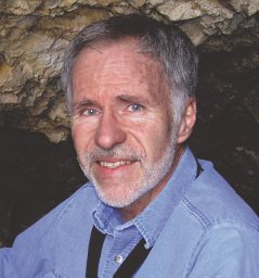 Anthony R. Kampf合作论文数Mineral Sciences Department, Natural History Museum of Los Angeles County6
 Stuart J Mills合作论文数Gallery of Natural Art;The Arkenstone;Museums Victoria6
 Francesco Radica合作论文数5Marcella Cadoni合作论文数5Roberta Oberti合作论文数5
 Luca Bindi合作论文数Department of Earth Sciences, University of Florence5
 Henrik Skogby合作论文数5Emanuele Costa合作论文数4
 P. Bonazzi合作论文数4
 Giovanni Ferraris合作论文数Dipartimento di Scienze della Terra, Università degli Studi di Torino4
 Elena Sokolova合作论文数4
 Federica Zaccarini合作论文数4Luigi Chiappino合作论文数3P. Ambrino合作论文数3Georges Favreau合作论文数3
 Giovanni Orazio Lepore合作论文数Dipartimento Sci Terra, Univ Firenze3
 Marcelo Andrade合作论文数3
 Daniel Atencio合作论文数Universidade de Sáo Paulo3Daniela Del Mauro合作论文数3
 Elena Bonaccorsi合作论文数Deparxnent of Earth Sciences;University of Pisa;Deparxnent of Earth Sciences, University of Pisa3
 Alberto Zanetti合作论文数Sezione di Pavia, CNR-IGG3
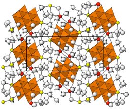 Barbara P Nash合作论文数Dept Geol & Geophys, Univ Utah3
 Federico Massimi合作论文数2Adrio Salvetti合作论文数2R. KAMPF合作论文数2Guenter Blass合作论文数2Alessandra Marengo合作论文数2Marco Marchesini合作论文数2
 Pierluigi Ambrino合作论文数2Fabrizio Castellaro合作论文数2
 Beatrice Celata合作论文数2Bruno Martini合作论文数2Roberto Cabella合作论文数Dipartimento per lo Studio del Territorio e delle sue Risorse, Università di Genova2
 Danilo Bersani合作论文数Department of Mathematical, Physical and Computer Sciences, University of Parma2
 Jakub Plášil合作论文数Department of Structure Analysis, Institute of Physics of the Czech Academy of Sciences2Günter Blass合作论文数2
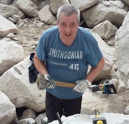 Pietro Vignola合作论文数Istituto per la dinamica dei processi ambientali (IDPA),, National Research Council (CNR),2




