ATRX interacts with H3.3 in maintaining telomere structural integrity in pluripotent embryonic stem cells Lee H. Wong, James D. McGhie, Marcus Sim, Melissa A. Anderson, Soyeon Ahn, Ross D. Hannan, Amee J. George, Kylie A. Morgan, Jeffrey R. Mann, and K.H. Andy Choo Chromosome and Chromatin Research, Murdoch Childrens Research Institute, Department of Paediatrics, University of Melbourne, Royal Children’s Hospital, Parkville, Victoria 3052, Australia; Growth Control and Differentiation Program, Peter MacCallum Cancer Centre, East Melbourne, Victoria 3002, Australia; Department of Biochemistry and Molecular Biology, University of Melbourne, Victoria 3052, Australia; Stem Cell Epigenetics, Murdoch Childrens Research Institute, Department of Paediatrics, University of Melbourne, Royal Children’s Hospital, Parkville, Victoria 3052, Australia
ATRX (alpha thalassemia/mental retardation syndrome X-linked) belongs to the SWI2/SNF2 family of chromatin remodeling proteins. Besides the ATPase/helicase domain at its C terminus, it contains a PHD-like zinc finger at the N terminus. Mutations in the ATRX gene are associated with X-linked mental retardation (XLMR) often accompanied by alpha thalassemia (ATRX syndrome). Although ATRX has been postulated to be a transcriptional regulator, its precise roles remain undefined. We demonstrate ATRX localization at the telomeres in interphase mouse embryonic stem (ES) cells in synchrony with the incorporation of H3.3 during telomere replication at S phase. Moreover, we found that chromobox homolog 5 (CBX5) (also known as heterochromatin protein 1 alpha, or HP1 alpha) is also present at the telomeres in ES cells. We show by coimmunoprecipitation that this localization is dependent on the association of ATRX with histone H3.3, and that mutating the K4 residue of H3.3 significantly diminishes ATRX and H3.3 interaction. RNAi-knockdown of ATRX induces a telomere-dysfunction phenotype and significantly reduces CBX5 enrichment at the telomeres. These findings suggest a novel function of ATRX, working in conjunction with H3.3 and CBX5, as a key regulator of ES-cell telomere chromatin.
Objective: To investigate the potential role of PArkin co-regulated gene (PACRG) in human male infertility.Design: Case-control study.Setting: Academic reproductive biology department.Patient(s): Blood samples were obtained from 610 patients and 156 normal control subjects.Intervention(s): Genomic DNA was used as template for polymerase chain reaction amplification of the PACRG promoter and coding exons. The amplified fragments were tested for DNA sequence variations by direct sequencing and restriction enzyme analysis.Main Outcome Measure(s): Gene structure and sequence alterations of PACRG in infertile male patients.Result(s): The structure of PACRG was determined to comprise 5 coding exons, generating a single transcript in the testis which encoded a predicted protein of 257 amino acids. No pathogenic mutations were identified; however, a variant in the promoter of PACRG was shown to be significantly associated with azoospermia, but not oligospermia, in the case-control cohort.Conclusion(s): Mutation of PACRG was not identified as a cause of male infertility, but variation in the promoter was demonstrated to be a risk factor associated with azoospermia. (Fertil Steril (R) 2010;93:2262-8. (C)2010 by American Society for Reproductive Medicine.)
Little is known about the telomere chromatin dynamics of embryonic stem (ES) cell. Here, we demonstrate localization of histone H3.3 at interphase telomeres and enrichment of Ser31-phosphorylated H3.3 at metaphase telomeres in pluripotent mouse ES cells. Upon differentiation, telomeric H3.3S31P signal decreases, accompanied by increased association of heterochromatin repressive marks and decreased micrococcal nuclease sensitivity at the telomeres. H3.3 is recruited to the telomeres at late S/G2 phase, coinciding with telomere replication and processing. RNAi-depletion of H3.3 induces telomere-dysfunction phenotype, providing evidence for a role of H3.3 in the regulation of telomere chromatin integrity in ES cells. The distinctive changes in H3.3 distribution suggests the existence of a unique and functionally essential telomere chromatin in ES cells that undergoes dynamic differentiation-dependent remodeling during the process of differentiation.
 Melissa Anderson合作论文数3
 Lee Hwa Wong合作论文数Monash Univ, Dept Biochem & Mol Biol, Clayton, Vic 3800, Australia3K H Andy Choo合作论文数3Ahn Soyeon合作论文数3McGhie James D合作论文数3
 Jeffrey R. Mann合作论文数Division of Biology, Beckman Research Institute of the City of Hope3Kylie A Morgan合作论文数2
 Ross Hannan合作论文数John Curtin School of Medical Research, College of Health and Medicine, Australian National University2
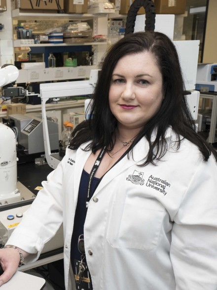 Amee George合作论文数John Curtin School of Medical Research, College of Health and Medicine, The Australian National University2Evan Williams合作论文数Department of Surgery, The Queen Elizabeth Hospital1Robert I McLachlan合作论文数1
 Paul J Lockhart合作论文数Department of Paediatrics, University of Melbourne1
 Moira O'Bryan合作论文数Faculty of Science, The University of Melbourne1
 Martin Delatycki合作论文数Bruce Lefroy Centre, Murdoch Children's Research Institute;Victorian Clinical Genetics Services1
 Kate Maree Brody合作论文数1Tam Angela合作论文数1Hua Ren合作论文数1
 Elizabeth D Earle合作论文数1Juliet M. Taylor合作论文数Bruce Lefroy Centre for Genetic Health Research, Murdoch Children's Research Institute1Gabrielle R. Wilson合作论文数1




