In viviparous teleosts, the lack of oviducts defines intraovarian gestation, with the ovary being the site for oogenesis but also the site for insemination, fertilization, and gestation. Consequently, intraovarian gestation is a complex and exceptional type of reproduction among vertebrates. The analysis of the morphological and physiological components of intraovarian gestation documents the evolutionary process of nutrition in viviparous species. Two types of embryonic nutrition may occur during gestation: (a) lecithotrophy, when most nutrients for the embryo come from the abundant yolk stored during oogenesis, and (b) matrotrophy, when nutrients for the embryo with scarce yolk must be obtained during gestation by additional maternal provisioning, developing a placenta. Then, investment of maternal nutrients for the embryo is greater during oogenesis in lecithotrophic species, and investment of maternal resources for the nutrition of the embryo is greater during gestation in matrotrophic species. Microscopic techniques allow for proper observation of maternal and embryonic structures involved in both types of nutrition during the development of embryos. Specifically, we focused on the morphology of placental structures associated with embryonic nutrition at different stages of development, which are the yolk sac and the pericardial sac. The oocytes of Poecilia latipinna contain a large amount of yolk (an average oocyte diameter of 1.9 mm); in contrast, the oocytes of Heterandria formosa contain extremely reduced amounts of yolk (an average oocyte diameter of 0.4 mm). Therefore, these species represent appropriate models for studying the strategy of two different types of embryonic nutrition, lecithotrophy and matrotrophy, in viviparous teleosts.
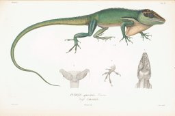
We used histological and morphometric methods to study the testis and associated glands, including the epididymis, ductus deferens, and renal sexual segment (RSS), of specimens of Basiliscus vittatus sampled from Tabasco, Mexico (17.5926° N, 92.5816° W). Samples were collected throughout 1 year, which included the dry (February to May) and rainy (June to January) seasons. Spermatogenesis in B. vittatus is active throughout the year, but a significant increase in the testicular volume, diameters of seminiferous tubules, height of the germinal epithelium, spermiogenesis, and released spermatozoa occur in the dry season. During the rainy season, all aforementioned parameters decreased except the secretory activity of the epididymis and the RSS, which increased concomitant with an increase of the spermatozoa population within the ductus deferens. These data strongly suggest that B. vittatus reproduce year-round, but males exhibit a peak in spermatogenic activity during the dry season and a peak in insemination and/or copulation at the beginning of the rainy season. We highlight the importance of analyzing not only the testis but also accessory ducts and glands when determining the reproductive cycles of reptiles. The reproductive cycle of B. vittatus is discussed in relation to the environmental conditions of Southern Mexico and is compared to that of other squamates.
Teleosts show unique features of the female reproductive system compared with the rest of vertebrates. Teleosts lack oviducts and therefore the ovary of viviparous teleosts is responsible for not only oogenesis, but also the reception of spermatozoa, fertilization, and intraovarian gestation which is unique among the vertebrates. The gestation may be intrafollicular, when the embryo remains in the follicle throughout embryogenesis, as in poeciliids; or intraluminal, when the embryo moves during early development from the follicle into the lumen where embryogenesis continues, as in goodeids. In viviparous teleosts, the embryonic and maternal tissues form placentas, mediating all exchanges: trophic, excretory, respiratory, immunological, and metabolic waste. These adaptations occur according to lecithotrophy, when the eggs may store enough yolk for the nutrition of the embryo until birth, and matrotrophy, when the eggs store scarce yolk, insufficient for the nutrition of all embryogenesis, which has to be complemented by transfer of maternal nutrients during gestation. This paper focuses primarily on the ovarian structure and embryonic traits of poeciliids and goodeids to illustrate the morphological and physiological elements that characterize viviparity in teleosts.
This study aimed to explore the reproductive histology and oocyte differentiation of the longnose seahorse Hippocampus trimaculatus (Leach, 1814) in captivity. Five mature healthy females were histologically observed. The reproductive systems of the five specimens exhibited similar morphological characteristics with a pair of saccular creamy white ovaries merging caudally into a single gonoduct. There were two germinal ridges lined with a layer of germinal epithelium (GE). The ovarian maturation of this species was considered asynchronous. The oogenic cells were classified into oogonia and oocytes at several developmental phases based on their size and characteristics. Oogonia were identified among the connective tissue in the middle area of the GE. The stromal compartment contained oocytes that were classified into four distinct phases: the primary growth (PG) phase having two steps (perinucleolar and oil droplets-cortical alveolar steps) and the secondary growth (SG) phase with three oocyte types, including early SG oocytes, late SG oocytes, and fully grown oocytes. The atretic oocytes (AO) were observed in all stages of oogenesis. Postovulatory follicles were also seen among the ovarian connective tissue. The occurrence of postovulatory follicles suggested that the specimens analysed in this study were in the spawning period. This research provides new insights into the identification of the reproductive cycles and morphological characteristics of the ovary of H. trimaculatus.
In viviparous teleosts, intraovarian gestation occurs intrafollicularly, as in poeciliids, or intraluminally, as in goodeids and anablepids. Furthermore, there are two different forms of embryonic nutrition: lecithotrophy and matrotrophy; depending on the species, these can be exclusive or coexist during gestation. In matrotrophic species, nutrients are transmitted from the mother to the embryo and are especially important in species with intraluminal gestation. Jenynsia lineata is a South American viviparous teleost with intraluminal gestation, characterized by eggs with scarce yolk, which is resorbed when embryos are 6 mm long, thus developing a branchial placenta. Using histological, histochemical, and immunohistochemical techniques, the present study describes the characteristics and changes of the ovarian mucosa in J. lineata during gestational and nongestational phases, and analyzes the embryonic pharyngeal epithelium in the branchial placenta. The ovaries of 30 adult female specimens were processed using histological techniques and stained with hematoxylin-eosin, Masson's trichrome, and Alcian Blue pH 2.5/periodic acid Schiff reagent. To detect cell proliferation, we used antiproliferating cell nuclear antigen antibody. In nonpregnant females, eosinophilic granular cells (EGCs) and lymphocytes were identified in the lamina propria of the tunica mucosa, and melanomacrophage centers (MMCs) and fibroblasts were identified adjacent to tissue debris in the ovarian folds'. In the cellular debris, an embryo in resorption was observed. In pregnant females, the ovarian mucosa has thin vascularization branches entering the opercular chamber of the embryos, in close contact with the forming gill processes, thereby establishing a branchial placenta. Active cell replacement was observed in these ovarian branches. The identification of fibroblasts, lymphocytes, EGCs, and MMCs adjacent to tissue debris could indicate that these cell types are involved in the embryonic resorption process. Considering the new data obtained in this study on the branchial placenta of J. lineata, we conclude that cell proliferation could be involved in the development of maternal-embryonic interaction.
The intraovarian gestation, occurring in teleosts, makes this type of reproduction a such complex and unique condition among vertebrates. This type of gestation of teleosts is expressed in special morphological and physiological characteristic where occurs the viviparity and it is an essential component in the analysis of the evolutionary process of viviparity in vertebrates. In viviparous teleosts, during embryogenesis, there are not development of Müllerian ducts, which form the oviducts in the rest of vertebrates, as a result, exclusively in teleosts, there are not oviducts and the caudal region of the ovary, the gonoduct, connects the ovary to the exterior. The lack of oviducts defines that the embryos develop into the ovary, as intraovarian gestation. The ovary forms the oocytes which may develop different type of oogenesis, according with the storage of diverse amount of yolk, variation observed corresponding to the species. The viviparous gestation is characterized by the possible intimate contact between maternal and embryonic tissues, process that permits their metabolic interchanges. So, the nutrients obtained by the embryos could be deposited in the oocyte before fertilization, contained in the yolk (lecithotrophy), and may be completed during gestation by additional provisioning from maternal tissues to the embryo (matrotrophy). Then, essential requirements for viviparity in poeciliids and goodeids are characterized by: a) the diversification of oogenesis, with the deposition of different amount of yolk in the oocyte; b) the insemination, by the transfer of sperm to the female gonoduct and their transportation from the gonoduct to the germinal region of the ovary where the follicles develop; c) the intrafollicular fertilization; d) the intraovarian gestation with the development of embryos in intrafollicular gestation (as in poeciliids), or intraluminal gestation (as in goodeids); and, e) the origin of embryonic nutrition may be by lecithotrophy and matrotrophy. The focus of this revision compares the general and specific structural characteristics of the viviparity occurring into the intraovarian gestation in teleosts, defining this reproductive strategy, illustrated in this review with histological material in a poeciliid, of the species Poecilia latipinna (Lesueur, 1821) (Poeciliidae), and in a goodeid, of the species Xenotoca eiseni (Rutter, 1896) (Goodeidae).
During embryogenesis, teleost females do not develop Müllerian ducts, which form the oviducts in all other vertebrates. Thus, when they reach sexual maturity they do not have oviducts. In viviparous teleosts, the lack of oviducts means that the development of the embryos occurs as an intraovarian gestation, unique among vertebrates. The ovary is an unpaired hollow organ whose cavity is continuous with the caudal portion, the gonoduct, characterized by the absence of germinal cells, which opens to the exterior at the gonopore. The gonoduct attains essential function as a barrier between the germinal region of the ovary and the exterior during all reproductive stages. This study describes the functional morphology of the gonoduct in the viviparous teleost Cnesterodon decemmaculatus during non‐gestation (previtellogenesis and vitellogenesis) and gestation. The ovaries were processed using histological techniques and stained with hematoxylin–eosin, and periodic acid Schiff. The gonoduct has two regions: cephalic and caudal, and is formed by three histological layers, which are, from inside to the periphery: (a) tunica mucosa; (b) tunica muscularis; and (c) tunica serosa. In the cephalic region there are mucosal folds extending into the lumen and forming a structure similar to a cervix. The histology of the gonoduct indicates essential functions, that is, (a) the control of the luminal diameter in the limit to the germinal region of the ovary by the presence of a cervix; (b) during insemination the gonoduct receives the spermatozoa, may store and transport them to the germinal region; (c) the presence of melano‐macrophage centers indicates support of immunological processes, especially during gestation when these centers increase in size; (d) production of exocrine secretions; and (e) it is the birth canal, internally lined by an ciliated epithelium and surrounded by smooth musclesboth tissues supposedly supporting the birth process.
The common snook (C. undecimalis) is a highly valuable commercial and sport fishing species in marine, estuarine, and freshwater habitats. Despite its abundance in freshwater ecosystems, the information on the reproductive biology comes mainly from studies in the marine habitat. In this work, the reproductive cycle at the gametogenic and hormonal level is addressed in specimens captured in contrasting environments interconnected by the Grijalva-Usumacinta fluvial system. Adult common snook presented a similar temporal pattern in sex steroid concentrations in both environments. Likewise, females and males were observed undergoing advanced maturity simultaneously in both environments. However, females in the freshwater environment did not reach final maturation, and no post-ovulatory follicles were found, indicating that spawn did not happen. Therefore, organisms are synchronized with gonads developing in both habitats, implying migrations of at least 300 km from the freshwater environment to the spawning grounds in the Gulf of Mexico.
We used histological techniques to describe the morphology of the yolk and pericardial sacs in developing embryos of the lecithotrophic species Girardinus creolus, Gambusia puncticulata, Limia vittata, and Quintana atrizona, in comparison with the extreme matrotrophic Heterandria formosa. In lecithotrophic species, the yolk sac was enlarged and lasted until the final stages of development, while in H. formosa it was completely absorbed soon after fertilization. Lecithotrophic poeciliids showed a pericardial sac with a single layer of blood vessels covering the dorsal surface of the cephalic region only, while H. formosa showed a more complex largely vascularized pericardial sac covering the entire dorsal surface, except the caudal region. In advanced gestation of G. creolus, a vascular plexus of the yolk sac reaches the pharyngeal region, behind the gills, suggesting that the pharynx may play a role in embryonic nutrition in lecithotrophic species. These morphological evidences suggest that matrotrophy derives from lecithotrophy.
In viviparous Mexican fishes of the family Goodeidae, embryos develop in the maternal ovarian lumen. They typically absorb maternal nutrients during gestation by means of "trophotaeniae," that is, specialized, elongated extensions of the hindgut that are exposed to the fluids, which occupy the ovarian lumen. The sole exception is Ataeniobius toweri, whose embryos lack trophotaeniae but are nevertheless matrotrophic. Thus, how its embryos obtain maternal nutrients is unclear. We studied a series of non-pregnant and pregnant ovaries of A. toweri using histology to identify the mechanism of maternal-embryo nutrient transfer. By early-gestation, embryos have depleted their yolk supplies. Yolks are released into the ovarian lumen and are ingested by the developing embryos, as shown by yolk material in their digestive tracts. The embryonic gut is lined by an epithelium consisting of columnar cells with apical microvilli, providing a means for nutrient absorption. Contrary to statements in the literature, embryos develop minuscule trophotaenial rudiments that extend slightly into the ovarian lumen. These structures are formed of an absorptive epithelium that overlies a vascular stroma, similar to the trophotaeniae of other goodeids. Through late gestation, vitellogenic follicles form and oocytes are discharged into the ovarian lumen, contributing to embryonic nutrition. Thus, histological evidence suggests that embryos chiefly obtain nutrients from ingestion of yolk and maternal secretions released into the ovarian lumen. This function possibly is supplemented by uptake via the small hindgut protrusions and other absorptive surfaces (e.g., the skin and the gill epithelium). Our observations are consistent with two evolutionary interpretations of the hindgut protrusions: (a) that they are rudimentary, evolutionary precursors of trophotaeniae formed by exteriorized hindgut; and (b) that they are vestigial remnants of trophotaeniae that were lost during a switch to a form of matrotrophy involving nutrient ingestion.
Female teleosts fishes do not develop Müllerian ducts; consequently, they do not have oviducts, and uniquely, the ovary forms the reproductive system. Therefore, the ovary is divided into two zones: germinal and gonoduct. The gonoduct of Poecilia reticulata is divided into three regions: anterior, middle, and posterior. The histological components of the gonoduct are epithelium, connective tissue, longitudinal smooth muscle, and visceral peritoneal epithelium at the periphery. The connective tissue contains macrophage, eosinophils, lymphocytes, and melanomacrophage centers. To conclude, the gonoduct is involved in essential aspects of reproduction such as secretory and immunological activities, receives the sperms during the insemination, and is the passage of embryos during birth.
The development of viviparity, live-bearing, is an ill-defined evolutionary event that is considered to have emerged in several lineages of reptiles. Therefore, the knowledge from a wide variety of viviparous species will offer better insight into understanding the reproductive adaptations associated with the development of viviparity. The dog-faced water snake, Cerberus rynchops, belongs to the family Homalopsidae that is considered to be viviparous, but detailed reproductive features are still poorly known for this species. In this study we investigated the female reproductive system of mature C. rynchops using histological techniques. A paired ovary was anchored to the kidney in this species. Eight to nine ovarian follicles were observed and classified as previtellogenic follicles, vitellogenic follicles and atretic follicles. The oviductal structure of C. rynchops was divided into four regions, namely infundibulum, uterine tube, uterus and vagina, and all oviductal regions were covered with ciliated and non-ciliated cuboidal epithelial cells and surrounded by connective tissue of submucosa, a thick layer of the muscularis and serosa. In the uterus we observed the uterine glands, which are commonly present in viviparous snakes.
The pea crab Pinnotheres cyclinus is a parasite of cultured mollusks.However, the information on reproductive histology of this crab has remained limited.Therefore, the present study investigated the ovarian structure and oogenesis of Pinnotheres cyclinus during maturation using histological techniques.Histologically, the ovary of this crab was found to be surrounded by a thin epithelium and connective tissue of the ovarian wall.Different phases of oogenesis were observed in the germinal area and could be classified into four phases: oogonial proliferation, the primary growth phase, the secondary growth phase, and the atretic oocyte phase.An oogonium was located in the ovarian cyst of the ovarian lobe, which was surrounded by a layer of pre-follicular cells.During the primary growth phase, oogonia continued to develop in the ovarian cyst, accumulating lipids and cortical alveoli.The appearance of spherical yolk granules related to changes in follicular cells during the secondary growth phase was also observed.These yolk granules reacted positively to Masson's trichrome and Mollary's trichrome staining, implying the presence of mucopolysaccharides and glycoproteins.Atretic oocytes were also found.Stages of embryonic development were also observed, including the formation of egg membranes covering embryos.Consequently, a fertilized egg was then filled with yolk granules, which all gradually combine to become one within the egg.
Heterandria formosa is a viviparous poeciliid native of the southeastern of United States of America. H. formosa exhibits unique reproductive features as: (a) production of extremely small eggs with scarce quantity of yolk (microlecithal eggs), (b) consequently, a high level of matrotrophy and development of a complex follicular placenta, (c) ovarian sperm storage that allows the continuous fertilization of oocytes and production of offspring and (d) development of high degree of superfetation. The degree of superfetation refers to the number of broods in different simultaneous stages of gestation. Morphological evidence of the degree of superfetation in H. formosa has not been documented. Therefore, and because of the general interest in the complex process of superfetation, the goal of this study is to morphologically define the degree of superfetation of H. formosa through two procedures: (a) histological analysis of entire ovaries in gestation and (b) dissection of visible embryos and the histological analysis of the remaining ovarian tissue. Results indicate that H. formosa can gestate up to seven broods at the same time.
The dynamics of cellular development and homeostasis of the ovary depend on the balance between proliferation and cell death throughout the reproductive cycle. Millerichthys robustus is an annual fish whose ovarian follicles develop asynchronously, allowing daily reproduction from sexual maturity until death. The objective of this research is to describe, histologically, the processes of follicular atresia and regression of postovulatory follicular complexes (POC) throughout a reproductive cycle of M. robustus. Patterns of cell death were documented by apoptosis in atretic follicles and POC, and necrosis in the POC after ovulation with an associated inflammatory response. Atretic follicles were seen from the onset of sexual maturity, during week three post-hatching (PH), both in primary growth (from the Cortical alveoli step, with folliculogenesis completed) and secondary growth Stages, with a higher prevalence in the latter. POCs were observed in different stages of regression from week four PH until the death of the fish. The apoptotic characteristics found were: (i) fragmentation of the nuclear membrane and zona pellucida, and liquefaction of the cortical alveoli and yolk; (ii) follicular cells becoming phagocytic, increasing their size, and migrating within the oocyte; and (iii) formation of an intrafollicular lumen, a product of phagocytosis of the oocyte constituents and dispersed pigments that remain after the digestion of yolk and cortical alveoli. The morphological changes of the follicular cells of the POC, from a squamous morphology after ovulation to columnar during its regression with PAS+ contents, was documented, suggesting a secretory activity.
Most teleosts' ovaries are paired, saccular structures with a central lumen and they do not have oviducts because of the lack of Müllerian ducts. Consequently, the communication of the ovary to the exterior occurs through the gonoduct. From the ovarian wall, lamellae project into the lumen, which are lined by the germinal epithelium and contain stroma with numerous ovarian follicles. The germinal epithelium contains oogonia among somatic epithelial cells. The lack of oviducts in viviparous teleosts leads to a unique type of intraovarian gestation. Folliculogenesis is completed when the oogonia initiate meiosis and are enclosed by follicle cells and a vascularized theca, becoming an oocyte. Oogenesis involves the morphological and functional processes by which oogonia transform into fertilizable eggs. There are six stages of oogenesis: 1) Oogonial proliferation, 2) The beginning of meiosis forms the chromatin-nucleolus stage, developing lampbrush chromosomes, 3) During previtellogenesis the oocyte has a single nucleolus becoming soon multinucleolar; cortical alveoli and lipid droplets appear in the ooplasm leading to a significant increase in oocyte diameter. 4) Vitellogenesis is characterized by the deposition of yolk in the ooplasm; the follicle reaches its maximum size, becoming a full-grown oocyte. 5) During maturation the germinal vesicle becomes eccentric and migrates to the animal pole, followed 6) by ovulation. Postovulatory follicles contains follicle cells. Atresia is observed in any stage especially at the end of a breeding season. The diversity of ovarian features in teleosts is a main aspect for understanding their essential function, such as: type of spawning pattern; oviparity and viviparity that implicate modifications for intraovarian gestation; hermaphroditism that reveals ovotestis; the change of sex in protogynous and protandric species.
Ichthyology & Herpetology (formerly Copeia) publishes work on the biology of fishes, amphibians, and reptiles, or work using those organisms as models for testing hypotheses of broad significance.
We describe the histological characteristics of the testis and spermatogenesis of the cave molly Poecilia mexicana, a viviparous teleost inhabiting a sulfur spring cave, Cueva del Azufre, in Tabasco, Southern Mexico. P. mexicana has elongate spermatogonial restricted testes with spermatogonia arranged in the testicular periphery. Germ cell development occurs within spermatocysts. As spermatogenesis proceeds, the spermatocysts move longitudinally from the periphery of the testis to the efferent duct system, where mature spermatozoa are released. The efferent duct system consists of short efferent duct branches connected to a main efferent duct, opened into the genital pore. Spermatogenesis consisted of the following stages: spermatogonia (A and B), spermatocytes (primary and secondary), spermatids, and spermatozoa. The spermatozoa are situated within spermatocysts, with their heads oriented toward the periphery and flagella toward the center. Once in the efferent duct system, mature spermatozoa are packaged as unencapsulated sperm bundles, that is, spermatozeugmata. We suggest that the histological characteristics of the testis and spermatogenesis of P. mexicana from the Cueva del Azufre, and the viviparous condition where the spermatozoa enter in the female without been in the water, have allowed them to invade sulfurous and/or subterranean environments in Southern Mexico, without requiring complex morphofunctional changes in the testis or the spermatogenetic process.
Cellular aspects of oocyte development of the Mexican rivulus Millerichthys robustus were morphologically described in order to analyze ovarian function and the cellular recruitment dynamics associating it with life history strategies of annual killifishes. Millerichthys is an iteroparous batch spawner with continuous oocyte recruitment and indeterminate fecundity with asynchronous development of the follicles. It has two ovaries of cystovarian type, with a central lumen, which communicates with the outside through the caudal region of the ovary, that is, the gonoduct. From the walls of the ovary, irregular lamellae composed of germinal epithelium and vascularized stroma project. Oogenesis starts with oogonial proliferation, found alone or in nests within the germinal epithelium. The oogonia come into meiosis becoming oocytes and advancing to the chromatin nucleolus stage and to early primary growth stage. Folliculogenesis is completed in the primary growth stage and cortical alveoli step. Follicles moves toward the stroma, but they continue to be attached to the germinal epithelium through the basement membrane until ovulation. The inclusion of fluid yolk in the follicles during the secondary growth stage was observed. During ovulation, the follicle collapsed, the oocyte was released into the lumen, and the constitutive elements of the post-ovulatory follicle complex remained in the stroma.
 Harry J. Grier合作论文数15Hernández-Franyutti Arlette合作论文数Laboratorio: Acuicultura Tropical, Universidad Juárez Autónoma de Tabasco9Adriana García Alarcón合作论文数7
 Lynne Parenti合作论文数Department of Vertebrate Zoology, National Museum of Natural History, Smithsonian Institution7Omar Domínguez-Castanedo合作论文数Consejo Nacional de Ciencia y Tecnología6
 Aaron Torres-Martinez合作论文数5De la Rosa-Cruz Gabino合作论文数4
 Sinlapachai Senarat合作论文数Dept Marine Sci & Environm, Rajamangala Univ Technol Srivijaya3Campuzano-Caballero Juan Carlos合作论文数3
 Wilfrido M. Contreras-Sanchez合作论文数3A. M. Rosales-Torres合作论文数Departamento de Producción Agrícola y Animal Unidad Xochimilco, Universidad Autónoma Metropolitana Bioquímica de la Reproducción3Lamai Thongboon合作论文数2wilfrido miguel contreras sanchez合作论文数2Jes Kettratad合作论文数2
 Lozano Abraham合作论文数Centro de Investigaciones Biológicas, Universidad Autónoma del Estado de Hidalgo;CIIDIR Unidad Durango, Instituto Politécnico Nacional;Instituto Politécnico Nacional, Universidad Autónoma del Estado de Hidalgo2
 Plaul S E合作论文数Cátedra de Histología y Embriología, UNLP. La Plata2
 Wannee Jiraungkoorskul合作论文数2
 Aurelio Ramirez-Bautista合作论文数Centro de Investigaciones Biológicas;Universidad Autónoma;Plaza Juárez;Centro de Investigaciones Biológicas, Universidad Autónoma|Plaza Juárez2
 Kevin M Gribbins合作论文数Department of Biology, Wittenberg University2
 Koraon Wongkamhaeng合作论文数Department of Marine Science;Faculty of Science;Chulalongkorn University;Faculty of Science, Chulalongkorn University2
 Claudio Barbeito合作论文数Facultad de Ciencias Veterinarias, National University of La Plata;National Scientific and Technical Research Council2
 Gen Kaneko合作论文数Graduate School of Agricultural and Life Sciences, The University of Tokyo2juan manuel vidallopez合作论文数1José L. Ponce de León合作论文数1Otilio Méndez-Marin合作论文数1
 Peerapon Sornying合作论文数1
 Jose Luis Ponce de Leon合作论文数1
 Luca Di Cesare合作论文数1Monasterio de Gonzo G合作论文数1Martínez Vh合作论文数1Paola‐Ivonne Cerda‐Jardón合作论文数1Tessy M Muñoz-Campos合作论文数1Piyamat Kongtueng合作论文数1
 Sansareeya Wangkulangkul合作论文数1
 Martin Acosta合作论文数1
 Chanyut Sudtongkong合作论文数Dept Marine Sci & Environm, Rajamangala Univ Technol Srivijaya1
 Carlos Alfonso Álvarez González合作论文数Laboratory of Physiology in Aquatic Resources, Academic Division of Biological Science, Universidad Juárez Autónoma de Tabasco1
 Rodet Rodriguez-Silva合作论文数Instituto de Ecología y Sistemática. Carretera Varona 11835 e/ Oriente y Lindero, La Habana 19, CP 11900, Calabazar, Boyeros, La Habana, Cuba.;Email: rodetrodriguezsilva@gmail.com.;Instituto de Ecología y Sistemática. Carretera Varona 11835 e/ Oriente y Lindero, La Habana 19, CP 11900, Calabazar, Boyeros, La Habana, Cuba.; Email: rodetrodriguezsilva@gmail.com.1Hernández-Vidal Ulises合作论文数1Blackburn Daniel G合作论文数1
 Martin Montes合作论文数1
 Atsuo Iida合作论文数High Energy Accelerator Research Organization, KEK,^ High Energy Accelerator Research Organization (KEK)1
 Manfred Schartl合作论文数Medical School, University of Würzburg;Xiphophorus Genetic Stock Center, Texas State University1
 Xavier Chiappa-Carrara合作论文数Instituto de Ciencias del Mar y Limnología;Apartado, Universidad Nacional Autónoma de México;Instituto de Ciencias del Mar y Limnología, Universidad Nacional Autónoma de México1
 Anan Kenthao合作论文数Faculty of Science, Khon Kaen University1
 Steven D. Mims合作论文数1
 Fabiana L. Lo Nostro合作论文数Laboratorio de Embriologı;a Animal, Departamento de Biodiversidad y Biologı́Facultad de Ciencias Exactas y Naturales, Universidad de Buenos Aires;Laboratorio de Embriologı́a Animal, Universidad de Buenos Aires1
 Theerakamol Pengsakul合作论文数Xiamen University, Xiamen University1
 Korakot Nganvongpanit合作论文数Faculty of Veterinary Medicine, Chiang Mai University1




