Objective: We showed that a tailored strengthening intervention based on the size of motor evoked potentials (MEPs) in the affected arm was effective in improving function in chronic stroke survivors. Here, we investigated whether the short-term gains in arm function were maintained at 1-year follow-up. Subjects: Twenty-five participants at the chronic stage of a stroke. Methods: Participants were classified in the light (LI; MEPs 50–120 μV, n = 8) and high (HI; MEPs > 120μV, n = 17) intensity training groups. The strengthening protocol consisted of adjusted exercises for the affected arm (3X/week; 4 weeks). The Fugl-Meyer Stroke Assessment (FMA), Grip strength (GS) and Box and Block test (BBT) were assessed at baseline, post-intervention and at 1-year follow-up. Changes in clinical measures were compared using repeated-measures ANOVA. Results: A significant effect of time was noted on all outcome measures [FMA: p < 0.001; BBT: p = 0.05; GS: p < 0.001], but the LI group improved more on the FMA (p = 0.003) and maintained their gains at 1-year follow-up (p = 0.527) than the HI group. Conclusion: The size of MEPs in the affected arm could be a significant factor in influencing responses to strengthening exercises post-stroke and allow gains to be maintained up to 1 year post-intervention.
Accurately predicting post-stroke motor impairment remains a challenge due to the complexity of functional recovery and its association with neuroimaging biomarkers. This study presents a deep learning (DL) framework that integrates Magnetic Resonance Imaging (MRI)-based measures such as Diffusion Tensor Imaging (DTI) metrics-fractional anisotropy (FA), mean diffusivity (MD), radial diffusivity (RD), and axial diffusivity (AD)-along with white matter (WM) and gray matter (GM) intensities to classify upper limb motor function. Unlike previous approaches, the proposed model directly extracts wholebrain volumetric features without predefined region-of-interest constraints. Feature representation is enhanced using residual connections, attention mechanisms, and Global Average Pooling (GAP), improving classification performance while maintaining computational efficiency. The ensemble framework combines six independently trained models to optimize multi-modality integration. The results demonstrate that the WM + FA combination achieved the highest accuracy (0.97), outperforming the full ensemble model (0.96). These findings exceed the performance reported in prior studies, emphasizing the effectiveness of microstructural and structural biomarkers in motor recovery prediction. This optimized DL framework has the potential to improve post-stroke motor impairment classification, supporting early rehabilitation planning, and personalized treatment strategies.
Objective To evaluate how the COVID-19 pandemic affected rehabilitation outcomes and care delivery processes in Physical Medicine and Rehabilitation programs of COVID-19-designated rehabilitation facilities. Design Retrospective cohort study comparing care processes and outcomes between prepandemic and pandemic periods and identifying factors that influenced rehabilitation efficiency using the total score of the Functional Independence Measure (FIM) instrument. Setting Four designated subacute rehabilitation facilities. Participants Patients (N=507) from the Physical Medicine and Rehabilitation program (PMR), with a mean age of 78.3 years (range: 28-99y) and 60% women (ranging from 58% to 64% across groups). The cohort included prepandemic patients (33%), non-COVID-19 patients during the pandemic (33%), patients who were COVID-19-positive before admission (23%), and those who acquired COVID-19 during rehabilitation (11%). Interventions Not applicable. Main Outcome Measures Average daily gain in functional independence using the FIM total score (FIM efficiency), clinical and demographic profiles at admission, functional outcomes, and indicators of care delivery processes. Results While functional outcomes were mostly similar across groups (FIM total score at discharge, P≥.05), patients who acquired COVID-19 during rehabilitation experienced longer stays (mean difference=32.2d, P<.001) and higher rehospitalization rates (48%, P<.001). In contrast, those infected before admission had shorter stays (mean difference=−9.5d, P<.001) and less multidisciplinary involvement (mean difference=1 discipline, P<.001). Linear mixed effects modeling, with rehabilitation site as random effects, demonstrated that COVID-19 acquisition during rehabilitation and increased multidisciplinary care were significant predictors of reduced FIM efficiency (P<.001). Conclusions The COVID-19 pandemic affected rehabilitation care delivery processes more than functional outcomes. Patients who acquired COVID-19 during rehabilitation and those requiring more diverse multidisciplinary care showed reduced FIM efficiency, highlighting the importance of infection control measures in rehabilitation settings. These insights will help health care professionals and decision makers optimize future crisis preparedness plans for rehabilitation services.
Objectives During the COVID-19 pandemic, designated rehabilitation centres were established in the province of Québec, where strict sociosanitary measures such as isolation and mandatory personal protection equipment requirements were followed. This study aimed to describe the impact of the pandemic on rehabilitation care indicators for poststroke users with (COV+) and without (COV−) COVID-19 infection in designated rehabilitation centres compared with those admitted in the previous year (pre-COV).Method A retrospective analysis of 292 medical files was performed in 3 rehabilitation centres. Demographic characteristics were collected, as well as indicators routinely collected in acute care and rehabilitation such as length of stay (LOS), the Functional Independence Measure and a number of physical/occupational therapy (PT/OT) sessions. Non-parametric statistical tests were used to compare variables among the three groups.Results COV+ users were older than COV− and pre-COV ones (p<0.01) and were more disabled on admission to a rehabilitation centre (p<0.01). They also exhibited longer LOS in acute care prior to rehabilitation (p<0.001) and were more often rehospitalised (p<0.002) during the course of their stay in the rehabilitation centre. Despite longer rehabilitation stays (p<0.001) and more PT/OT sessions, COV+ users remained more disabled at discharge (p<0.002). COV− users showed rehabilitation care indicators resembling the ones of pre-COV despite spending less time in rehabilitation.Conclusions Patients who had a stroke infected with COVID-19 exhibited greater vulnerability on admission to rehabilitation. They required more care and services during their rehabilitation period. However, this additional support did not enable them to achieve the same level of recovery as COV− and pre-COV users. This underscores the added impact of the disease on already impaired patients and highlights the specific needs of COV+ users undergoing rehabilitation.
Background/Objectives: We showed that a tailored strengthening intervention based on the size of motor evoked potentials (MEPs) elicited by transcranial magnetic stimulation (TMS) in the affected hemisphere resulted in an improved affected arm function, regardless of stroke severity. Also, adding anodal transcranial direct stimulation (atDCS) during training did not alter the results as participants receiving real or sham stimulation showed similar gains. The goal of this study was to report on the changes in basic measures of corticomotor excitability in response to the intervention and to determine whether these changes were influenced by tDCS and correlated with those measured in arm function. Methods: The TMS measures consisted of the resting motor threshold (rMT), MEP amplitude at rest, and the silent period (SP) duration. Clinical outcomes included the Box and Block test (BBT) and grip strength (GS). Results: Post-intervention, regardless of atDCS (p > 0.62), no significant change in corticomotor excitability was noted (p > 0.15), as well as no association between the changes in TMS measures and arm function gains (p > 0.06). Conclusions: As observed for clinical measures, atDCS did not influence corticomotor excitability. The absence of an increase in the excitability of the affected hemisphere and important associations between changes in corticomotor excitability and clinical gains suggest that factors other than brain plasticity could mediate gains in arm function. Further investigations are required regarding the role of tDCS in stroke rehabilitation.
This pilot study evaluated feasibility (retention/adherence) of a 7-visit museum-based art therapy intervention (1-hour guided artworks visit followed by a 2-hour workshop), for 7 chronic stroke survivors and investigated its impact on their psychosocial well-being (depression/self-esteem/body image/community integration). Pre/post-intervention, questionnaires were used to assess the psychological variables and post-intervention, semi-structured individual interviews and a focus group were conducted. The findings showed that the intervention was feasible, with a retention of 77% and adherence of 84%. Participants reported positive effects of the intervention on psychosocial variables such as self-esteem and mood but less so on body image. Clinical implications include initial evidence of the feasibility and potential benefits of a museum-based art therapy intervention in improving post-stroke psychosocial well-being.
Functional brain connectivity measures extracted from resting-state functional magnetic resonance imaging (fMRI) scans have generated wide interest as potential noninvasive biomarkers. In this context, performing global signal regression (GSR) as a preprocessing step remains controversial. Specifically, while it has been shown that a considerable fraction of global signal variations is associated with physiological and motion sources, GSR may also result in removing neural activity. Here, we address this question by examining the fundamental sources of resting global signal fluctuations using simultaneous electroencephalography (EEG)-fMRI data combined with cardiac and breathing recordings. Our results suggest that systemic physiological fluctuations account for a significantly larger fraction of global signal variability compared to electrophysiological fluctuations. Furthermore, we show that GSR reduces artifactual connectivity due to heart rate and breathing fluctuations, but preserves connectivity patterns associated with electrophysiological activity within the alpha and beta frequency ranges. Overall, these results provide evidence that the neural component of resting-state fMRI-based connectivity is preserved after the global signal is regressed out. ### Competing Interest Statement The authors have declared no competing interest.
Background: In the last few years, transcranial alternating current stimulation (tACS) has attracted attention as a promising approach to interact with ongoing oscillatory cortical activity and, consequently, to enhance cognitive and motor processes. While tACS findings are limited by high variability in young adults’ responses, its effects on brain oscillations in older adults remain largely unexplored. In fact, the modulatory effects of tACS on cortical oscillations in healthy aging participants have not yet been investigated extensively, particularly during movement. This study aimed to examine the after-effects of 20 Hz and 70 Hz High-Definition tACS on beta oscillations both during rest and movement. Methods: We recorded resting state EEG signals and during a handgrip task in 15 healthy older participants. We applied 10 min of 20 Hz HD-tACS, 70 Hz HD-tACS or Sham stimulation for 10 min. We extracted resting-state beta power and movement-related beta desynchronization (MRBD) values to compare between stimulation frequencies and across time. Results: We found that 20 Hz HD-tACS induced a significant reduction in beta power for electrodes C3 and CP3, while 70 Hz did not have any significant effects. With regards to MRBD, 20 Hz HD-tACS led to more negative values, while 70 Hz HD-tACS resulted in more positive ones for electrodes C3 and FC3. Conclusions: These findings suggest that HD-tACS can modulate beta brain oscillations with frequency specificity. They also highlight the focal impact of HD-tACS, which elicits effects on the cortical region situated directly beneath the stimulation electrode.
Background: Stroke can lead to lasting sensorimotor deficits of the upper limb (UL) persisting into the chronic phase despite intensive rehabilitation. A major impairment of reaching after stroke is a decreased range of active elbow extension, which in turn leads to the use of compensatory movements. Retraining movement patterns relies on cognition and motor learning principles. Implicit learning may lead to better outcomes than explicit learning. Error augmentation (EA) is a feedback modality based on implicit learning resulting in improved precision and speed of UL reaching movements in people with stroke. However, accompanying changes in UL joint movement patterns have not been investigated. The objective of this study is to determine the capacity for implicit motor learning in people with chronic stroke and how this capacity is affected by post-stroke cognitive impairments. Methods: Fifty-two subjects who have chronic stroke will practice reaching movements 3x/wk. for 9 wk. in a virtual reality environment. Participants will be randomly allocated to 1 of 2 groups to train with or without EA feedback. Outcome measures (pre-, post-and follow-up) will be: endpoint precision, speed, smoothness, and straightness and joint (UL and trunk) kinematics during a functional reaching task. The degree of cognitive impairment, lesion profile, and integrity of descending white matter tracts will be related to training outcomes. Conclusions: The results will inform us which patients can best benefit from training programs that rely on motor learning and utilize enhanced feedback. Trial status: Ethical approval for this study was finalized in May 2022. Recruitment and data collection is actively in progress and is planned to finish in 2026. Data analysis and evaluation will occur subsequently, and the final results will be published.
The degree of motor impairment and profile of recovery after stroke are difficult to predict for each individual. Measures obtained from clinical assessments, as well as neurophysiological and neuroimaging techniques have been used as potential biomarkers of motor recovery, with limited accuracy up to date. To address this, the present study aimed to develop a deep learning model based on structural brain images obtained from stroke participants and healthy volunteers. The following inputs were used in a multi-channel 3D convolutional neural network (CNN) model: fractional anisotropy, mean diffusivity, radial diffusivity, and axial diffusivity maps obtained from Diffusion Tensor Imaging (DTI) images, white and gray matter intensity values obtained from Magnetic Resonance Imaging, as well as demographic data (e.g., age, gender). Upper limb motor function was classified into "Poor" and "Good" categories. To assess the performance of the DL model, we compared it to more standard machine learning (ML) classifiers including k-nearest neighbor, support vector machines (SVM), Decision Trees, Random Forests, Ada Boosting, and Naïve Bayes, whereby the inputs of these classifiers were the features taken from the fully connected layer of the CNN model. The highest accuracy and area under the curve values were 0.92 and 0.92 for the 3D-CNN and 0.91 and 0.91 for the SVM, respectively. The multi-channel 3D-CNN with residual blocks and SVM supported by DL was more accurate than traditional ML methods to classify upper limb motor impairment in the stroke population. These results suggest that combining volumetric DTI maps and measures of white and gray matter integrity can improve the prediction of the degree of motor impairment after stroke. Identifying the potential of recovery early on after a stroke could promote the allocation of resources to optimize the functional independence of these individuals and their quality of life.
There is increasing evidence that the effects of non-invasive brain stimulation can be maximized when the applied intervention matches internal brain oscillations. Extracting individual brain oscillations is thus a necessary step for implementing personalized brain stimulation. In this context, different methods have been proposed for obtaining subject-specific spectral peaks from electrophysiological recordings. However, comparing the results obtained using different approaches is still lacking. Therefore, in the present work, we examined the following methodologies in terms of obtaining individual motor-related EEG spectral peaks: fast Fourier Transform analysis, power spectrum density analysis, wavelet analysis, and a principal component based time-frequency analysis. We used EEG data obtained when performing two different motor tasks - a hand grip task and a hand opening- and-closing task. Our results showed that both the motor task type and the specific method for performing the analysis had considerable impact on the extraction of subject-specific oscillation spectral peaks.Clinical Relevance-This exploratory study provides insights into the potential effects of using different methods to extract individual brain oscillations, which is important for designing personalized brain-machine-interfaces.
GOAL:Transcranial alternating current stimulation (tACS) is a non-invasive technology for modulating brain activity, with significant potential for improving motor and cognitive functions. To investigate the effects of tACS, many studies have used electroencephalographic (EEG) data recorded during brain stimulation. However, the large artifacts induced by tACS make the analysis of tACS-EEG recordings challenging, which in turn has prevented the implementation of closed-loop brain stimulation schemes. Here, we propose a novel combination of blind source separation (BSS) and wavelets to achieve removal of tACS-EEG artifacts with improved performance.METHODS:We examined the performance of several BSS methods both applied individually, as well as combined with the empirical wavelet transform (EWT) in terms of denoising realistic simulated and experimental tACS-EEG data.RESULTS:EWT combined with BSS yielded considerably improved performance compared to BSS alone for both simulated and experimental data. Overall, independent vector analysis (IVA) combined with EWT yielded the best performance.SIGNIFICANCE:The proposed method yields promise for quantifying the effects of tACS on simultaneously recorded EEG data, which can in turn contribute towards understanding the effects of tACS on brain activity, as well as extracting reliable biomarkers that may be used to develop closed-loop tACS strategies for modulating the underlying brain activity in real time.
Being able to accurately quantify the hemodynamic response function (HRF) that links the blood oxygen level dependent functional magnetic resonance imaging (BOLD-fMRI) signal to the underlying neural activity is important both for elucidating neurovascular coupling mechanisms and improving the accuracy of fMRI-based functional connectivity analyses. In particular, HRF estimation using BOLD-fMRI is challenging particularly in the case of resting-state data, due to the absence of information about the underlying neuronal dynamics. To this end, using simultaneously recorded electroencephalography (EEG) and fMRI data is a promising approach, as EEG provides a more direct measure of neural activations. In the present work, we employ simultaneous EEG-fMRI to investigate the regional characteristics of the HRF using measurements acquired during resting conditions. We propose a novel methodological approach based on combining distributed EEG source space reconstruction, which improves the spatial resolution of HRF estimation and using block-structured linear and nonlinear models, which enables us to simultaneously obtain HRF estimates and the contribution of different EEG frequency bands. Our results suggest that the dynamics of the resting-state BOLD signal can be sufficiently described using linear models and that the contribution of each band is region specific. Specifically, it was found that sensory-motor cortices exhibit positive HRF shapes, whereas the lateral occipital cortex and areas in the parietal cortex, such as the inferior and superior parietal lobule exhibit negative HRF shapes. To validate the proposed method, we repeated the analysis using simultaneous EEG-fMRI measurements acquired during execution of a unimanual hand-grip task. Our results reveal significant associations between BOLD signal variations and electrophysiological power fluctuations in the ipsilateral primary motor cortex, particularly for the EEG beta band, in agreement with previous studies in the literature.
 Georgios D. Mitsis合作论文数Department of Bioengineering, Faculty of Engineering, McGill University20
 Marie-Helene Milot合作论文数Faculté de Médecine et des Sciences de la Santé, Université de Sherbrooke13
 Nick Ward合作论文数Department of Clinical and Motor Neuroscience, UCL Queen Square Institute of Neurology;National Hospital for Neurology and Neurosurgery10
 Xuanteng Yan合作论文数8
 Alba Xifra-Porxas合作论文数McGill University8
 Francois Tremblay合作论文数7
 Johanne Higgins合作论文数6
 Michalis Kassinopoulos合作论文数6Paul D. Cheney合作论文数6
 Stephania Palimeris合作论文数5
 Sara Lariviere合作论文数Multimodal Imaging and Connectome Analysis Laboratory, McConnell Brain Imaging Centre, Montreal Neurological Institute and Hospital, McGill University5
 Chang-Hyun Park合作论文数Neuro-X Institute (INX) and Brain Mind Institute (BMI),, Clinique Romande de Réadaptation5
 Dal Maso Fabien合作论文数Laboratoire de Simulation et Modélisation du Mouvement, Université de Montréal5
 Helene Corriveau合作论文数5
 Arna Ghosh合作论文数Mila - Quebec Artificial Intelligence Institute4
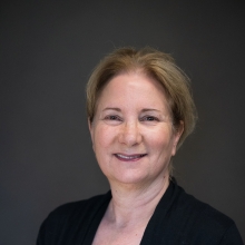 Mindy F. Levin合作论文数Faculty of Medicine and Health Sciences, School of Physical & Occupational Therap, McGill University4Louis-David Beaulieu合作论文数Neuroscience Division of the Centre de recherche du CHU de Québec, Université Laval4
 Holly E. Rossiter合作论文数4Prokopis C. Prokopiou合作论文数3
 Alexander Thiel合作论文数Department of Neurology & Neurosurgery, Faculty of Medicine and Health Sciences, McGill University;Lady Davis Institute for Medical Research, Jewish General Hospital3
 Sylvain Baillet合作论文数Department of Neurology and Neurosurgery, Faculty of Medicine and Health Sciences, McGill University;Université de Montréal;Centre de Recherche du CHUM;Centre Hospitalier de l'Université de Montréal;The Montreal Neurological Institute3
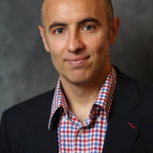 Marc Roig合作论文数School of Physical & Occupational Therapy, Faculty of Medicine and Health Sciences, McGill University3Cereb Cortex合作论文数3
 Claude Vincent合作论文数3Theodore H. Wein合作论文数3
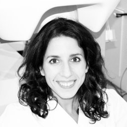 Guiomar Niso合作论文数3Sang-Pil Lee合作论文数2H Corriveau合作论文数2
 Rukiye Karakis合作论文数Faculty of Technology, Sivas Cumhuriyet University2Emma M. Davis合作论文数2
 Charlotte Stagg合作论文数Nuffield Department of Clinical Neurosciences, Medical Sciences Division, University of Oxford2
 Remaud Anthony合作论文数Bruyère Research Institute2
 Diana Zidarov合作论文数2Feldman Anatol G合作论文数2
 Heidi Johansen-Berg合作论文数Wellcome Centre for Integrative Neuroimaging, University of Oxford;Plasticity Group, Nuffield Department of Clinical Neurosciences, Medical Sciences Division, University of Oxford;Department of Clinical Neurology, University of Oxford2
 Shawn Frost合作论文数2Penelope Talelli合作论文数2S. Svojanovsky合作论文数2Damon Hoad合作论文数1M Deliano合作论文数1
 Yekta Ansari合作论文数1Myriam Villeneuve合作论文数1
 Yacine Mahdid合作论文数1Ball Joanna合作论文数1
 Anouk Lamontagne合作论文数1Erika Renda合作论文数1Bennet Desormeau合作论文数1Karen Barratt合作论文数1Zachary Legault合作论文数1




