Camacho-Hubbard, Isabella MD; Ota, Koji MD; Biro, Mark MD; Deng, Min MD Author Information
Supplementary Methods include description of: 1) "Puzzle"-fit" analysis, 2) Intensity-based objective sensitivity and specificity analysis, 3) Blinded physician "Reader Study" analysis, and 4) Evaluation of reader responses regarding intensity.
Figure S3D shows histology of false positive (FP) skin samples with presence of healing area and inflammation.
BackgroundNonmelanoma skin cancer (NMSC) is the most common cancer worldwide and is frequently treated with Mohs micrographic surgery (MMS); however, data regarding characteristics of Hispanic patients undergoing MMS for NMSC are limited.ObjectiveTo review the characteristics of Hispanic patients undergoing MMS for NMSC in the United States.MethodsA systematic review of PubMed articles from inception through September 2022 providing data for Hispanic patients undergoing MMS for NMSC was completed.ResultsOverall, six publications met inclusion criteria and provided data for 2,856 Hispanic patients that underwent MMS for 2,955 NMSCs. Results demonstrate 60% of Hispanic patients were male, and the majority of NMSCs were basal cell carcinoma (BCC) (71%), followed by squamous cell carcinoma (SCC) (21%). Additionally, a larger percentage of pigmented BCC was found in the Hispanic population. While there is conflicting data in the literature, Hispanic patients may also have larger MMS defects when controlled for additional variables. Finally, over 64% of NMSCs in Hispanic patients were in high-risk locations.ConclusionLiterature regarding the characteristics of Hispanic patients undergoing MMS for NMSC demonstrates most patients were male, BCC was the most common tumor subtype, and the majority of NMSCs were in high-risk locations.
Table S1 shows cancer type and histologic detection status following excision; Table S2 characterizes patients and specimens
BACKGROUND Perioperative anxiety is associated with negative patient outcomes in Mohs micrographic surgery (MMS). Both pharmacologic and nonpharmacologic therapies have been used to alleviate perioperative anxiety in MMS. OBJECTIVE To systematically evaluate the efficacy of therapies aimed at reducing perioperative anxiety in MMS. METHODS AND MATERIALS Eligible articles were identified using PubMed MEDLINE, Cochrane Central Register of Controlled Trials, metaRegister of Controlled Trials, ClinicalTrials.gov, and World Health Organization International Clinical Trials Registry Platform. All available studies investigating interventions to reduce perioperative anxiety during MMS were considered. RESULTS Of the 183 abstracts identified and screened, 5 studies met inclusion criteria. Three studies reported a postintervention reduction in patient anxiety (midazolam, educational video, and personalized music). Two studies reporting on similar interventions did not find an effect. CONCLUSION There is currently limited evidence to support either pharmacologic or nonpharmacologic therapy for alleviation of perioperative patient anxiety in MMS. Midazolam may provide patients a short-term benefit, though any estimate of the effect is very uncertain. Personalized music may be a promising nonpharmacologic intervention for future research.
Erythroderma is a problem frequently encountered by dermatologists working on an inpatient consultative service. There are few modern studies examining the etiologies and outcomes of patients admitted to the hospital with erythroderma. Dermatology consultation is associated with improved outcomes in patients with inflammatory skin conditions. We reviewed the charts of 38 patients in whom dermatologic consultation was obtained specifically for erythroderma between 2015 and 2019 and included 35 patients with complete records. Psoriasis was the most frequent etiology in our cohort, accounting for 34% of cases. Among psoriasis patients, hospital admission was precipitated by drug discontinuation in 7/12 patients. Corticosteroids were the most frequent culprits of erythrodermic flares (5/7 cases). Interestingly, in the 5 patients who were admitted for erythrodermic flares of their psoriasis due to steroid withdrawal, 4 out of 5 patients recently received systemic steroids from a dermatologist. Drug rashes accounted for an additional 34%, with 6 cases of drug-induced hypersensitivity syndrome, and one case of toxic epidermal necrolysis. Overall, the diagnosis suspected by the primary team was confirmed in 11 cases and changed in 6. The diagnosis was narrowed appreciably in the remainder of cases. The diagnosis remained unknown despite long-term follow-up in only 2 cases. A life-threatening diagnosis (including DIHS, TEN, Sézary syndrome, and hypereosinophilic syndrome) was made in 9 instances. Dermatology consultation remains instrumental in the evaluation of patients admitted with erythroderma as consultants can frequently identify serious underlying diseases that are not initially suspected.
Abstract Keratinocyte carcinomas, including basal and squamous cell carcinomas, are the most common human cancers worldwide. While 75% of all keratinocyte carcinoma (4 million annual cases in the United States) are treated with conventional excision, this surgical modality has much lower cure rates than Mohs micrographic surgery, likely due to the bread-loaf histopathologic assessment that visualizes <1% of the tissue margins. A quenched protease-activated fluorescent probe 6qcNIR, which produces a signal only in the protease-rich tumor microenvironment, was topically applied to 90 specimens ex vivo immediately following excision. “Puzzle-fit” analysis was used to correlate the fluorescent images with histology. Probe-dependent fluorescent images correlated with cancer determined by conventional histology. Point-of-care fluorescent detection of skin cancer had a clinically relevant sensitivity of 0.73 and corresponding specificity of 0.88. Importantly, clinicians were effectively trained to read fluorescent images within 15 minutes with reliability and confidence, resulting in sensitivities of 62%–78% and specificities of 92%–97%. Fluorescent imaging using 6qcNIR allows 100% tumor margin assessment by generating en face images that correlate with histology and may be used to overcome the limitations of conventional bread-loaf histology. The utility of 6qcNIR was validated in a busy real-world clinical setting, and clinicians were trained to effectively read fluorescent margins with a short guided instruction, highlighting clinical adaptability. When used in conventional excision, this approach may result in higher cure rates at a lower cost by allowing same-day reexcision when needed, reducing patient anxiety and improving compliance by expediting postsurgical specimen assessment. Significance: A fluorescent-probe-tumor-visualization platform was developed and validated in human keratinocyte carcinoma excision specimens that may provide simple, rapid, and global assessment of margins during skin cancer excision, allowing same-day reexcision when needed.
Introduction: Distal tibial osteotomy is commonly used for surgically correcting tibial torsion. While the effect of purposeful oblique osteotomies are well described, minimal data exists regarding how variation of distal tibial osteotomy cut angle may inadvertently affect final ankle alignment. Aim: To assess changes in orientation of the cut angle in distal tibial osteotomies on ankle alignment in specimens with existing rotational deformities. Materials and Methods: Using preserved human tibia specimens; we identified ten specimens with the greatest degree of rotational deformity. Surface scanning produced a three-dimensional image of each tibia. Digitally, we created transverse and 10 degrees angled cuts, five centimeters above the distal tibial articular surface. Specimens were three-dimensionally printed and cross-pinned at 0 degrees, 20 degrees and 40 degrees of internal or external rotation. Anterior-posterior and lateral radiographs were obtained at each degree of rotation and the Anterior Distal Tibial Angle (ADTA) and Lateral Distal Tibial Angle (LDTA) were calculated. Results: No significant differences in LDTA or ADTA measurements was found based on degree of rotation in internally and externally rotated specimens using a transverse osteotomy. In angled osteotomies, LDTA and ADTA measurements were significantly different based on the degree of angulation in internally rotated (p=0.02, p=0.007; respectively), but not externally rotated (p=0.74, p=0.40; respectively) specimens. Conclusion: Distal tibia osteotomy cuts parallel to the distal joint surface can result in unanticipated coronal and sagittal plane deformity particularly with correction of internal tibial torsion. Osteotomies made perpendicular to the axis of the tibial shaft maintain native ankle alignment.
Abstract. In an effort to increase the efficiency and cure rate of nonmelanoma skin cancer (NMSC) excisions, we have developed a point-of-care method of imaging and evaluation of skin cancer margins. We evaluate the skin surgical specimens using a smart, near-infrared probe (6qcNIR) that fluoresces in the presence of cathepsin proteases overexpressed in NMSC. Imaging is done with an inverted, flying-spot fluorescence scanner that reduces scatter, giving a 70% improved step response as compared to a conventional imaging system. We develop a scheme for careful comparison of fluorescent signals to histological annotation, which involves image segmentation, fiducial-based registration, and nonrigid free-form deformation on fluorescence images, corresponding color images, “bread-loafed” tissue images, hematoxylin and eosin (H&E)-stained slides, and pathological annotations. From epidermal landmarks, spatial accuracy in the bulk of the sample is ∼500 μm, which when extrapolated with a linear stretch model, suggests an error at the margin of ∼100 μm, within clinical reporting standards. Cancer annotations on H&E slides are transformed and superimposed on the fluorescence images to generate the final results. Using this methodology, fluorescence cancer signals are generally found to correspond spatially with histological annotations. This method will allow us to accurately analyze molecular probes for imaging skin cancer margins.
Background: Perioperative patient anxiety in Mohs micrographic surgery (MMS) is associated with increased postoperative pain and decreased satisfaction. Objective: To determine whether a 3-dimensionally printed MMS model with standardized education (SE) improves perioperative patient understanding and anxiety. Methods: An unblinded, randomized controlled trial was conducted, with patients randomly assigned to receive the MMS model plus SE or SE alone. Baseline and poststage understanding and anxiety were evaluated with the Visual Analog Scale (VAS) and State-Trait Anxiety Inventory (STAI). Additionally, patients completed a 6-item knowledge assessment. Results: Eighty-two patients were enrolled, 42 in the MMS model and 40 in the SE group, with similar group mean age (67.8 years), sex (59.8% male), and previous MMS experience (47.6%). Both groups experienced significant reductions in VAS anxiety and State-Trait Anxiety Inventory scores and significant increases in VAS understanding. Compared with SE alone, the MMS model group had larger VAS anxiety reduction (change, -1.31; approaching significance) than the SE group (change, -0.52; P = .052) and 5.59 (93.25%) correct responses versus 5.15 (85.83%) correct responses in the SE group (P < .028). Limitations: Overestimations of baseline patient anxiety in our population and 91.1% recruitment of the intended study population limited study power. Conclusion: A 3-dimensionally printed MMS model with SE may improve patient understanding of MMS and decrease perioperative anxiety.
We have developed a point-of-care imaging method for non-melanoma skin cancer surgery whereby excised tissues are imaged with a smart near infrared quenched protease probe (6qcNIR) that fluoresces in the presence of cathepsin proteases in non-melanoma skin cancer (NMSC) to rapidly identify the presence of cancer within the surgical margins. Reported here is the imaging system and data validation method to confirm the efficacy of the technology. Skin specimens were imaged with an inverted, flying spot fluorescence scanner (LI-COR Odyssey CLx). The scatter in Odyssey system was greatly reduced giving an 80% improvement in the step response as compared to a previously used macroscopic imaging system when utilizing a fluorescence phantom1. We developed a validation scheme for careful comparison of fluorescent cancer signal to histology annotation, the current gold standard to analyze NMSC. This involved image segmentation, fiducial based registration and non-rigid free-form deformation, to coregister LI-COR fluorescence images, corresponding color images, "bread-loafed" tissue images, H&E slide sections and pathologist annotation. Spatial accuracy in the bulk of the sample was similar to 500 mu m. Extrapolated with a linear stretch model suggests an error at the margin of <100 mu m that meets clinical reporting standards. Cancer annotations on H&E slides were transformed and superimposed on the probe fluorescence to generate the final result. Using this methodology, the fluorescence cancer signal corresponded accurately with histological annotation.
 Daniel Popkin合作论文数10
 Sukanya Raj Iyer合作论文数Case Western Reserve Univ8
 Walker Ethan合作论文数Case Center for Imaging Research, Case Western Reserve University8
 Bo Zhou合作论文数Advanced AI in Medicine and Physics Lab, Department of Radiology, Feinberg School of Medicine, Northwestern University8
 Matthew Bogyo合作论文数Department of Pathology, Stanford School of Medicine, Stanford University;Department of Microbiology and Immunology, Stanford School of Medicine, Stanford University;Akrotome Imaging, Inc.8
 James P. Basilion合作论文数Department of Biomedical Engineering, School of Engineering, Case Western Reserve University;Department of Radiology, School of Medicine, Case Western Reserve University8
 Brian Straight合作论文数7
 Margaret W Mann合作论文数7
 Yiqiao Liu合作论文数Department of Biomedical Engineering, Case Western Reserve University6
 Joshua J. Yim合作论文数Boyce Thompson Institute and Department of Chemistry and Chemical Biology, Cornell University6Merati Miesha合作论文数6Ezaldein Harib H合作论文数6
 David L. Wilson合作论文数Case Center for Imaging Research, School of Medicine, Case Western Reserve University;Department of Radiology, School of Medicine, Case Western Reserve University6
 Jeffrey F Scott合作论文数Department of Otolaryngology-Head and Neck Surgery, School of Medicine, Johns Hopkins University3
 Jeffrey Scott合作论文数Department of Entomology, College of Agriculture and Life Sciences, Cornell University3Helena Pasieka合作论文数2Min Deng合作论文数2
 Derrick Knapik合作论文数1Angie Y Wan合作论文数1Kaeleen A Boden合作论文数1InYoung Kim合作论文数1
 Alison Hill合作论文数1McBride Michael合作论文数1Isabella Camacho合作论文数1Kristen Russomanno合作论文数Medstar Washington Hosp Ctr, Medstar Georgetown Univ Hosp1Michael A Cardis合作论文数1
 Mary M. Stevenson合作论文数Department of Microbiology and Immunology, Faculty of Medicine and Health Sciences, McGill University1Saardi Karl M合作论文数1
 Rambhia Pooja合作论文数Department of Obstetrics and Gynecology, Cleveland Clinic1Sowash Madeleine G合作论文数1
 Kevin D. Cooper合作论文数Case Comprehensive Cancer Center, Case Western Reserve University;Department of Dermatology, School of Medicine, Case Western Reserve University;Department of Pathology, School of Medicine, Case Western Reserve University;Department of Dermatology, University Hospitals Cleveland Medical Center;University Hospitals Rainbow Babies & Children’s Hospital1
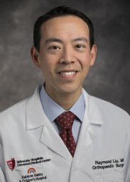 Raymond W. Liu合作论文数1
 Pingfu Fu合作论文数Department of Population and Quantitative Health Sciences, School of Medicine, Case Western Reserve University;Case Comprehensive Cancer Center, Case Western Reserve University1




